RE S EAR CH | R E P O R T S
24. A. Atkinson, V. Siegel, E. Pakhomov, P. Rothery, Nature 432,
100–103 (2004).
25. A. Atkinson et al., Mar. Ecol. Prog. Ser. 362, 1–23 (2008).
26. L. J. Grange, C. R. Smith, PLOS ONE 8, e77917 (2013).
27. E. Rignot, J. Mouginot, M. Morlighem, H. Seroussi, B. Scheuchl,
Geophys. Res. Lett. 41, 3502–3509 (2014).
28. A. H. Orsi, T. Whitworth III, W. D. Nowlin Jr., Deep Sea Res. I 42,
641–673 (1995).
13. S. Schmidtko, G. C. Johnson, J. M. Lyman, J. Geophys. Res.
Oceans 118, 1658–1672 (2013).
14. S. G. Purkey, G. C. Johnson, J. Clim. 26, 6105–6122 (2013).
15. P. R. Holland, R. Kwok, Nat. Geosci. 5, 872–875 (2012).
16. R. Bintanja, G. J. van Oldenborgh, S. S. Drijfhout, B. Wouters,
C. A. Katsman, Nat. Geosci. 6, 376–379 (2013).
17. A. L. Stewart, A. F. Thompson, Geophys. Res. Lett. 39, L18604
(2012).
18. P. Dutrieux et al., Science 343, 174–178 (2014).
19. H. H. Hellmer, F. Kauker, R. Timmermann, J. Determann,
J. Rae, Nature 485, 225–228 (2012).
20. M. S. Dinniman, J. M. Klinck, E. E. Hofmann, J. Clim. 25,
4799–4816 (2012).
21. C. Heuzé, K. J. Heywood, D. P. Stevens, J. K. Ridley, Geophys.
Res. Lett. 40, 1409–1414 (2013).
22. S. R. Rintoul, Geophys. Res. Lett. 34, L06606 (2007).
23. W. Zenk, E. Morozov, Geophys. Res. Lett. 34, L14607 (2007).
Federal Ministry for Education and Research (BMBF) project
MiKlip (S.S.); NERC Antarctic Funding Initiative research
grant GENTOO NE/H01439X/1 (S.S. and K.J.H.); a Grant-in-Aid
for Scientific Research of the Ministry of Education, Culture,
Sports, Science and Technology, Japan, and a Daiwa Foundation
Small Grant (S.A.); and NSF award OPP-1246460 (A.F.T.).
SUPPLEMENTARY MATERIALS
All data used in this manuscript are publicly available; data
sources are detailed in the supplementary materials. We thank
all officers, crews, and scientists involved in collecting and
calibrating the data in the often harsh Southern Ocean
environment, as well as everyone involved in making the data
publicly available. We thank three anonymous reviewers for
helpful discussions and feedback. Supported by the German
ANIMAL PHYSIOLOGY
The shocking predatory strike
of the electric eel
Kenneth Catania
Electric eels can incapacitate prey with an electric discharge, but the mechanism of the
eel’s attack is unknown. Through a series of experiments, I show that eel high-voltage
discharges can activate prey motor neurons, and hence muscles, allowing eels to remotely
control their target. Eels prevent escape in free-swimming prey using high-frequency
volleys to induce immobilizing whole-body muscle contraction (tetanus). Further, when
prey are hidden, eels can emit periodic volleys of two or three discharges that cause massive
involuntary twitch, revealing the prey’s location and eliciting the full, tetanus-inducing volley.
The temporal patterns of eel electrical discharges resemble motor neuron activity that
induces fast muscle contraction, suggesting that eel high-voltage volleys have been
selected to most efficiently induce involuntary muscle contraction in nearby animals.
T
he electric eel (Electrophorus electricus) is
one of just a few species that uses electrical discharges to capture prey and defend
against predators. It is the most powerful
electrogenic fish, with most of its body composed of electrocytes (muscle-derived biological
batteries), providing a combined discharge of up
to 600 V (1). Early attempts to understand electricity made use of electric eels (2), and more
recently, eels were important for identifying acetylcholine receptors (3) and for providing insights
into the evolution of electric organs (4), but little
is known about how the eel’s electrical discharge
affects prey. In this study, I designed a set of expe-
www.sciencemag.org/content/346/6214/1227/suppl/DC1
Materials and Methods
Figs. S1 to S11
Table S1
References (29, 30)
15 May 2014; accepted 27 October 2014
10.1126/science.1256117
riments to explore the impacts of the electric eel
discharges on potential prey and the mechanism
that operates during such attacks.
Electric eels emit three distinct types of electric
organ discharges: (i) low-voltage pulses for sensing
their environment, (ii) pairs and triplets of highvoltage pulses given off periodically while hunting
in complex environments, and (iii) high-frequency
volleys of high-voltage pulses during prey capture
or defense (movie S1) (5–9). Under most conditions, eels attack free-swimming prey with the latter strategy, using high-voltage volleys combined
with a suction-feeding strike. To explore this more
common behavior, I simultaneously recorded eel
behavior and electric organ discharges in a naturalistic experimental environment (10). Eels began
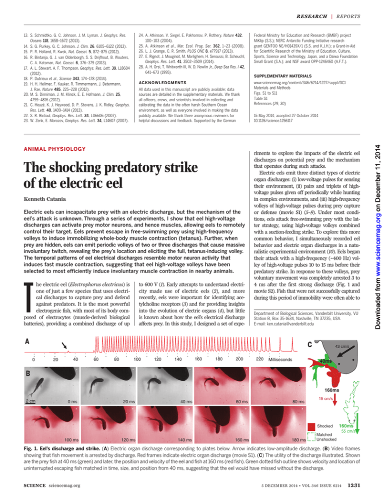
their attack with a high-frequency (~400 Hz) volley of high-voltage pulses 10 to 15 ms before their
predatory strike. In response to these volleys, prey
voluntary movement was completely arrested 3 to
4 ms after the first strong discharge (Fig. 1 and
movie S2). Fish that were not successfully captured
during this period of immobility were often able to
Department of Biological Sciences, Vanderbilt University, VU
Station B, Box 35-1634, Nashville, TN 37235, USA.
E-mail: ken.catania@vanderbilt.edu
43 cm/s
0
20
40
60
80
100
120
140
160
180
200
220
Milliseconds
40ms
160ms
2 cm
0 ms
20 ms
40 ms
60 ms
80 ms
15 cm/s
Shocked
100 ms
120 ms
140 ms
160 ms
180 ms
160ms
Matched
Unshocked
55 cm/s
Fig. 1. Eel’s discharge and strike. (A) Electric organ discharge corresponding to plates below. Arrow indicates low-amplitude discharge. (B) Video frames
showing that fish movement is arrested by discharge. Red frames indicate electric organ discharge (movie S1). (C) The utility of the discharge illustrated. Shown
are the prey fish at 40 ms (green) and later, the position and velocity of the eel and fish at 160 ms (red fish). Green dotted fish outline shows velocity and location of
uninterrupted escaping fish matched in time, size, and position from 40 ms, suggesting that the eel would have missed without the discharge.
SCIENCE sciencemag.org
5 DECEMBER 2014 • VOL 346 ISSUE 6214
1231
Downloaded from www.sciencemag.org on December 11, 2014
ACKN OWLED GMEN TS
R ES E A RC H | R E PO R TS
return to previous movement patterns and escape
(movie S2).
To characterize the mechanism by which highvoltage volleys cause this remote immobilization
of prey (10), anesthetized fish were pithed (to destroy the brain), the hole was sealed with cyanoacrylate, and the fish was attached to a force
transducer. An eel in the aquarium was separated from the fish by an electrically permeable
Force
Transducer
Agar Barrier
agar barrier (Fig. 2A) (11) and fed earthworms,
which it attacked with volleys of its high-voltage
discharge. The discharge directed at the earthworms induced strong muscular contractions in
the fish preparation, precisely correlated in time
with the volley (no tension developed during the
weak discharge). A steep rise in fish tension occurred with a mean latency of 3.4 ms (n = 20
trials) after the first strong pulse (Fig. 2B), which
Force
Transducer
is similar to the 2.9-ms mean immobilization
latency (n = 20 trials) observed in free-swimming
fish. Tension induced by the eel in the fish preparation was similar to the maximum that could
be induced experimentally (fig. S1) (10). This result
indicates that fish are immobilized by massive, involuntary muscle contraction.
To further investigate the fidelity of prey muscle contractions relative to the electric organ
20 ms
3.7 ms
2
3 45 6 7 8 9
1
Fish
Tension
Eel
EOD
Pithed
Fish
1
Eel
EOD
10 ms
23 4 5 6 7 8 9
100 ms
Fish Tension
Time 2:40
1
3:14
6:29
9:16
12:04
14:55
17:58
21:03
23:10
EOD
253g
Fish 1
Tension
SHAM
429g
2
CURARE
Fish 2
Tension
Fig. 2. Paradigm for investigating strong electric organ discharge. (A) An agar barrier separated eels from pithed fish. Eels shocked earthworms
while fish tension was recorded. (B) All eels induced whole-body tension, occurring 2 to 4 ms after strong discharge onset. No tension was developed
from weak discharge. At low frequencies, individual twitches emerged for each discharge (top right) (fig. S2). (C) Two pithed fish (fish 1, 19 g; fish 2, 21 g)
preparation. (D) Effect of curare. Red trace indicates strong electric organ discharge matched in time to unnormalized fish tension (green). Arrows
indicate time of injections (fig. S3). Bar in (D) = 500 ms.
Voltage Tension
90g
200 ms
Doublet
Expanded Doublet
5 ms
Weak EOD
Fish
Twitch
Doublet
Attack
Volley
Agar Barrier
Prey Movement
1
2 3
4
5
6 7
8
1
2
Prey Movement
1
2
3 4
1
5 6
3
7
9
8
2
3
9
10
4
5
6
7
8
9
25 ms
10
4
5
6
7
8
9
10
10
Fig. 3. Doublets during hunting. (A) Examples of doublets and corresponding tension responses. (B) Expansion of the first doublet and corresponding
tension trace (off-scale peaks were estimated). (C) Schematic of attack sequence. (D) Example of high-voltage electric organ discharge for an attack
preceded by a doublet. (E) Video frames from volley shown in (D). Numbers correspond to numbers in (D). (F) Timing of the high-voltage discharge for
attack preceded by a triplet. (G) Video frames for volley shown in (F).
1232
5 DECEMBER 2014 • VOL 346 ISSUE 6214
sciencemag.org SCIENCE
RE S EAR CH | R E P O R T S
Carbon Electrode
Agar Barrier
EOD Recording
PowerLab 8/35
Stimulator
Plastic Bag
Conditions “B” through “G” Below Agar Barrier
Eel
Stimulator
Off
Stimulator
(Movie S6 Clip 6)
Plastic Bag
Eel Attack
Eel
Stimulator
Triggered
By Doublet
Stimulator
Plastic Bag
Fish Twitch
(Movie S6 Clip 1-2)
Eel Attack
Stimulator
Triggered
By
Experimenter
Eel
Stimulator
Plastic Bag
Fish Twitch
(Movie S6 Clip 3-4)
Eel
Stimulator
Triggered
By Doublet
Stimulator
Plastic Bag
Stimulator
Triggered
By Doublet
Freeze-Thawed
Fish
Eel
Stimulator
(Movie S6 Clip 5)
Plastic Bag
Eel
Freeze-Thawed
Fish
No Plastic Bag
Below Plexiglas
Stimulator
Triggered
By
Experimenter
Eel
Stimulator
Fish Twitch
Plexiglas
(Movie S6 Clip 7)
Fig. 4. Paradigm and controls showing eels attack doublet-generated movements. (A) Movement
in electrically isolated pithed fish (below agar) was generated through stimulator. (B) Without fish twitch,
eels did not follow doublets with attack (10 trials each for two eels). (C) When stimulator triggered fish
twitch after doublets, eels attacked (10 trials each for two eels). (D) Without doublets, fish twitches also
elicited attack volleys (10 trials each of two eels). (E) Doublets that triggered stimulator leads in bag did
not elicit attack (10 trials for each of two eels). (F) Likewise, no attack volleys were elicited after stimulation
of a freeze-thawed fish (10 trials each of two eels). (G) Doublets directed at a freeze-thawed fish under
agar without the plastic bag or stimulator did not elicit eel attack volleys or strikes (10 trials each of two
eels). These latter conditions, along with (H) trials with Plexiglas barrier, show that visual cues did not
generate eel attacks. Examples are provided in movie S6 and (10).
SCIENCE sciencemag.org
discharge, and the mechanism of the contractions’
induction, two pithed-fish preparations were stationed side by side (Fig. 2C). The high-voltage
discharge reliably created muscle tension with
similar form and time course in both fish (fig. S2).
As the discharge frequency decreased, individual
fish twitches often emerged on the tension trace,
each corresponding to a single discharge (Fig. 2B
and fig. S2). To determine whether the discharge
induced muscle contractions by initiating action
potentials directly in prey muscles or through activation of some portion of fish motor neurons, one
of two similarly sized fish was injected with curare
(an acetylcholine antagonist) so as to block the
acetylcholine gated ion channels at the neuromuscular junction, whereas the other fish was
sham-injected (Fig. 2D). In each of four cases,
tension responses in the curarized fish dropped to
near zero, whereas the sham-injected fish continued to respond (fig. S3). These findings indicate
that fish motor neuron activation is required to
induce tetanus in prey. To determine whether this
activation of prey motor neurons was the result of
central nervous system (spinal) activity or activity in
efferent branches of motor neurons, the dual tension experiment was repeated twice with extensively double-pithed fish (in which both the brain
and spinal cord were destroyed, but the branches
of motor efferents were left intact within the fish
body) and compared with a brain-pithed fish. No
diminution in contractile response, or difference
in contractile response latency, was observed for
the double-pithed fish relative to the brain-pithed
fish (fig. S2). These experiments suggest that the
electric eel’s strong electric organ discharge remotely activates motor neuron efferents of its
prey, although this activation could occur anywhere between the spinal cord and the presynaptic side of the neuromuscular junction. Given
that the eel’s strong electric organ discharge
remotely activates prey motor neurons, it was
useful to consider the form of this pulse train in
the context of prey muscle activation. Analysis of
the first 11 impulses from strong discharge volleys from each of four eels showed that each begins with a doublet—two pulses with a shorter
interpulse interval (fig. S4). Doublets at the onset of
motor neuron trains have been shown to induce
high rates of muscle tension (12–15). Moreover, the
overall distribution of pulses in the eel’s strong
discharge resembles motor neuron trains found
to be near optimal for muscle tension development (16, 17). These observations raise the possibility that eel volleys have been selected to
efficiently induce rapid muscle tension.
As described above, hunting eels often pause
and give off isolated high-voltage doublets (9),
particularly in complex environments, when seeking hidden prey or when exploring conductors
(movie S3). In the course of the present study,
eels stationed behind the agar barrier in the fish
tension experiments occasionally emitted such
isolated doublets or triplets and then attempted
to break through the barrier to reach the fish preparation (movie S4). This suggested that eels were
able to detect fish movements through the thin
agar barrier, which was not designed to mask
5 DECEMBER 2014 • VOL 346 ISSUE 6214
1233
R ES E A RC H | R E PO R TS
mechanosensory cues. To identify the function
of this additional behavior, eels were presented
with prey hidden below a thin agar barrier (Fig.
3C). In some cases, eels detected prey through
the barrier and attacked directly, but in other
cases, the eel investigated the agar surface with a
low-amplitude electric organ discharge and then
produced a high-voltage doublet. The doublet invariably caused prey movement. Stimulated prey
movement was closely followed (in 20 to 40 ms)
by a full predatory strike consisting of a strong
electric discharge volley and directed attack
(Fig. 3 and movie S5), as characterized in the first
experiments. The distinct form of the discharge
trace in these trials consisted of a doublet (or
triplet) followed by a 20- to 40-ms pause (during
which prey moved) and then a full discharge
volley (Fig. 3, D and F).
The results of the doublet experiment suggest
that the eels may use doublet and triplet discharges to detect cryptic prey by inducing movement. To test this hypothesis, a pithed fish was
placed in a thin plastic bag to isolate it from the
eel’s discharge. The electrically isolated fish was
positioned below an agar barrier, with electrical
leads embedded in the head and tail region (10)
that allowed production of artificial fish twitch by
the experimenter. Artificial fish twitch was triggered remotely through a stimulator (Fig. 4A), allowing control over its timing and occurrence.
When the stimulating electrodes were inactive, eel
doublets caused no response in the pithed fish
and eels did not attack the preparation (Fig. 4B
and movie S6). However, when the stimulator was
configured to trigger fish twitch when the eel produced a doublet, the eel’s full “doublet attack” behavior was replicated (Fig. 4C and movie S6). The
attack pattern consisted of a doublet, followed by
a short pause, during which the prey moved (resulting from the triggered stimulator), followed by
a high-voltage volley and strike. This key experiment showed that eels never (10 of 10 trials for
each of two eels) followed a doublet with an attack
volley without a “mechanosensory echo” from the
prey, but attacked in response to the stimulatorgenerated fish twitch (10 of 10 trials for each of
two eels; P < 0.0001, binomial test). Experimentertriggered twitches, in the absence of eel hunting
doublets, also generated attacks (movie S6) with
the time course observed above (Fig. 4D and supplementary materials). Thus, prey movement,
whether doublet-generated or independently generated, elicited short latency (20 to 40 ms) attacks. Eels also appeared to use either active or
passive electrolocation to detect live prey under
agar and often attacked without a preceding doublet. But in no case did an attack volley follow a
doublet in the absence of prey response. Thus, the
doublet appears to answer the question, “Are you
living prey?” when information is limited. Preliminary observations suggest that “doublet hunting”
is most common in complex environments (movie
S7). A range of controls confirmed that eels were
responding to twitch-generated mechanosensory
cues in this paradigm (Fig. 4 and movie S6).
Together, the results of these experiments show
that high-voltage discharges of electric eels re1234
5 DECEMBER 2014 • VOL 346 ISSUE 6214
motely activate motor neuron efferents in nearby
animals. Prey that have been detected can be immobilized and captured. Hidden prey can be induced to twitch, revealing their location. The
latter strategy, which often triggers an escape response, depends on the eel’s short reaction time.
An eel can discharge its high-voltage train 20 ms
after a mechanosensory stimulus, allowing it to
cancel the very escape response it has generated.
Overall, this study reveals that the electric eel has
evolved a precise remote control mechanism for
prey capture, one that takes advantage of an organisms’ own nervous system.
RE FERENCES AND NOTES
1. H. Grundfest, Prog. Biophys. Biop. Chem. 1957, 1–85 (1956).
2. S. Finger, M. Piccolino, The Shocking History of Electric Fishes:
From Ancient Epochs to the Birth of Modern Neurophysiology
(Oxford Univ. Press, Oxford, 2011), p. 5.
3. J. Keesey, J. Hist. Neurosci. 14, 149–164 (2005).
4. J. R. Gallant et al., Science 344, 1522–1525 (2014).
5. R. Bauer, Behav. Ecol. Sociobiol. 4, 311–319 (1979).
6. C. W. Coates, R. T. Cox, W. A. Rosemblith, M. B. Brown,
Zoologica 25, 249 (1940).
7. S. Hagiwara, T. Szabo, P. S. Enger, J. Neurophysiol. 28,
775–783 (1965).
8. T. H. Bullock, Brain Behav. Evol. 2, 85–101 (1969).
9. G. M. Westby, Behav. Ecol. Sociobiol. 22, 341–354 (1988).
10. Materials and methods are available as supplementary
materials on Science Online.
11. A. J. Kalmijn, J. Exp. Biol. 55, 371–383 (1971).
12. R. Hennig, T. Lømo, Nature 314, 164–166 (1985).
13. K. K. Pedersen, O. B. Nielsen, K. Overgaard, Physiol. Rep.
1, e00026 (2013).
14. A. J. Cheng, N. Place, J. D. Bruton, H. C. Holmberg,
H. Westerblad, J. Physiol. 591, 3739–3748 (2013).
15. J. Celichowski, K. Grottel, Acta Neurobiol. Exp. (Warsz.) 58,
47–53 (1998).
16. F. E. Zajac, J. L. Young, J. Neurophysiol. 43, 1206–1220 (1980).
17. F. E. Zajac, J. L. Young, J. Neurophysiol. 43, 1221–1235 (1980).
AC KNOWLED GME NTS
I thank E. Catania for suggesting experimental designs and
manuscript comments. This work was supported by a Pradel
Award from the National Academy of Sciences, a Guggenheim
Fellowship, and NSF grant 0844743. Raw data are available in
the supplementary materials.
SUPPLEMENTARY MATERIALS
www.sciencemag.org/content/346/6214/1231/suppl/DC1
Materials and Methods
Supplementary Text
Figs. S1 to S4
Movies S1 to S7
4 September 2014; accepted 13 November 2014
10.1126/science.1260807
INFLAMMATION
Neutrophils scan for activated
platelets to initiate inflammation
Vinatha Sreeramkumar,1 José M. Adrover,1 Ivan Ballesteros,2 Maria Isabel Cuartero,2
Jan Rossaint,3 Izaskun Bilbao,1,4 Maria Nácher,1,5 Christophe Pitaval,1 Irena Radovanovic,1
Yoshinori Fukui,6 Rodger P. McEver,7 Marie-Dominique Filippi,8 Ignacio Lizasoain,2
Jesús Ruiz-Cabello,1,4 Alexander Zarbock,3 María A. Moro,2 Andrés Hidalgo1,9*
Immune and inflammatory responses require leukocytes to migrate within and through
the vasculature, a process that is facilitated by their capacity to switch to a polarized
morphology with an asymmetric distribution of receptors. We report that neutrophil
polarization within activated venules served to organize a protruding domain that engaged
activated platelets present in the bloodstream. The selectin ligand PSGL-1 transduced signals
emanating from these interactions, resulting in the redistribution of receptors that drive
neutrophil migration. Consequently, neutrophils unable to polarize or to transduce signals
through PSGL-1 displayed aberrant crawling, and blockade of this domain protected mice
against thromboinflammatory injury. These results reveal that recruited neutrophils scan for
activated platelets, and they suggest that the neutrophils’ bipolarity allows the integration of
signals present at both the endothelium and the circulation before inflammation proceeds.
N
eutrophils are primary effectors of the immune response against invading pathogens
but are also central mediators of inflammatory injury (1). Both functions rely on
their remarkable ability to migrate within and through blood vessels. The migration of
neutrophils is initiated by tethering and rolling
on inflamed venules, a process mediated by endothelial selectins (2). Selectin- and chemokinetriggered activation of integrins then allows firm
adhesion, after which leukocytes actively crawl
on the endothelium before they extravasate or
return to the circulation (3). A distinct feature
of leukocytes recruited to inflamed vessels is the
rapid shift from a symmetric morphology into
a polarized form, in which intracellular proteins and receptors rapidly segregate (4). In this
way, neutrophils generate a moving front or
leading edge where the constant formation of
lamellipodia (actin projections) guides movement, and a uropod or trailing edge where highly glycosylated receptors accumulate (5, 6). We
deemed it unlikely that this dramatic reorganization served to exclusively generate a frontto-back axis for directional movement, and we
explored the possibility that neutrophil polarization functions as an additional checkpoint
during inflammation.
sciencemag.org SCIENCE
The shocking predatory strike of the electric eel
Kenneth Catania
Science 346, 1231 (2014);
DOI: 10.1126/science.1260807
If you wish to distribute this article to others, you can order high-quality copies for your
colleagues, clients, or customers by clicking here.
Permission to republish or repurpose articles or portions of articles can be obtained by
following the guidelines here.
The following resources related to this article are available online at
www.sciencemag.org (this information is current as of December 11, 2014 ):
Updated information and services, including high-resolution figures, can be found in the online
version of this article at:
http://www.sciencemag.org/content/346/6214/1231.full.html
Supporting Online Material can be found at:
http://www.sciencemag.org/content/suppl/2014/12/03/346.6214.1231.DC1.html
A list of selected additional articles on the Science Web sites related to this article can be
found at:
http://www.sciencemag.org/content/346/6214/1231.full.html#related
This article cites 15 articles, 7 of which can be accessed free:
http://www.sciencemag.org/content/346/6214/1231.full.html#ref-list-1
This article appears in the following subject collections:
Physiology
http://www.sciencemag.org/cgi/collection/physiology
Science (print ISSN 0036-8075; online ISSN 1095-9203) is published weekly, except the last week in December, by the
American Association for the Advancement of Science, 1200 New York Avenue NW, Washington, DC 20005. Copyright
2014 by the American Association for the Advancement of Science; all rights reserved. The title Science is a
registered trademark of AAAS.
Downloaded from www.sciencemag.org on December 11, 2014
This copy is for your personal, non-commercial use only.


