Current Biology, Vol. 13, R482–R484, June 17, 2003, ©2003 Elsevier Science Ltd. All rights reserved. DOI 10.1016/S0960-9822(03)00408-1
RNA Repair: Damage Control
Alfonso Bellacosa and Eric G. Moss
RNA in a cell is subject to many of the same insults
as DNA. RNA damage can induce apoptosis and
may be exploited for anti-cancer chemotherapy. It is
a surprise, however, to learn that cells may repair
RNA damage, suggesting a far greater significance
of RNA in genotoxic stress.
Damage to DNA is a significant issue for all cells,
particularly in cancer where DNA repair commonly
fails. It is not widely appreciated that many agents that
cause damage to DNA, such as radiation and certain
cancer chemotherapy drugs, also damage RNA. Given
that there is at least as much RNA in a cell as DNA,
wherever DNA is damaged by such agents, RNA is
surely damaged as well. When the damage to RNA is
substantial, apoptosis is induced, which is the desired
affect of anti-cancer chemotherapy [1]. Nevertheless,
RNA has not been the major focus in investigating
how cells cope with such insults (until very recently,
‘RNA damage’ and ‘RNA repair’ did not crop up in the
PubMed index). There have been scattered insights
over the years into the significance of RNA to
genotoxic stress, but a recent report by Aas and
colleagues [2] suggests that the cell has at least one
specific mechanism to repair RNA damage, indicating
a greater investment in the protection of RNA than
previously suspected.
AlkB Reverses Alkylation Damage
Alkylating agents are endogenous and environmental
compounds that cause mutations, tumors and
neurotoxicity. Chemically, they add alkyl groups, like
methyl or ethyl groups, to organic macromolecules, in
particular to the ring nitrogens and oxygens of bases of
nucleic acids. Cells have developed a host of repair
systems to deal with alkylation damage of DNA. These
include the removal of the damaged residues by DNA
glycosylases, followed by replacement of the nucleotide
by DNA polymerases using the opposite strand as template. Another mechanism is the direct reversal of the
methylation damage, which does not require a template
to make the repair. For example, the Ada and Ogt
enzymes of Escherichia coli restore the normal base of
an alkylated DNA by directly removing the offending
chemical group in a suicidal reaction. The E. coli
enzyme AlkB has long been known to act in alkylation
damage repair, but until recently its mechanism of
action was not clear, as biochemical assays failed to
detect in the AlkB protein any of the enzymatic activities
known to occur in DNA repair enzymes.
In a remarkable display of the power of bioinformatics applied to genome sequences, Aravind and Koonin
[3] predicted, based on its relationship to another
Fox Chase Cancer Center, Philadelphia, Pennsylvania 19111,
USA. E-mail: a_bellacosa@fccc.edu, eg_moss@fccc.edu
Dispatch
family of enzymes, that AlkB would cause hydroxylation of the methyl group on damaged DNA bases, and
thus directly reverse alkylation damage. The Seeberg
[4] and Sedgwick [5] labs independently confirmed
this prediction and showed that, indeed, AlkB
enzymatically demethylates the DNA bases adenine
and cytosine, unlike the suicidal Ada and Ogt proteins,
by oxidative demethylation: the methyl group is
converted to an hydroxymethyl group which then
leaves the base as formaldehyde.
Now there has been another twist to the plot. From
the large number of AlkB homologues in plants and
their RNA viruses, Aravind and Koonin [3] predicted
that some members of this family might act on RNA
bases modified by alkylating agents. Indeed, in their
recent article, Krokan and colleagues [2] describe two
human AlkB homologues, hABH2 and hABH3, and
show that AlkB and hABH3 have RNA repair activity.
Recombinant AlkB and hABH3 proteins demethylate
methylated bases in RNA in vitro. Furthermore, expression of AlkB and hABH3 in an alkB mutant strain of E.
coli reactivated a methylated RNA virus, providing
functional proof of the relevance of RNA repair activity.
While AlkB and hABH3 prefer single-strand DNA,
hABH2 prefers double-strand DNA substrates and
lacks significant RNA repair activity. It remains to be
seen, however, whether hABH2 has activity on doublestrand RNA.
Functional specialization is also reflected in the
different subcellular localization of the two human
enzymes [2,6]. hABH2 localizes to the nucleoplasm
with some accumulation in nucleoli, indicating a
possible involvement in the repair of ribosomal RNA,
but in S phase it colocalizes with replication foci.
hABH3 has a predominantly nucleoplasmic localization, with nucleolar exclusion and some cytoplasmic
staining. This differential compartmentalization raises
the intriguing question of what are the physiological
targets of hABH2 and hABH3 in eukaryotic cells. This
may be important, as some of the modifications
induced by alkylating agents are within the spectrum
of normal physiological RNA modifications [2].
Coping with RNA Damage
Cells may have mechanisms of dealing with RNA
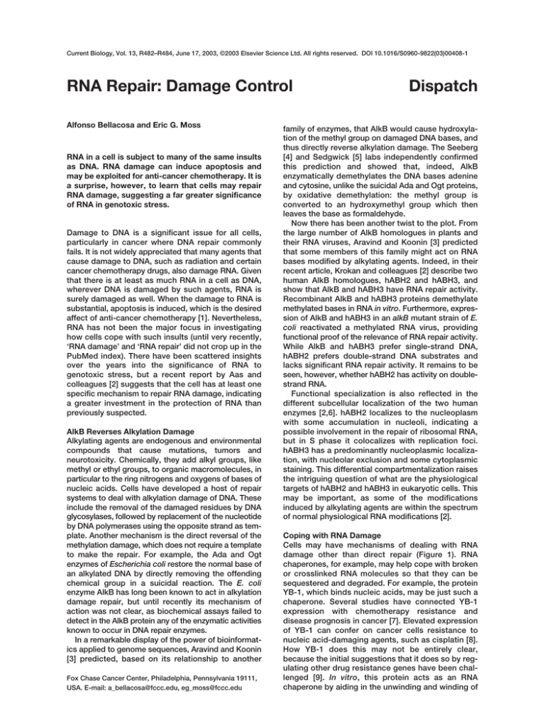
damage other than direct repair (Figure 1). RNA
chaperones, for example, may help cope with broken
or crosslinked RNA molecules so that they can be
sequestered and degraded. For example, the protein
YB-1, which binds nucleic acids, may be just such a
chaperone. Several studies have connected YB-1
expression with chemotherapy resistance and
disease prognosis in cancer [7]. Elevated expression
of YB-1 can confer on cancer cells resistance to
nucleic acid-damaging agents, such as cisplatin [8].
How YB-1 does this may not be entirely clear,
because the initial suggestions that it does so by regulating other drug resistance genes have been challenged [9]. In vitro, this protein acts as an RNA
chaperone by aiding in the unwinding and winding of
Current Biology
R483
RNA duplexes [10]. Perhaps YB-1 is part of a general
RNA handling machinery that helps move the molecules from one complex to another.
Another gene with a potential role in RNA damage
control is LSM1 of budding yeast. Deletion of LSM1
causes resistance to ultraviolet radiation [11]. LSM1
encodes a protein involved in mRNA decapping and
protection against trimming of 3′′ untranslated regions
[12], suggesting that regulation of mRNA stability may
affect the response to exogenous RNA and DNA
damage. Interestingly, the human LSM1 orthologue is
associated with the invasive potential of prostate
cancer and perhaps other neoplasms [13].
Additional isolated suggestions that other general
RNA binding proteins contribute to a cell’s ability to
deal with nucleic acid damaging agents have appeared
[14]. There are many RNA binding proteins encoded in
the genome with no known function. Perhaps one
question to be asked of these is whether they play
some caretaking role for the cell’s RNA.
Anti-Cancer Chemotherapy and RNA Damage
It is well established that RNA damage caused by anticancer chemotherapy agents is an important component of their action. For example, the incorporation of
5-fluorouracil (5-FU) into newly synthesized RNA
appears to be the primary determinant of its cytotoxicity [15]. Cisplatin, which forms adducts on both DNA
and RNA, inhibits translation in cancer cells by
crosslinking of mRNA to ribosomal RNA or ribosomal
RNA to itself [16]. Adriamycin (doxorubicin), an anthracycline drug which intercalates between base pairs of
double-helical nucleic acids, has been shown to bind
RNA helices and inhibit RNA helicase, an activity
essential for RNA synthesis, processing, transport and
turnover [17].
Considering that there is generally more RNA in a
cell than DNA, most of it in the form of ribosomes that
turn over at relatively low rate, it is likely that there will
be significant damage to cellular RNA when cells are
treated with these drugs. Furthermore, the ‘information content’ of cellular RNA is greater than that of the
chromosomal DNA: whereas only 3% of the human
DNA has coding potential, almost all RNA sequences
in the cytoplasm have functional significance, whether
coding for proteins (mRNA) or performing translation
(ribosomal RNA and tRNA). Therefore an agent that
can affect RNA or DNA equally well from a chemical
perspective is more likely to cause significant RNA
damage in a living cell.
Importantly, a new class of anticancer agents that
specifically damage RNA has been shown to be
effective and is currently undergoing clinical trials.
This cytotoxic ribonuclease — called Onconase —
cleaves tRNA, and in so doing, induces apoptosis via
a p53-independent mechanism [1]. Drugs that interfere with the synthesis of RNA and DNA precursors —
for example hydroxyurea and methotrexate — or that
block RNA polymerase — for example actinomycin D
and α-amanitin — are also known to induce p53 and
cause cell cycle arrest [18]. These examples show that
RNA damage can lead to cell-cycle arrest and cell
death, much as DNA damage does. The precise
Proteins guarding the RNome
RNA chaperones
YB-1
RNA processing factors
RNA repair enzymes
LSM1
hABH
RNA quality control factors
UPF
Current Biology
Figure 1. Proteins at all stages of an RNA’s life may passively
or actively protect it from damage.
Damaged RNA may simply interfere with a cell’s normal activities, and/or it may induce checkpoints leading to apoptosis, as
DNA damage does.
mechanisms by which apoptosis is induced by RNA
damage deserves further study.
RNome Stability: A Link to Cancer?
Alterations of genome stability have an important
role in tumorigenesis, particularly in the inherited
predisposition to cancer. Typically, DNA repair genes
mutated in sporadic and hereditary tumors have a
double function: they effect DNA repair and also
regulate cell cycle and apoptosis checkpoints.
Inactivation of these genes increases the mutation rate
and at the same time prevents engagement of checkpoints, resulting in a growth advantage. Is it possible
that an ability of RNA damage to induce similar checkpoints means that defects in RNome stability might
also play a role in tumorigenesis? One suggestion that
this is so comes from the study of inherited predisposition to cancer. For example, the hereditary prostate
cancer gene HPC2/ELAC2 encodes a protein that
shows homology to the mRNA cleavage and
polyadenylation specificity factor CPSF73 [19], indicating a potential link between mRNA biogenesis and
cancer predisposition.
A cell has a great investment in its RNAs — they are
working copies of genomic information. The study of
mRNA biogenesis in the last few years has revealed
an elaborate surveillance mechanism involving factors
such as the UPF proteins that culls aberrantly spliced
mRNAs and mRNAs with premature termination
codons. There might be a hint that such RNA quality
control mechanisms go awry in cancers, just as DNA
quality control mechanisms do, where aberrantly
spliced transcripts accumulate in a tumor [20]. Now
that the gates are open, we may have a flood of
studies on RNome stability and cancer.
References
1. Iordanov, M.S., Ryabinina, O.P., Wong, J., Dinh, T.H., Newton, D.L.,
Rybak, S.M. and Magun, B.E. (2000). Molecular determinants of
apoptosis induced by the cytotoxic ribonuclease onconase: evidence for cytotoxic mechanisms different from inhibition of protein
synthesis. Cancer Res. 60, 1983–1994.
Dispatch
R484
2. Aas, P.A., Otterlei, M., Falnes, P.O., Vagbo, C.B., Skorpen, F.,
Akbari, M., Sundheim, O., Bjoras, M., Slupphaug, G., Seeberg, E.
and Krokan, H.E. (2003). Human and bacterial oxidative demethylases repair alkylation damage in both RNA and DNA. Nature 421,
859–863.
3. Aravind, L. and Koonin, E.V. (2001). The DNA-repair protein AlkB,
EGL-9, and leprecan define new families of 2-oxoglutarate- and
iron-dependent
dioxygenases.
Genome
Biol
2,
research0007.1–0007.8.
4. Falnes, P.O., Johansen, R.F. and Seeberg, E. (2002). AlkB-mediated
oxidative demethylation reverses DNA damage in Escherichia coli.
Nature 419, 178–182.
5. Trewick, S.C., Henshaw, T.F., Hausinger, R.P., Lindahl, T. and Sedgwick, B. (2002). Oxidative demethylation by Escherichia coli AlkB
directly reverts DNA base damage. Nature 419, 174–178.
6. Duncan, T., Trewick, S.C., Koivisto, P., Bates, P.A., Lindahl, T. and
Sedgwick, B. (2002). Reversal of DNA alkylation damage by two
human dioxygenases. Proc. Natl. Acad. Sci. U.S.A. 99,
16660–16665.
7. Janz, M., Harbeck, N., Dettmar, P., Berger, U., Schmidt, A., Jurchott, K., Schmitt, M. and Royer, H.D. (2002). Y-box factor YB-1
predicts drug resistance and patient outcome in breast cancer
independent of clinically relevant tumor biologic factors HER2, uPA
and PAI-1. Int. J. Cancer 97, 278–282.
8. Ohga, T., Koike, K., Ono, M., Makino, Y., Itagaki, Y., Tanimoto, M.,
Kuwano, M. and Kohno, K. (1996). Role of the human Y box-binding
protein YB-1 in cellular sensitivity to the DNA-damaging agents cisplatin, mitomycin C, and ultraviolet light. Cancer Res. 56,
4224–4228.
9. Hu, Z., Jin, S. and Scotto, K.W. (2000). Transcriptional activation of
the MDR1 gene by UV irradiation. Role of NF-Y and Sp1. J. Biol.
Chem. 275, 2979–2985.
10. Skabkin, M.A., Evdokimova, V., Thomas, A.A. and Ovchinnikov, L.P.
(2001). The major messenger ribonucleoprotein particle protein p50
(YB-1) promotes nucleic acid strand annealing. J. Biol. Chem. 276,
44841–44847.
11. Birrell, G.W., Giaever, G., Chu, A.M., Davis, R.W. and Brown, J.M.
(2001). A genome-wide screen in Saccharomyces cerevisiae for
genes affecting UV radiation sensitivity. Proc. Natl. Acad. Sci. U.S.A.
98, 12608–12613.
12. He, W. and Parker, R. (2001). The yeast cytoplasmic LsmI/Pat1p
complex protects mRNA 3’ termini from partial degradation. Genetics 158, 1445–1455.
13. Takahashi, S., Suzuki, S., Inaguma, S., Cho, Y.M., Ikeda, Y.,
Hayashi, N., Inoue, T., Sugimura, Y., Nishiyama, N., Fujita, T., et al.
(2002). Down-regulation of Lsm1 is involved in human prostate
cancer progression. Br. J. Cancer 86, 940–946.
14. Yang, C. and Carrier, F. (2001). The UV-inducible RNA-binding
protein A18 (A18 hnRNP) plays a protective role in the genotoxic
stress response. J. Biol. Chem. 276, 47277–47284.
15. Glazer, R.I. and Lloyd, L.S. (1982). Association of cell lethality with
incorporation of 5-fluorouracil and 5-fluorouridine into nuclear RNA
in human colon carcinoma cells in culture. Mol. Pharmacol. 21,
468–473.
16. Heminger, K.A., Hartson, S.D., Rogers, J. and Matts, R.L. (1997).
Cisplatin inhibits protein synthesis in rabbit reticulocyte lysate by
causing an arrest in elongation. Arch. Biochem. Biophys. 344,
200–207.
17. Zhu, K., Henning, D., Iwakuma, T., Valdez, B.C. and Busch, H.
(1999). Adriamycin inhibits human RH II/Gu RNA helicase activity by
binding to its substrate. Biochem. Biophys. Res. Commun. 266,
361–365.
18. Chernova, O.B., Chernov, M.V., Agarwal, M.L., Taylor, W.R. and
Stark, G.R. (1995). The role of p53 in regulating genomic stability
when DNA and RNA synthesis are inhibited. Trends Biochem. Sci.
20, 431–434.
19. Tavtigian, S.V., Simard, J., Teng, D.H., Abtin, V., Baumgard, M.,
Beck, A., Camp, N.J., Carillo, A.R., Chen, Y. and Dayananth, P. et al.
(2001). A candidate prostate cancer susceptibility gene at chromosome 17p. Nat. Genet. 27, 172–180.
20. Kelsall, S.R., Le Fur, N. and Mintz, B. (1997). Qualitative and quantitative catalog of tyrosinase alternative transcripts in normal murine
skin melanocytes as a basis for detecting melanoma-specific
changes. Biochem. Biophys. Res. Commun. 236, 173–177.


