Educational Tools
1
Enhancing the Safety of Medical Suction
Through Innovative Technology
Patricia Carroll, RN,BC, CEN, RRT, MS
Abstract: Medical suctioning is essential for patient care.
However, few clinicians receive training on the principles of
physics that govern the safe use of medical suction. While all
eight manufacturers of vacuum regulators sold in North
America require occlusion of the tube before setting or changing vacuum levels, anecdotal evidence reveals that clinicians
are not aware of this requirement or skip this step when
pressed for time. This white paper summarizes the physics
relating to medical suction, the consequences of damaged
mucosa, the risks to patient safety when suction levels are not
properly set and regulated, and technology advances that
enhance patient safety.
Medical suction is an essential part of clinical practice. Since
the 1920s, it has been used to empty the stomach, and in the
1950s, airway suction levels were first regulated for safety.
Today, medical suction is used for newly born babies and seniors, and in patients weighing between 500 grams and 500
pounds. Medical suction clears the airway, empties the stomach, decompresses the chest, and keeps the operative field
clear. It is essential that clinicians have reliable equipment
that is accurate and easy to use.
Why a Safety Mindset is Important
The current focus on patient safety extends to suction procedures and routines. When suction pressures are too high,
mucosal damage occurs, both in the airway1 and in the stomach. If too much negative pressure is applied through a chest
tube, lung tissue can be drawn into the eyelets of the thoracic
catheter2. Researchers are examining the connection between
airway mucosal damage and ventilator-associated pneumonia. In pediatrics, airway suction catheters are inserted to a
pre-measured length that avoids letting the suction catheter
come in contact with the tracheal mucosa distal to the endotracheal tube3. Mucosal damage can also be mitigated with
appropriate suction techniques, and every effort should be
made to reduce this insult to the immune system of patients
who are already compromised. Damaged airway mucosa
releases nutrients that support bacterial growth4, and P.
aeruginosa and other organisms are drawn to damaged epithelium5, 6. Mucosal damage in the stomach can result in bleeding and anemia as well as formation of scar tissue.
secretions are removed from the patient. Ideally, clinicians
need the best flow rate out of a vacuum system at the lowest
negative pressure. Three main factors affect the flow rate of a
suction system:
• The amount of negative pressure (vacuum)
• The resistance of the suction system
• The viscosity of the matter being removed
The negative pressure used establishes the pressure gradient
that will move air, fluid, or secretions. Material will move
from an area of higher pressure in the patient to an area of
lower pressure in the suction apparatus. The resistance of the
system is determined primarily by the most narrow part of the
system — typically, a tubing connector — but the length of
tubing in the system can increase resistance as well. Watery
fluids such as blood will move through the suction system
much more quickly than thick substances such as sputum. At
one time, it was thought that instilling normal saline into an
artificial airway would thin secretions, enhancing the flow of
secretions out of the airway. However, research shows no
thinning occurs and that patients’ oxygenation drops with
saline installation. Thus, the practice should be abandoned7, 8.
Increasing the internal diameter of suction tubing or catheters
will increase flow better than increasing the negative pressure
or shortening the length of the tube. However, in most clinical applications the size of the patient will be the key factor
determining the size of the catheter that can be safely used.
Researchers at the Madigan Army Medical Center explored
factors affecting evacuation of the oropharynx for emergency
airway management. They tested three substances — 90 mL
of water, activated charcoal, and Progresso vegetable soup —
with three different suction systems, progressing from a standard 0.25-inch internal diameter to a 0.625-inch internal
diameter at its most restrictive point. All systems evacuated
water in three seconds. The larger diameter tubing removed
the soup 10 seconds faster and the charcoal mixture 40 seconds faster than the traditional systems. The researchers note
that this advantage in removing particulate material can speed
airway management and reduce the risk or minimize the complications from aspiration9, 10, 11.
Video 1: Clinical Animation Video
“Avoiding the Hazards of Inadvertent
Administration of High Suction Pressures”
Occlude to Set for Safety
Physics of Suction
Flow rate is the term used to describe how fast air, fluid, or
Vacuum regulators are ever-present in the hospital setting.
Clinicians use them daily and may not be as attentive to this
Reference Neonatal Intensive Care,
May-June 2008 Issue, Article: Enhancing
the safety of Medical Suction Through
Innovative Technology,
by Patricia Carroll, RN, BC, CEN,
RRT, MS
Video 2: Clinical Video
“Understanding the Hazards of Tracheal
Oversuctioning”
The Principles of Vacuum and Clinical
Application in the Hospital Environment
Safe Suction
Advisory Care ProgramTM
A comprehensive analysis and educational program created for clinicians,
physicians, quality managers and hospital staff.
Medical Leadership
Education
In-Service Training
Patient
Safety
Clinical Audits
Inventory
Assessment
Reference Information
1.
Excerpt from American Association of Respiratory Care® 2010 published Guidelines Endotracheal Suctioning of Mechanically Ventilated Patients with
Artificial Airways; Section 2.3
2.
Smeltzer, Suzanne, Brenda Bare, Janice Hinkle, and Kerry Cheever. Brunner and Suddarth’s Textbook of Medical-Surgical Nursing: 12th edition (2009),
page 1022.
3.
Prevention of Endotracheal Suctioning-induced Alveolar De-recruitment in Acute Lung Injury; Salvatore M. Maggiore, Francois Lellouche, Jerome Pigeot,
Solenne Taille, Nicolas Deye, Xavier Durrmeyer, Jean-Christophe Richard, Jordi Mancebo, Francois Lemaire, Laurent Brochard. Published in Feb. 2003
American Journal of Respiratory and Critical Care Medicine.
Ohio Medical Corporation®, 1111 Lakeside Drive, Gurnee, IL 60031
1.866.549.6446 | www.ohiomedical.com
255489 (Rev.5) 01/2015
Ohio Medical Corporation is a registered trademark and Push-to-Set and Safe Suction Advisory Care Program are trademarks of Ohio Medical Corporation.
American Association for Respiratory Care is a registered trademark of American Association for Respiratory Care.
© Copyright 2015. All Rights Reserved.
This document contains information that is proprietary and confidential to Ohio Medical Corporation. Use of this information is under license from Ohio Medical Corporation.
Any use other than that authorized by Ohio Medical Corporation is prohibited.
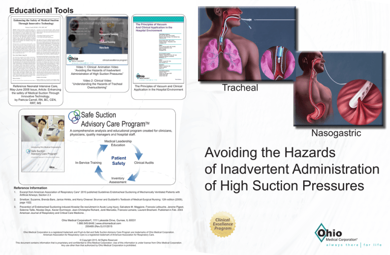
Tracheal
Nasogastric
Avoiding the Hazards
of Inadvertent Administration
of High Suction Pressures
There are two
most commonly overlooked key aspects
of clinical suctioning that could lead to over-suctioning and
various clinical complications:
1
2
Choosing the appropriate
suction pressure:
Recommended Guidelines for setting
Suction Pressures
Setting the suction
pressure correctly:
Are your Vacuum Regulators helping you to avoid
over suctioning your patients?
OHIO MEDICAL’S Push-To-SetTM Technology
is an integrated passive safety system designed
to prevent inadvertent over-suctioning.
Always Occlude-To-Set for proper
suction levels
THE TECHNOLOGY:
The negative pressure must be checked
by occluding the end of the suction tubing
before attaching it to the suction catheter,
and prior to each suctioning event[2]*
Endotracheal [1]*
Adults
<150 mmHg
Infants
-60 to -80 mmHg
When the vacuum adjustment knob is depressed,
the vacuum flow path is “automatically occluded”
and will accurately reflect maximum suction
pressure.
Nasogastric drainage [2]*
Adult:
-30 to -40 mmHg (Intermittent)
Always follow your institution’s recommended
guidelines for clinical suctioning.
?
If not occluded . . .
and/or not properly set . . .
Suction-induced
When this happens the clinician
Lung De-recruitment
inadvertently sets the minimum
vacuum pressure. This may expose
the patient to high suction pressure
that may cause suction-induced lung
de-recruitment associated with acute
lung injury and Acute Respiratory
Distress Syndrome (ARDS) patients.[3]*
If the flow path is not occluded,
the pressure setting may be as
high as -635 mmHg.
tubing left open
Because the patented PTS technology occludes
the flow path when the knob is depressed, the
clinician is not required to occlude the flow path to
set maximum pressure.
tubing
occluded
pressure set
too high
DID YOU KNOW......
The results could be . . .
inadvertent high suction pressures.
Hazards during suctioning are as follows:
During tracheal suctioning:
During nasogastric suctioning:
• Hypoxia/Hypoxemia
• Atelectasis [1]*
• Mucosal tissue tears [1]*
• Bleeding [1]*
• Increased risk of infection [1]*
• Mucosal tissue tears [2]*
• Bleeding [2]*
• Increased risk of infection [2]*
[1]*
One Simple, One Handed Step
Push-To-SetTM technology enhances setting safe suction
pressures through automatic occlusion.
Digital technology enhances display of accurate suction
pressure (± 1%) providing unsurpassed safety.
*See back cover for reference information
255489 (Rev.5) 01/2015
255489 (Rev.5) 01/2015


