ProSound α10 - Olympus
advertisement

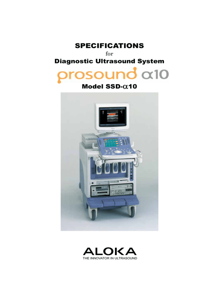
SPECIFICATIONS for Diagnostic Ultrasound System ProSound α10 Model SSD-α10 THE INNOVATOR IN ULTRASOUND ALOKA ProSound α10 Striving to eliminate undesirable sound components from the transmitted ultrasound beam itself and with the ever-evolving ProSound technologies ALOKA has created the ProSound α10 platform. With its Ultimate Compounding Technologies, stress-free operation and combination of versatility and performance, the ProSound α10 provides the capability for a wide range of sophisticated diagnoses. ・ Compound Pulse Wave Generator allows us to actually design the transmission waveform for individual application. The clear waveform enhances focus accuracy, spatial and contrast resolution, while reducing artifacts. ・ Thanks to Precise Delay Timing Control, the accuracy of reception/transmission delay is four to eight times higher than conventional systems, providing high-resolution beams. ・ Compound Array Probes enhance focus precision in the elevation direction and enable beams to be focused homogeneously from superficial to deep areas of interest. ・ ProSound Unlimited Unlimited expandability via a flexible and scalable system architecture allows for future hardware and software upgrades ・ ProSound Usability With ergonomic system architecture, the α10 ensures higher examination efficiency. ・ ProSound Utility The α10 offers various scan methods and probes with a wide range of storage media. Scanning Method ・ ・ ・ ・ Electronic Convex Sector Electronic Linear Electronic Phased Array Sector Mechanical sector/radial* * Option (EU-9109) Image Display Modes *1 ・ ・ ・ ・ ・ ・ ・ ・ ・ ・ ・ ・ ・ 2 ・ ・ ・ ・ ・ ・ ・ ・ Quad B (PowerFlow) Dual B (eFlow*3) Quad B (eFlow) M (Flow) M (PowerFlow) B (Flow) and M (Flow) B (Flow) and D B (Flow) and D simultaneous real-time display (Triplex mode) ・ B and B (Flow) simultaneous real-time display (DDD: Dual Dynamic Display) ・ B and B (PowerFlow) simultaneous real-time display (DDD) ・ ・ ・ ・ B and B (eFlow) simultaneous real-time display (DDD) B (Flow), M (Flow), and D Intermittent trigger mode *2 Monitor mode *2 (Fundamental image/Intermittent C.H.E. image, side by side display) ・ TDI (Tissue Doppler Imaging) ・ Real-time 3D mode (Option: EU-9012 + SOP-ALPHA10-4) Request function: In multi-mode display, it is possible to select one mode for full screen display. *1 Probe dependent. *2 Option: CHM-ALPHA10 *3 eFlow is optional: SOP-ALPHA-12 Beamformer Transmission CPWG (Compound Pulse Wave Generator) Programmable waveform transmission Reception Multi processing high-speed digital beam former 12-bit A/D converter (4096 gray levels) Delay precision: 1/64λ at minimum in both transmission and reception Focusing Lateral direction B: gray-scale imaging Transmission: Multi-stage transmission focus of up to 4 M stages out of 16 stages D: Spectral Doppler (PW, HPRF PW, and CW) Reception: PixelFocusTM Dual B Quad B B and M B and D B, M, and D B (Flow) B (Power) Dual B (Flow) Quad B (Flow) Dual B (PowerFlow) Elevation direction Compound Dual Focusing (when Compound Array probe is used) Beam signal processing Dynamic 4D apodization Frame rate Max. 926 frames/s* * Depends on probe and various settings B-mode ・ Display Gray Scale: 256 levels ・ Scanning area: 100% to 25%, continuously variable Write zoom (magnification of real-time image): Max. 6 times (probe dependent) Read zoom (magnification of frozen image): ・ Depth range selections: 0.5-30 cm (probe dependent), changeable by 1 cm Longitudinal and lateral inversion Rotation by 90 degrees (probe dependent), HPRF (High Pulse Repetition Frequency) PW Doppler ・ Reference frequencies (probe dependent): PW: 2.14, 2.5, 3, 3.75, 5, 6, and 7.5 MHz CW: 2.14, 3, 3.75, 5MHz ・ Analysis rate: Frame rate (Line density): 3 selections (-1, 0, +1) PW: 0.5 to 20 kHz Gain*: 30 to 90 dB CW: 0.5 to 42 kHz STC (sensitivity time control) — gain versus depth curve control: 8 slide controls Angle gain: Gain versus angle curve control: 8 sectors (probe dependent) ・ Contrast*: 16 steps ・ AGC—Suppression of brightness saturation and Edge Enhancement: 16 steps ・ ・ ・ ・ ・ PW (Pulsed Wave) Doppler CW (Continuous Wave) Doppler Max. 16 times ・ * Option: SOP-ALPHA10-3 ・ Doppler methods: ・ Zoom ・ ・ ・ ・ ・ Spectral Doppler: ・ Display: Power spectrum ・ Real-time Doppler Auto Trace* Relief: 4 steps FTC: On/Off Frame correlation: ・ Max. velocity range: PW: –6.37 to 0 or 0 to +6.37 m/sec (2.14 MHz reference freq., 0 degree, with base line shift) CW: –15.90 to 0 or 0 to +15.90 m/sec (2.14 MHz reference freq., 0 degree, with base line shift) ・ Base line shift: Possible up to double velocity (changeable after freezing) ・ Steerable CW Doppler: Possible (probe dependent) ・ Steered linear scanning: Max. ±30 degrees changeable in 5 degrees step 16 steps (Auto/Manual) Smoothing: 16 steps Post Processing Echo enhance curve: 5 kinds Rejection: 64 steps ・ View gamma: 5 kinds * Gain and contrast can be changed after freezing ・ Spectrum inversion: Possible ・ Angle correction: Available up to 80 degrees, presetable (changeable after freezing) ・ Sample volume size for PW Doppler: 0.5 – 20 mm, changeable in 0.5 –1.0 mm step ・ Wall motion filter: Manual: M-mode: ・ Sweep method: Moving bar ・ Sweep speed*: 17.5, 11.6, 8.7, 5.8, 4.4, 2.9, 2.2 cm/sec. ・ Gain*: B-gain ±30 dB ・ Contrast*: 16 steps ・ AGC—Suppression of brightness saturation: 16 steps 50, 100, 200, 400, 800 or 1600 Hz, Auto: varies in 12 steps ・ Doppler gain: 0 - 50 dB ・ Contrast: 16 steps (changeable after freezing) ・ Black-and-white inversion: possible (changeable after freezing) ・ Audio output: Stereo (including relief processing) ・ Relief: 4 steps ・ FTC: On/Off ・ FAM* (Free Angular M-mode) Up to 3 M-mode cursors can be set omni-directionally on real-time at any position on a B-mode image. * Gain, contrast and sweep speed can be changed after freezing * FAM is optional (SOP-ALPHA10-5) 3 Color Flow Imaging ・ Gradation: ±32 levels for velocity (red and blue) ・ Display patterns: Velocity (derived from mean Doppler frequency shift), Velocity + variance, Variance, Power, TDI (Tissue 16 levels for variance (green) ・ Color Polarity: Normal, Inver Doppler Imaging) ・ Color area size: Variable from 100% to 5% continuously Cine Memory • Cine search and loop display (in B mode): ・ Steered linear scanning: Max. ±30 degrees *, 5 degrees step changeable * Probe dependent ECG time phase display possible • Max. approximately 1000 seconds ・ Line density: 9 steps ・ Image Select: 3 selections • Smoothing: 16 selections Flow filter: 6 selections Frame correlation: 16 selections Wall Motion Reduction: 16 selections Average: 3 levels Color coding (Possible to make with color coding editor) Abdomen : 5 kinds PV : 5 kinds Cardiology : 5 kinds Other : 5 kinds User : 5 kinds PowerFlow ・ ・ ・ ・ Gradation: 32 levels Color coding: 5 kinds Non-display of B/W image: Possible Smoothing: 16 levels Directional PowerFlow: Possible eFLOW*: One of the Color Flow imaging functions that can display blood flow information in a high spatial and temporal resolution. Directional eFLOW*: Possible * eFlow is optional: SOP-ALPHA-12 Color Doppler ・ Reference frequency: (Probe dependent) 2.14, 2.5, 3, 3.75, 5, 6, and 7.5 MHz ・ Pulse repetition frequency: 0.5 to 10.0 kHz ・ Maximum velocity range: - 1.23 to 0, or 0 to +1.23 m/sec (at 2.14 reference frequency, with baseline shifted) ・ Color base line shift: Possible up to double velocity (±31 steps) 4 Capacity B mode: Resolution, Standard, Penetration ・ ・ ・ ・ ・ ・ Cine scroll (in M or D mode): Max. 16,320 frames (Possible to store a maximum of 404 seconds of 30 frames/s sector images.) M and D modes: Max. approx. 1000 seconds. • High-speed mutual data transfer between Cine Memory and hard disk is possible. Note: The number of storable images in a loop depends on probe type, scanning angle and other conditions. Data Management • CD-R 1. Image data 1-1. Format Multiple-frame (moving) image Line data (DICOM) Image data (DICOM M-JPEG)* * Option: EU-9102 + SOP-ALPHA10-2 Single-frame (still) image DICOM(Pallet, RGB, JPEG) Tiff, Bmp, JPEG • Network interface: 10 BASE/T or 100 BASE/TX, 1-2. Image acquisition mode • Real-time multi-frame image acquisition (Line, Image) Post ECG: Max. 4 cardiac cycles (R-R) Pre ECG: Max. 4 cardiac cycles (R-R) Post TIME: Max. 16 seconds Pre TIME: Max. 16 seconds Manual: Line data: Up to the capacity of the Cine Memory Image data: Max. 180 seconds • Cine loop high-speed data transfer (Line, Image) (automatically switched) 5. DICOM network communication* ・ Conformity to DICOM service class: Ultrasound image storage SCU Ultrasound multi-image storage SCU Storage media FSC/FSR Print management SCU Modality worklist management SCU (For details, please refer to the DICOM Conformance Statement issued by Aloka.) Modality performed procedure step (MPPS) SCU ・ Storage: Possible to store patient information directly to DICOM file server ・ Print: Possible to printout images with DICOM compatible printer directly ・ Work list management: Retrieval of patient and reservation information from hospital information system (HIS) It is possible to selectively store data of arbitrary NOTE: The HIS needs to be compatible with DICOM standard section in the Cine Memory. supplement 10. The HIS network and the DICOM network • Multiple media simultaneous output It is possible to output still image data to multiple of storage media and printer at the touch of a button. 1-3 Image data management tool Image viewer Thumbnail display of stored images (1-36 images) Image zoom, rotation, inversion 1:1 replay (main unit HDD or DICOM storage data) CD-R writing Re-storing to media, transfer need to be linked. ・ Router setting: possible ・ IHE (Integrated Healthcare Enterprise) SWF (Scheduled Work Flow) PIR (Patient Information Reconciliation) * Option: SOP-ALPHA10-10 2. Measurement data It is possible to store measurement data in the main unit hard disk 3. Patient data Displayed information* Patient information: ID (up to 64 characters), Name (up to 64 characters), Birthday, Sex Study information: Study ID, Age, Height, Weight, Accession, Referring physician, Study description, Operator Series information: Application * Conforms to DICOM 3.0 standard 4. Data storage media • Main unit hard disk Usable space: up to about 32 GB • Floppy disk • MO disk 5 Measurement and Analysis: ● General measurements On B-mode image Distance Area and Circumference (Trace, Ellipse, Circle) Volume (Spheroidal, Prolate, Area-length, BP Simpson, SP Simpson)⎯Automatic heart cavity trace is possible. (3-point designation method) Index (general purpose) Histogram Angle, Hip joint angle On M-mode image Velocity Distance (amplitude) Time interval Heart rate Index (general purpose) On spectral Doppler Velocity, Acceleration, Mean flow velocity, Pressure gradient RI: Resistance index, PI: Pulsatility index Pressure half time Heart rate Dop Caliper measurement Index (general purpose) Time interval Stenotic flow measurement Regurgitant flow measurement D. Trace Doppler auto trace*: Possible * Option: SOP-ALPHA10-3 On B/D mode Flow Volume SV/CO On B(Flow) mode Flow Profile* * Option: SOP-ALPHA10-7 ● Obstetrical measurements & calculations Gestational age, Fetal weight Fetal Doppler measurements Fetal cardiac function measurements AFI (Amniotic fluid index) Cervical length Compatible with multiple pregnancy Growth analysis function (display of past measurement data) ● Gynecological measurements & calculations Uterus measurements Endometrial thickness Cervical measurements Ovary measurements Follicular measurements Urinary bladder measurements Uterine artery, Ovarian artery measurements 6 ● Cardiac analysis B mode LV Volume measurements Area-length, BP-ellipse, Simpson (Disc), Modified Simpson, Bullet, Pombo, Teichholz, Gibson) ⎯Automatic heart cavity trace is possible. (3-point Valve area measurements (AVA, MVA) LA/AO Ratio Right ventricle measurements LV myocardial mass IVC (inferior vena cava) measurement M mode Pombo (wall), Teichholz (wall) Gibson (wall) Mitral valve measurements LA/Ao measurements Tricuspid valve measurements Pulmonary valve measurements IVC (inferior vena cava) measurement Doppler mode LVOT (left ventricle outflow tract) flow RVOT (right ventricle outflow tract) flow Trans-mitral flow Regurgitant flow (AR, PR, MR, TR) Stenotic flow (AS, PS, MS, TS) Pulmonary vein Coronary flow TDI PW IMP (Index of Myocardial Performance)* * Option: SOP-ALPHA10-8 B(Flow)/D mode PISA measurement TDI PowerFlow mode BETA (B and B/M (longitudinal) modes) ● Vascular analysis Carotid artery: CCA (common carotid artery) ICA (internal carotid artery) ECA (external carotid artery) BIFUR (Bifurcation of carotid artery) VERT (Vertebral artery) % Stenosis area % Stenosis diameter IMT (Intima-media thickness) Measurements of arteries in extremities: Lower extremity artery flow Upper extremity artery flow Stenotic rate: % Stenosis area % Stenosis diameter Measurements of veins in extremities: Lower extremity venous flow Upper extremity venous flow Trans-cranial blood flow measurement ● Urological measurements & calculations Prostate volume: PSA volume, PRS Slice volume Bladder volume Seminal vesicle Testicle volume Renal volume Cortical thickness Adrenal volume Renal artery Doppler measurements (pulsatility index, resistance index) ● Abdominal measurements Stenotic rate (diameter, area) Gall bladder Common bile duct Pancreas Kidney Spleen SOL (Space Occupying Lesion) Abdominal aortic diameter Portal vein diameter Renal artery blood flow Abdominal blood flow Shunt flow Flow volume Physiological Signal Display*1 ・ Displayed information: ECG, PCG, Pulse wave*2, breathing waveform ・ ECG synchronized display: Available for one phase ・ Display position: Continuously variable (both in B and M modes) *1 Option: PEU-ALPHA10B *2 Pulse wave transducer (TY-307A) is optional. Brachytherapy grid display* It is possible to display grid for prostate gland brachytherapy. * Available when UST-678 is connected. ● Report Functions • Obstetrical report • Gynecological report • Cardiac function report • Vascular report • IMT (Intima-Media Thickness) report • Urological report • Abdominal measurements report It is possible to recall past measurement reports. Examination data history can be plotted on the report. Direct printout of each report is possible with an optional PC printer. Output of measurement values in CSV file is possible. ● Hot Key function; It is possible to assign measuring functions to the alphabet keys on the keyboard ● Measurement on VCR playback image: Possible (manual calibration) ● User’s calculation 6 equations can be set for each application BETA (Backscattered Energy Temporal Analysis) Backscattered wave from the cardiac muscle is frequency-analyzed to obtain instantaneous energy. It is possible to capture cyclic variation of the myocardium. 7 Optional Functions PC printer* It is possible to printout report of OB/GYN, cardiology, PV, and urology including ultrasound images directly with an external PC printer. * HP Deskjet 5740 and CANON PIXUS iP4000 printers have been verified for operation. For detailed information on compatible printers, please inquire of Aloka. RT-3D (Real-time 3D)* Trans-abdominal 3D probe (ASU-1010) and trans-vaginal 3D probe (ASU-1012): • Scanning rate: up to 10 volumes/sec • It is possible to display 3 arbitrary sections simultaneously • Omnidirectional rotation (360 degrees in any direction) • 5 kinds of rendering selectable • Detail scan of the ROI (Region of interest) is possible • B-mode measurements are possible on an arbitrary plane Transthoracic cardiac 3D probe (ASU-1011): • Scanning rate: Max 15 volumes/sec (60 deg. X 60 deg.) Optional Analysis Functions Comprehensive Cardiac Analysis* A-SMA* (Automated Segmental Motion Analysis) The A-SMA can automatically detect the boundary between the cardiac cavity and the endocardium to calculate the area of the cavity in each frame, enabling quantification of endocardial segmental motion. • FAC (Fractional Area Change) can be displayed in line graph mode. • In histogram mode, FAC of each segment can be displayed in real time as a bar graph. KI* (Kinetic Imaging) The KI can automatically detect the boundary between the cardiac cavity and the endocardium based on the brightness information of the gray scale image and displays the temporal change of the boundary with change of color or gradation. CQ* (Cardiac Quantification) It is possible to display variation of LV function indexes in real time as a line graph. Indexes: EF (Ejection Fraction), Volume, etc. * Option: EU-9102 + SOP-ALPHA10-4 WT* (Wall Thickness) It is possible to display variation of myocardial thickness in real time as a line graph. EFV (Extended Field of View)* * Option (EU-9100) + PEU-ALPHA10B (Physiological It is possible to display an image of an extensive range of Signal Display unit) • Scanning angle: Max. 90 deg. X 60 deg. the body by moving the probe. An area wider than the scanning width of the probe can be displayed. * Option: EU-9102 + SOP-ALPHA10-1 CHE (Contrast Harmonic Echo)* Contrast agent such as Levobist generates abundant second harmonics when disrupted, which eases detection by Harmonic Echo. In Subtraction mode, difference from the reference image is displayed to clarify the distribution of the contrast agent. • Monitor mode In the Monitor mode, images are available with a low sound pressure during the intermission of intermittent high sound pressure transmission. • Possible with UST-9130 Option: CHM-ALPHA10 8 eTRACKING (Echo Tracking) * It is possible to precisely measure displacement of blood vessel to obtain indexes of stiffness of the vessels such as pressure-strain elastic modulus (Ep), stiffness parameter (β), arterial compliance (AC), one-point pulse wave velocity (PWVβ), and augmentation index (AI). * Option: SOP-ALPHA10-11 TDI analysis* B-mode Temporal Velocity Profile Velocity, time, acceleration, ratio Regional Velocity Profile Velocity, distance TDI-Myocardial Thickness (Wall thickness) Distance, time, velocity Strain rate Time, strain rate Strain Time, strain M-mode Velocity trace Velocity, time, acceleration, ratio, velocity difference TDI-Myocardial Thickness (wall thickness) Distance, time, velocity Velocity Profile Velocity, distance CSV output of analyzed data is possible. CSV is a file format that can be taken into Excel file directly. * Option: SOP-ALPHA10-13B Contrast Echo analysis* • Image Subtraction Fixed Reference: Subtraction of reference frame from all frames Any 2 Frame: Subtraction between 2 selected frames Display modes: All images, arbitrary images • Time-Intensity Curve display for subtraction images: available Series: Graphic display in frame sequence or time sequence By Group: Graphic display with the time of one sequence of intermittent acquisition as the horizontal scale (Graphs of multiple sequences are overlapped.) Display mode: Image, Graph ROI type: Square, Draw, Arc, and Circle * CSV output of analyzed data is possible. * Option: SOP-ALPHA10-14 Stress Echo analysis* Image acquisition methods: • ECG synchronized acquisition • Compatible frame rate: Up to 75 Hz • Recalled screen Playback speed: Variable Image allocation: Possible Scoring: Possible Automatic registration: On/Off Protocol: • Exercise stress protocols: Exercise Stress Echo Treadmill Exercise Bicycle Exercise • Pharmacological stress protocols: DSE High-Dose DSE Low-Dose DSE Arbutamine Dipyridamole • User's protocol: The user can make a protocol within 8 images X 12 stages in 1 stage. • Full disclosure (Multi acquisition): possible for 180 seconds • Scoring screen Playback speed: Variable Comparison with the reference image is possible Image playback range is selectable Systolic image • Report screen Display format Chart/Stage overview/View overview * Option: SOP-ALPHA10-15B (Physiological Signal Display unit PEU-ALPHA10B is also necessary.) Brachytherapy* It is possible to display grid for prostate grand brachytherapy. * Option: SOP-ALPHA10-17 9 Viewing Monitor Acoustic Power ・ 17-inch diagonal multi-sync display 0 to 100%, continuously changeable ・ SVGA non-interlaced monitor Preset Function ・ 45 separate programs for specific clinical applications and/or users ・ User programmable and/or factory default settings ・ Factory default settings: 33 kinds ・ Preset contents storable in a floppy disk ・ Tilt and swivel are possible. ・ Height adjustment together with operation panel: Possible Safety Regulation ・ Characters and graphic displays Complies with IEC 60601-1 Class I, Type BF Environmental Requirements ・ Character input area: In Operation ID, name, age, sex, retained text ・ Automatic Annotation Labeling: 120 words or more (User registration is possible.) ・ Temperature: +10 to +40 degrees C ・ Relative Humidity: 30 to 75% (non condensing) ・ Body mark: 47 kinds Body mark editor to create user’s body mark: 24 ・ Atmospheric pressure: kinds In Storage/transportation ・ Temperature: Menu control 700 to 1060 hPa -10 to +50 degrees C (0 to +50 degrees C for mechanical probes) 8.4-inch color TFT LCD touch panel ・ Relative Humidity: Number of Probe Connectors 10 to 90% (non condensing) • For electronic scanning probes: 4 ・ Atmospheric pressure: 700 to 1060 hPa 1 • For mechanical scanning probes* : 1 • For independent probes*2: 1 Power Requirement 1 * Option: EU-9109 ・ *2 Option: EU-9110 115/ 200 to 240V ±10%, 50 or 60 Hz, Max. 1500 VA (with optional recorders connected) Max. 800 VA (main unit only) Video Signals (for printer, VCR, DVD) Dimensions • Input: ・ Y/C Weight Audio (L/R), 1 channel each ・ • Output: Color composite (BNC): 1 channel B/W composite (BNC): 1 channel Audio (L/R): 1 channel Y/C color: 1 channel Y/C B/W: 1 channel • Resolution: 640 x 480 pixels No conversion of the displayed image. Output of trimmed image is possible. • High-resolution DV output*: 800 x 600 pixels (for DV-800) * Option: DV-800 main unit and DV-800 adaptation kit PM-A10-H001 are necessary. 10 58 cm (W) × 109 cm (D) × 144 – 157 cm (H) Approx. 210 kg (main unit only) System Configuration ProSound α10 main unit (including 17-inch monitor) Optional Software Optional Recorders/Printers/Units Contrast Harmonic Echo VCR Physiological Signal Display unit CHM-ALPHA10 MITSUBISHI NTSC: HS-MD3000U PAL: HS-MD3000E * RS232C interface and 34-pin interface are optional PEU-ALPHA10B Real Time DOP Auto Trace SOP-ALPHA10-3 FAM (Free Angular M-mode) SOP-ALPHA10-5 Flow Profile SOP-ALPHA10-7 IMP (Index of Myocardial DVD recorder DVD-BD-X201M (VICTOR)*1 1 * Connection cable L-CABLE-756 is necessary for remote control from main unit. DVD-DVO-1000MD (SONY)*2 *2 Connection cable KRS-9F25F02K Performance) (SANWA SUPPLY) is necessary for remote control from main unit. SOP-ALPHA10-8 DVD-DV-800 DICOM communication SOP-ALPHA10-10 eTRACKING SOP-ALPHA10-11 eFLOW SOP-ALPHA10-12 TDI/Strain Analysis SOP-ALPHA10-13B CHE analysis SOP-ALPHA10-14 Stress Echo function SOP-ALPHA10-15B 3 (TEAC)*3 * PM-A10-H001 is necessary for connection of this recorder. B/W printer SONY NTSC: UP-895MD, UP-D897MD PAL: UP-895CE, UP-D897CE Worldwide: UP-895MD/SYN UPD-897MD/SYN Pulse wave transducer: TY-307A Comprehensive Cardiac Analysis unit (KI, A-SMA, CQ, WT) EU-9100 Footswitch MP-2345B, MP-2614B Mechanical scanning probe connection unit EU-9109 Independent probe connection unit EU-9110 MITSUBISHI NTSC: P93W PAL: P93E Color Printer SONY (NTSC/PAL): UP-21MD (CED) UP-D23MD MITSUBISHI NTSC: CP900UM PAL: CP900E Host Interface Unit EU-9102 Extended Field of View SOP-ALPHA10-1 Motion JPEG SOP-ALPHA10-2 Real Time 3D SOP-ALPHA10-4 LT 11 OPTIONAL PROBES Electronic convex sector probes THE: Tissue Harmonic Echo, CHE: Contrast Harmonic Echo Application (description) General abdomen, Model UST-9130 Ultrasound Frequencies (MHz) B and M modes 3.0/3.75/5.0/6.0 OB/GYN Doppler/Flow Flow: Scanning angle (degrees) Radius of curvature (mmR) 60 60 2.14/2.5/3.0/3.75 (THE and CHE ) THE: 2.14/2.5 PW: 2.5 (Compatible with CHE:1.88/2.14 CHE:1.88/2.5 Puncture adapter: MP-2473 EFV and eFLOW) General abdomen, OB/GYN Optional accessories UST-9115-5 3.75/5.0/6.0/7.5 Flow: 3.0/3.75/5.0 60 60 120 14 PW: 3.75 (Compatible with EFV and eFLOW) General abdomen, UST-9128 intercostal scanning (THE and CHE) Small part, UST-9120 3.0/3.75/5.0/6.0 THE: 2.14 CHE: 1.88/2.14 5.0/6.0/7.5/10.0 Flow: 2.5/3.0 PW: 2.5 Flow: 3.75/5.0/6.0/7.5 (Compatible with eFLOW) PW: 5.0 Endo-cavity UST-9118 3.75/5.0/6.0/7.5 MP-2474 70 Neonatal head Flow: 5.0/6.0 20 UST-675P 3.75/5.0/6.0/7.5 Flow: 5.0/6.0 9 180 9 82 20 65 20 - 65 20 - PW: 5.0 (Compatible with eFLOW) Intraoperative, UST-9133 Flow: 2.14/2.5/3.0 /3.75 Abdominal biopsy THE: 2.14 (THE and CHE) PW: 2.5/3.0 CHE: 1.88/2.14 (Compatible with eFLOW) Intraoperative 3.0/3.75/5.0/6.0 Puncture adapter set MP-2748-SET Probe cover: RB-945BP-S (Sterilized*) RB-945BP-NS (Nonsterilized) Puncture adapter MP-2452 is attached as standard Probe cover: RB-665P-NS (Non-sterilized) RB-665P-S (Sterilized*) Puncture adapter : MP-2781 180 eFLOW) Endo-cavity Puncture adapter: MP-2458 PW: 5.0 (Compatible with Puncture adapter: UST-9132I 5.0/6.0/7.5/10.0 Flow: 3.75/5.0/6.0 /7.5 PW: Intraoperative UST-9132T 5.0/6.0/7.5/10.0 3.75/5.0 Flow: 3.75/5.0/6.0 /7.5 PW: 3.75/5.0 * Sterilized probe cover cannot be sold in EU member countries. 12 Electronic linear probes Application (description) Model Peripheral vessels/ Small parts (Steered linear) (EFV compatible) (eFLOW compatible) (Harmonic Echo) (Compound Array) UST-5411 Peripheral vessels/ Small parts (Steered linear) (EFV compatible) (eFLOW compatible) (Harmonic Echo) UST-5412 Small part UST-5712 Ultrasound Frequencies (MHz) Doppler, Flow B and M modes 5.0/7.5/10.0/13.0 Flow: 5.0/6.0/7.5 THE.: 5.0 PW: 5.0 5.0/7.5/10.0/13.0 Flow: 5.0/6.0/7.5 THE.: 5.0 PW: 5.0 5.0/6.0/7.5/10.0 Flow: 5.0/6.0/7.5 (Steered linear) (EFV compatible) Scanning width (mm) 38 - 38 - 60 Puncture adapter: MP-2456 Water path: MP-2463 60 Puncture adapter: MP-2448 PW: 6.0 Intraoperative UST-5713T 5.0/6.0/7.5/10.0 Intraoperative UST-547 7.5/10.0/13.0 Flow: 5.0/6.0/7.5 PW: 6.0 Flow: 5.0/6.0/7.5 Optional accessories 20 - 10 Handling tool T type: MP-2749 Handling tool I type: MP-2750 38 - 38 - 38 - PW: 6.0 UST-533 Microsurgery 7.5/10.0/12.0/13.0 Flow: 5.0/6.0/7.5 PW:6.0/7.5 Superficial tissue UST-5543 7.5/10.0/13.0 Flow: 5.0/6.0/7.5 (Steered linear) PW: 6.0 Peripheral vessels (Steered linear) (Compatible with eFLOW) (Harmonic Echo) UST-5548 Intraoperative (Flexible laparoscopic) UST-5550 3.75/5.0/6.0/7.5 THE:3.75 Flow: 3.0/3.75 /5.0/6.0 PW: 3.75/5.0 5.0/6.0/7.5/10.0 Flow: 5.0/6.0/7.5 PW: 5.0/6.0 (Steered linear) Electronic convex sector/linear combination probe Application Ultrasound Frequencies (MHz) Model number B and M modes Transrectal (Bi-plane: Convex sector + Linear) UST-678 Sector 3.75/5.0/6.0/ 7.5 Doppler & Flow Flow:3.0/3.75 /5.0/6.0 Scanning angle/width Radius of curvature (mmR) 120 deg. 9 60 mm - Optional accessories Puncture adapter: MP-2451 PW :3.75/5.0 Linear 5.0/6.0/7.5/ Flow:5.0/6.0 10.0 PW :5.0/6.0 Probe cover: BL-664-S (sterilized)*2 BL-664-NS(Non-sterile) Elastic band: FS5/16 Grip holder:MP-2447 *2 Sterilized probe cover cannot be sold in EU member countries. 13 Electronic phased array sector probes T.E.E.: Trans-esophageal Examination, T.H.E.: Tissue Harmonic Echo Application Ultrasound Frequencies (MHz) Model (description) B and M Doppler & Flow Cardiology (T.H.E.) UST-52101 2.5/3.0/3.75/5.0 T.H.E.: 1.88 Pediatric cardiology (T.H.E.) UST-52108 3.75/5.0/6.0/7.5 Rotary plane T.E.E. UST-5293-5 3.75/5.0/6.0/7.5 Neurosurgery (burr hole) UST-52114P 3.75/5.0/6.0/7.5 Neonatal cardiology UST-5296 5.0/6.0/7.5/10.0 Neonatal and pediatric T.E.E. UST-52110S 3.75/5.0/6.0/7.5 Pediatric T.E.E. (Bi-plane) UST-52111S 3.75/5.0/6.0/7.5 Motorized T.E.E. (T.H.E.) UST-52116 3.75/5.0/6.0 T.H.E.: 3.0 THE: 3.0 Scanning angle (degrees) Optional accessories Flow: 2.14/2.5/3.0/3.75 PW: 2.14/2.5/3.0/3.75 CW: 2.14 Flow: 3.0/3.75/5.0 PW: 3.75/5.0 CW: 3.75 90 Flow: 3.75/5.0 PW: 3.75 CW: 3.75 Flow: 3.75/5.0 PW: 3.75/5.0 90 Flow: 5.0 PW: 5.0 CW: 3.75 Flow: 3.0/3.75/5.0 PW: 3.75/5.0 CW: 3.75 Flow: 3.0/3.75/5.0 PW: 3.75/5.0 CW: 3.75 Flow:3.0/3.75/5.0 PW : 3.0/3.75 CW : 3.75 90 - 90 - 90 - 90 - - 90 - 90 - Includes adapter standard biopsy as 3D Probes* * EU-9102 and SOP-ALPHA10-4 are necessary. Application Trans-abdominal scanning Model ASU-1010 Ultrasound Frequencies (MHz) B, M 3.75/5.0/7.5/10 Flow: 2.14/2.5/3.0 THE: 2.14/2.5 /3.75 ASU-1012 scanning (Harmonic Echo) Cardiology ASU-1011 3.75/5.0/6.0/7.5 Flow: 5.0/6.0 THE: 2.5/3.0 PW: 5.0 2.5/3.0/3.75/5.0 Flow: 2.14/2.5 THE: 1.88 /3.0/3.75 PW:2.14/2.5/3.0 /3.75 CW: 2.14 * Sterilized probe cover cannot be sold in EU member countries. 14 Scanning angle (degrees) Radius of curvature (mmR) 60/60 40 140/90 10 90/60 - Optional accessories — PW: 2.5 (Harmonic Echo) Trans-vaginal Doppler/Flow Probe cover: RB-945BP-S (Sterilized*) RB-945BP-NS (Nonsterilized) — ANNULAR ARRAY MECHANICAL SECTOR PROBE* * Mechanical scanning unit EU-9109 is necessary. Application Small parts Ultrasound Frequencies (MHz) Model Doppler/Flow B, M 10.0 ASU-36WL-10 Scanning angle (degrees) - Optional accessories Puncture adapter: MP-2493 MECHANICAL RADIAL PROBES* * Mechanical scanning unit EU-9109 is necessary. Ultrasound Frequencies (MHz) Application Trans-rectal Model ASU-67 B, M Doppler/Flow 7.5/10.0 - Optional accessories Scanning angle (degrees) 360 Puncture adapter: MP-2493 Transurethral ASU-65B 7.5 - 360 Outer sheath adapters for STORZ #27040B: MP-2421 for Olimpus A3128, A2101: MP-2422 for A.C.M.I. #8414: MP-2423 for Wolf #8654.021: MP-2424 Independent CW Doppler Probes* * Independent probe connection unit EU-9110 is necessary. Application CW Doppler (for heart) CW Doppler (for peripheral vessels) Model UST-2265-2 UST-2266-5 Ultrasound Frequencies (MHz) B, M - Doppler CW: 2.14 CW: 5.0 Optional accessories - 15 ・ The specifications are subject to change without notice. ・ The standard components and optional items depend on the country. Not all the products are available in all countries. Please contact your local ALOKA distributors for details. ・ ProSound is a registered trademark of ALOKA CO., LTD. ・ Harmonic Echo is a trademark of ALOKA CO., LTD. We care, Ultrasound@Aloka 6-22-1 Mure, Mitaka-shi, Tokyo 181-8622 Japan Telephone: +81 422 45 6049, Facsimile: +81 422 45 4058 URL: http://www.aloka.com/ Printed in Japan, 2005-12 SP-A10LT-V40-10


