Hyaluronan inhibit the LPS induced Cathepsin K Expression via
advertisement

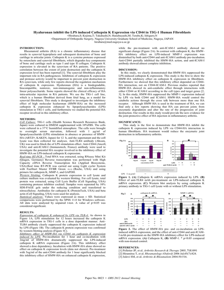
Hyaluronan inhibit the LPS induced Cathepsin K Expression via CD44 in TIG-1 Human Fibroblasts +Hirabara,S; Kojima,T; Takahashi,N; Hanabayashi,M; Terabe,K; Ishiguro,N +Department of Orthopedic Surgery, Nagoya University Graduate School of Medicine, Nagoya, JAPAN hirabara@med.nagoya-u.ac.jp RESULTS: Expression of cathepsin K enhanced by LPS via TLR-4: As shown in Figure 1A, LPS stimulation for 12 hours increased the cathepsin K mRNA expression in TIG-1 cells in a dose dependent manner. AntiTLR4 antibody clearly suppressed the cathepsin K expression induced by LPS (Figure 1B). The cathepsin K protein expression was confirmed by western blotting analysis (Figure 1C). Inhibitory effect of HMW-HA via CD44 on cathepsin K expression induced by LPS: Pre-incubation for 1 hour and co-incubation with 2mg/ml of HMW-HA dramatically suppressed the LPS-induced cathepsin K mRNA expression (Figure 2A). This inhibitory effect showed a dose dependency. Incubation with HMW-HA alone showed no effect on cathepsin K expression levels (data not shown). Pre-treatment with 5ug/ml of the anti-CD44 antibody for 1 hour significantly blocked this inhibitory effect of HMW-HA on enhanced cathepsin K expression, DISCUSSION: In this study, we clearly demonstrated that HMW-HA suppressed the LPS-induced cathepsin K expression. This study is the first to show the HMW-HA inhibitory effect on cathepsin K expression in fibroblasts. Furthermore we showed that this inhibitory effect depended on CD44HA interaction, not on CD44-ICAM-I. Previous studies reported that HMW-HA showed its anti-catabolic effect through interactions with either CD44 or ICAM-I according to the cell types and target genes [2, 3]. In this study, HMW-HA suppressed the MMP-1 expression induced by LPS via both CD44 and ICAM-I. HMW-HA would control the catabolic actions through the cross talk between more than one HA receptor. Although HMW-HA is used in the treatment of RA, we can find only a few reports showing that HA can prevent joints from enzymatic degradation and alter the rate of the progression of joint destruction. Our results in this study would provide the new evidence for the joint protective effect of HA injection in inflammatory arthritis. SIGNIFICANCE: This study is the first to demonstrate that HMW-HA inhibit the cathepsin K expression induced by LPS via CD44-HA interaction in human fibroblasts. HA treatment would reduce the enzymatic joint destruction in inflammatory arthritis. Fig 1A B 600 # 700 500 600 400 500 (% of control) (% of control) 300 200 100 400 300 200 100 0 0µg/ml 0.1µg/ml 1µg/ml concentration of LPS LPS - + anti TLR-4 mAb - - C LPS (1μg/ml) 0 10µg/ml - - + - + + Figure 1. (A) Cathepsin K mRNA expression induced by LPS. (B) Effect of anti-TLR4 mAb pre-treatment on LPS-induced cathepsin K mRNA expression. (C) Western blot analysis by using cathepsin K primary antibody in TIG-1 cell lysate with or without LPS stimulation. Fig 2 cathepsin K mRNA # # A MMP-1 mRNA # # # * B 300 250 * 250 * (% of control) METHODS: Cell Culture: TIG-1 cells (Health Science Research Resources Bank, Japan) were cultured in DMEM supplemented with 10%FBS. The cells were cultured in a 37°C humidified atmosphere. The cells were brought to overnight serum starvation, followed with 1 µg/ml of lipopolysaccharide (LPS) stimulation in absence or presence of HMWHA (ARTZ®, KAKEN, Japan) for 12 h, and the supernatant and the cell lysate was then collected for analyses. Anti-TLR4 antibody (Abcam, UK) was used to block the of LPS stimulation effect. Anti-CD44 (Ancell, USA) and anti-ICAM-I (Immunotech, France) antibody were used to investigate the potential HA receptor involved in the inhibitory effect of HMW-HA on LPS-induced cathepsin K expression. Real-time RT-PCR:Total RNA was extracted, using RNeasy Mini Kit (Qiagen, Germany) Reverse transcription was performed with High Capacity cDNA Reverse Transcription Kit (Applied Biosystems, USA).Real time RT-PCR was carried out using Light cycler System with FastStart Master SYBR Green PLUS (Roche, USA) and using primers for cathepsin K, MMP-1, and GAPDH. Western blotting: Cathepsin K protein expression in cell lysate and culture medium was evaluated by western blotting. For cell lysate, total protein was extracted, using Cell Lysis Buffer (Cell Signaling, USA) containing protease inhibitor cocktail. Samples were separated by 10% SDS-PAGE gels under the reducing condition and transferred to nitrocellulose. Antibodies for cathepsin K (ProteinTech, USA) and beta actin (Cell Signaling, USA) were used for analysis. Statistical analysis: Values were expressed as mean ± SD. Statistical comparisons were performed by the SPSS 11.0 for Windows software. All data were analyzed by unpaired t-test. A value of p<0.05 was considered significant. while the pre-treatment with anti-ICAM-I antibody showed no significant change (Figure 2A). In contrast with cathepsin K, the HMWHA inhibitory effect on LPS-induced MMP-1 expression was diminished by both anti-CD44 and anti-ICAM-I antibody pre-incubation. Anti-CD44 partially inhibited the HMW-HA action, and anti-ICAM-I antibody showed almost complete inhibition. (% of control) INTRODUCTION: Rheumatoid arthritis (RA) is a chronic inflammatory disease that results in synovial hyperplasia and subsequent destruction of bone and cartilage in articular joints. Cathepsin K is a cysteine protease expressed by osteoclasts and synovial fibroblasts, which degrades key components of bone and cartilage such as type I and type II collagen. Cathepsin K expression is elevated in the synovium of RA patients. The positive correlation between the extent of joint destruction and the cathepsin K expression level has been reported [1]. The synovial fibroblasts play the important role in RA pathogenesis. Inhibition of cathepsin K expression and protease activity would be important to prevent joint destruction in RA synovium, while only few reports showed the regulation mechanism of cathepsin K expression. Hyaluronan (HA) is a biodegradable, biocompatible, nontoxic, non-immunogenic and non-inflammatory linear polysaccharide. Some reports showed the clinical efficacy of HA intra-articular injection in RA patients. We use the TIG-1 cell line, which is a human fibroblast derived from fetal lung, as a model for synovial fibroblasts. This study is designed to investigate the inhibitory effect of high molecular hyaluronan (HMW-HA) on the increased cathepsin K expression enhanced by lipopolysaccharides (LPS) stimulation in TIG-1 cells, and furthermore we studied the potential HA receptor involved in this inhibitory effect. * 200 150 100 * 150 100 50 50 0 0 LPS - HA - anti-CD44 mAb - anti-ICAM-1 mAb - 200 + - - - + + - - + + + - + + - + - - - - + - - - + + - - + + + - + + - + Figure 2. The effect of HMW-HA pre- and co-incubation on LPSinduced mRNA expression, and the effect of anti-CD44 and anti-ICAMI mAb pre-treatment on the HMW-HA inhibitory effect for LPS-induced mRNA expression. (A) Cathepsin K, (B) MMP-1. * p<0.05 compared with non-treated control. REFERENCES: [1] Pelletier JP, et al. Arthritis Research & Therapy 2005, 7:R1091 [2] Hiramitsu T, et al. Rheumatology (Oxford) 2006 Jul;45(7):824. [3] Julovi SM, et al. Arthritis & Rheumatism 2004:50:516. Paper No. 0023 • ORS 2012 Annual Meeting


