(Pressure-Volume) Theory of Operation
advertisement
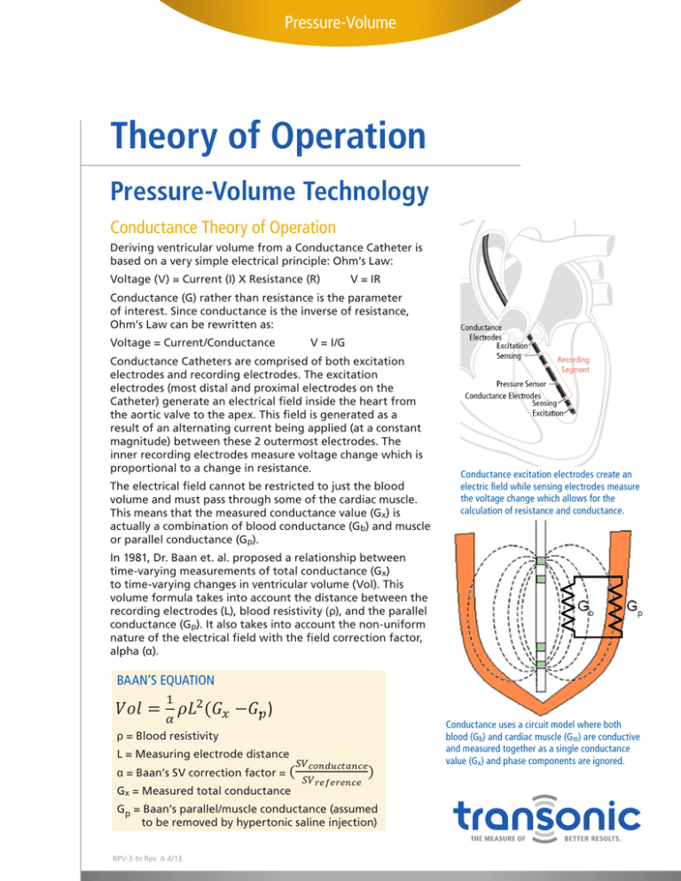
Pressure-Volume Theory of Operation Pressure-Volume Technology Conductance Theory of Operation Deriving ventricular volume from a Conductance Catheter is based on a very simple electrical principle: Ohm’s Law: Voltage (V) = Current (I) X Resistance (R) V = IR Conductance (G) rather than resistance is the parameter of interest. Since conductance is the inverse of resistance, Ohm’s Law can be rewritten as: Voltage = Current/Conductance V = I/G Conductance Catheters are comprised of both excitation electrodes and recording electrodes. The excitation electrodes (most distal and proximal electrodes on the Catheter) generate an electrical field inside the heart from the aortic valve to the apex. This field is generated as a result of an alternating current being applied (at a constant magnitude) between these 2 outermost electrodes. The inner recording electrodes measure voltage change which is proportional to a change in resistance. The electrical Scisense field cannot be restricted to just the blood Transonic Technical Note volume and must pass through some of the cardiac muscle. This means that the measured conductance value (Gx) is actually a combination of blood conductance (Gb) and muscle or parallel conductance (Gp). Conductance excitation electrodes create an electric field while sensing electrodes measure A. Henton and F. Konecny the voltage change which allows for the March, 2013 calculation of resistance and conductance. mathematical reviewaof SV correction InA1981, Dr. Baan et. al. proposed relationship between factors employed time-varying measurements of total conductance (Gx) equation and Wei’s equation: to time-varying changes in ventricular volume (Vol). This volume formula takes into account the distance between the recording electrodes (L), blood resistivity (ρ), and the parallel conductance (Gp). It also takes into account the non-uniform nature of the electrical field with the field correction factor, Baan’s equation: Where: alpha (α). ρ = blood resistivity L = measuring electrodes distance in Baan’s BAAN’S EQUATION 𝑆𝑆𝑆𝑆 α = SV correction factor = 𝑐𝑐𝑐𝑐𝑐𝑐𝑐𝑐𝑐𝑐𝑐𝑐𝑐𝑐𝑐𝑐𝑐𝑐𝑐𝑐𝑐𝑐 A. Henton and F. Konecny Transonic Scisense Technical Note 1 𝑆𝑆𝑆𝑆𝑟𝑟𝑟𝑟𝑟𝑟𝑟𝑟𝑟𝑟𝑟𝑟𝑟𝑟𝑟𝑟𝑟𝑟 March, 2013 𝑉𝑉𝑉𝑉𝑉𝑉 = 𝜌𝜌𝜌𝜌2 (𝐺𝐺𝑥𝑥 −G.𝑝𝑝 ) Gx = measured TOTAL conductance 𝛼𝛼 Conductance uses a circuit model where both conductance Gp = parallel/muscle ρ = Blood resistivity (Gb) equations and cardiac (Gm) are conductive Therefore, we can compare SV correction process between Baan’sblood and Wei’s bymuscle the following: measured saline together as a single conductance (assumed to be removed byand hypertonic injection) L = Measuring electrode distance value (G𝐺𝐺 and phase components are ignored. x)𝑏𝑏 𝑆𝑆𝑆𝑆𝑐𝑐𝑐𝑐𝑐𝑐𝑐𝑐𝑐𝑐𝑐𝑐𝑐𝑐𝑐𝑐𝑐𝑐𝑐𝑐𝑐𝑐 ) VS α = Baan’s SV correction factor = ( 𝑆𝑆𝑆𝑆𝑟𝑟𝑟𝑟𝑟𝑟𝑟𝑟𝑟𝑟𝑟𝑟𝑟𝑟𝑟𝑟𝑟𝑟 𝛾𝛾 Gx = Measured total conductance (1 − ) Note: for the purpose ofwethis document weBaan’s will ‘alpha’ not use a hypertonic saline processfactor to calculate However, must recognize that correction is a constant, whilebolus Wei’s correction is Gp = Baan’s parallel/muscle conductance (assumed dynamic and defined by the following: gamma, a non-linear constant, and Gb, the unique value of blood the average Vp to satisfy Baan’s equation. Instead we will employ the admittance correction process to be removed by hypertonic saline injection) measured throughout the cardiac cycle. Therefore, it is our opinion that to compare these reliant on phase conductance shift measurement. By doing this we can better compare resulting volume two correction processes it is best to use a sample data set and follow the process of each equation. measurements derived by each Baan’s and Wei’s equations and understand the effect of each unique SV RPV-3-tn Rev. A 4/13 The following guideline shows how to compare the SV correction processes using LabScribe2 software. correction factor. Therefore, after satisfying Vp by admittance we can understand Baan’s equation as: Pressure-Volume Pressure-Volume Technology Theory of Operation Cont. Conductance Theory of Operation Cont. However, Baan assumed alpha to be a constant with a value of one for a uniform current field distribution (in reality electrical field strength decreases non-linearly with distance). Alpha can be calculated from the SV conductance ratio (see equation on page 1) or by cuvette calibration. Both of these methods give a single constant value for alpha. Parallel or muscle conductance (Gp) is often determined by hypertonic saline injection which temporarily changes blood conductance but not myocardial conductance; allowing for the parallel conductance value to be determined from the graph of changing conductance. This produces a single constant value for parallel conductance. IMPACT OF PHYSIOLOGY ON CONDUCTANCE MEASUREMENTS At systole there is relatively little blood in the ventricle which means that a larger portion of the electrical field passes through the myocardium. Thus, myocardial resistance contributes more to the total measured conductance than blood at this time. However, because the hypertonic saline bolus method provides an average measurement of muscle contribution, it is typical for the derived volume to be overestimated at systole. At diastole there is a large quantity of blood in the ventricle and the heart walls have expanded. This means that most of the electrical field is passing though blood with a very small contribution from the myocardium. Thus, the measured conductance value is almost entirely At Systole blood conductance. However, the same value of parallel conductance is still subtracted from the total conductance which leads to an under estimation of blood volume. The electrical field strength decreases in a non-linear manner with increasing field size. This means measurements of blood conductance further from the Catheter do not have the same strength as those nearer to the Catheter. Without correction this leads to an under estimation of total volume. The larger the volume which is being measured, the greater the under estimation: volume measurements at diastole are thus more prone to under estimation than those at systole. Alpha attempts to correct some of this error but fails to address the non-linearity of the electric field or the varying strength the under estimation has at different phases of the heart cycle. At Diastole Pressure-Volume Transonic Scisense Technical Note A. Henton and F. Konecny March, 2013 Pressure-Volume Technology Theory of Operation Cont. A mathematical review of SV correction factors employed in Baan’s Admittance Theory of Operation equation and Wei’s equation: Admittance technique is an extension of the Conductance method which measures both resistive and capacitive properties of blood and muscle. In the electric field blood is purely resistive, but muscle has both capacitive and resistive properties. This allows separation of the muscle component of conductance from Baan’s equation: Where: Transonic Technical Note A. Henton and F. Konecny that of blood,Scisense using electric field theory. ρ = blood resistivity March, 2013 The capacitive property of muscle causes a time (phase) delay L = measuring electrodes distance in measured signal (see graph at bottom right). By tracking this𝑆𝑆𝑆𝑆 α = SV correction factor = 𝑐𝑐𝑐𝑐𝑐𝑐𝑐𝑐𝑐𝑐𝑐𝑐𝑐𝑐𝑐𝑐𝑐𝑐𝑐𝑐𝑐𝑐 delay known phase angle in real time and mathematically 1 as the 𝑆𝑆𝑆𝑆𝑟𝑟𝑟𝑟𝑟𝑟𝑟𝑟𝑟𝑟𝑟𝑟𝑟𝑟𝑟𝑟𝑟𝑟 2 relating to Correction: the resistance tissue, the ADV500 Wei’s=itSV 𝑉𝑉𝑉𝑉𝑉𝑉 𝜌𝜌𝜌𝜌 (𝐺𝐺𝑥𝑥 −G.of𝑝𝑝 )the myocardial Gx = measured TOTAL conductance 𝛼𝛼 continuous, non-invasive tracking System allows of muscle/ = parallel/muscle conductance GSV pheartbeat. The phase parallel conductance (Gm) throughout the To demonstrate the calculations for Wei’s correction factor, we will use two sample points in the (assumed be removed by hypertonic saline injection) angle reports heart tissueTransonic intrusion into the field to as the heart Note Scisense Technical Henton and F. K cardiac cycle (End-SystoleTransonic and End-Diastole as determined by dP/dt inflection points). This is required as A. Scisense Technical A. Henton and F. K contracts and expands, and as expected, this measurement is Note Transonic Scisense Technical Note A. Henton and F. K Marc Wei’s SVatcorrection is dynamic and therefore unique to any stage of the cardiac cycle. The same Marc greatest systole and lowest at diastole. This provides a great Marc advantage overabove classical conductance volumetry which treats variables used in Baan’s SV correction will apply: Note: for the purposeasofa this document wethan will not use a hypertonic saline bolus process to calculate parallel conductance constant, rather a dynamic Admittance uses a circuit model where blood Wei’s SV Correction: variable which changes throughout the cardiac cycle. L = 4.5 mm Vp to satisfy Baan’s the average Instead we will employ the admittance correction process Wei’sequation. SV Correction: Wei’s SV Correction: is conductive (Gb) and cardiac muscle is both The ADV500 system an equation developed by Dr. m) and capacitive (Cm). reliant on phase shiftemploys measurement. By doing this we can better compareconductive resulting(Gvolume To demonstrate the calculations SV correction factor, we will use two sample points in th ρ = 1.2 Ohm·m Chia-Ling Wei to convert conductance to volume instead for of Wei’s To demonstrate the calculations for Wei’s SV correction factor, we will use two points in th measurements derived byTo each Baan’s andthe Wei’s equationsfor and understand the effect of each unique SV sample demonstrate calculations Wei’s SV correction factor, we will use two sample points in th cardiac cycle (End-Systole and End-Diastole as determined by dP/dt inflection points). This is requ the traditional Baan’s equation. In Baan’s equation the Field cardiac cycle (End-Systole and End-Diastole as determined by dP/dt inflection points). This is requ correction factor. Therefore, after satisfying Vp by admittance we can understand Baan’s equation as: cardiac cycle (End-Systole and End-Diastole as determined by dP/dt inflection points). This is requ Correction alpha (α), is assumed to beisconstant despite SVreference =Factor, 32uL (as derived from hi-resolution echocardiography) Wei’s SV correction dynamic and therefore unique to any stage of the cardiac cycle. The same Wei’s SV correction is dynamic and therefore unique to any stage of the cardiac cycle. The same the non-linear nature of the electrical field. However, Wei’s Wei’s SV correction is dynamic and therefore unique to any stage of the cardiac cycle. The same variables used above in Baan’s SV correction will apply: variables used above in Baan’s SV correction will apply: equation corrects for the nonhomogeneous nature of the 1 Gb-ED = 855 uS =20.855 mS variables used above in Baan’s SV correction will apply: Catheter’s by assuming a non-linear 𝑉𝑉𝑉𝑉𝑉𝑉 = electrical 𝜌𝜌𝜌𝜌 (𝐺𝐺field 𝑏𝑏 ) distribution LL == 4.5 mm 𝛼𝛼 relationship between conductance and 4.5 mm Gb-ES = 465 uS = 0.465 mSL = 4.5 mm volume, gamma (γ), thus improving accuracy over a wider volume range. ρ = 1.2 Ohm·m 1.2 To measure blood volumeρ time values are needed for ρin== real 1.2 Ohm·m Ohm·m myocardial conductivity and permittivity (for σ/ε ratio/heart 2 −4𝑎𝑎𝑎𝑎 SV == 32uL (as derived from hi-resolution echocardiography) reference −𝑏𝑏 ±√𝑏𝑏 𝐺𝐺𝑏𝑏 Equation: SV 32uL (as from hi-resolution type), blood resistivity ( ρ), and reference stroke volume (SV). reference Wei’s (as derived derived from hi-resolution echocardiography) echocardiography) �1 − 𝛾𝛾 � , 𝑤𝑤ℎ𝑒𝑒𝑒𝑒𝑒𝑒 γ = SVreference = 32uL Where: Default values for ‘Heart Type’2𝑎𝑎 (S/E ratio) ρare provided and most = blood resistivity == 855 uS == 0.855 mS commonly used. However,Gb-ED researchers can study this value using Gb-ED mS measuring electrodes distance Gb-ED = 855 855 uS uS =L =0.855 0.855 mS a tetrapolar surface probe provided with the ADV500. Stroke BLOOD conductance G0.465 Gb-ES = 465 uS = mS b = measured volume can be 2measured via other technologies. Gb-ES = 465 uS 0.465 mS uS ==− 0.465 mSusing2 (0.855 𝑎𝑎 = 𝑆𝑆𝑆𝑆 − 𝜌𝜌𝐿𝐿 (𝐺𝐺𝑏𝑏−𝐸𝐸𝐸𝐸 −Gb-ES 𝐺𝐺𝑏𝑏−𝐸𝐸𝐸𝐸=)465 32 (1.2)(4.5) − 0.465) =involving 22.5230 (calculated admittance process,The removal of a “delay” compared output voltage shows parallel/muscle conductance using phase measurement) to the input voltage signal used to generate ρ = Blood resistivity WEI’S EQUATIONS 𝑏𝑏 = −𝑆𝑆𝑆𝑆 · (𝐺𝐺𝑏𝑏−𝐸𝐸𝐸𝐸 + 𝐺𝐺𝑏𝑏−𝐸𝐸𝐸𝐸 ) −32(0.855 0.465) =2 −4𝑎𝑎𝑎𝑎 - 42.2400 2 −4𝑎𝑎𝑎𝑎 −𝑏𝑏+±√𝑏𝑏 −𝑏𝑏 ±√𝑏𝑏 L = Measuring electrode distance 2 −4𝑎𝑎𝑎𝑎 1 ±√𝑏𝑏 2 �1 − 𝐺𝐺𝐺𝐺𝑏𝑏𝑏𝑏 � , 𝑤𝑤ℎ𝑒𝑒𝑒𝑒𝑒𝑒 2 γ = γ = −𝑏𝑏 −4𝑎𝑎𝑎𝑎 𝑉𝑉𝑉𝑉𝑉𝑉 = ) 𝐺𝐺𝛾𝛾𝑏𝑏��G,, 𝑤𝑤ℎ𝑒𝑒𝑒𝑒𝑒𝑒 2𝑎𝑎±√𝑏𝑏 �1𝑏𝑏− γγ == −𝑏𝑏 𝐺𝐺𝑏𝑏 𝜌𝜌𝜌𝜌 (𝐺𝐺 2𝑎𝑎 = Measured blood 𝛾𝛾 b𝑤𝑤ℎ𝑒𝑒𝑒𝑒𝑒𝑒 𝑐𝑐 = 𝑆𝑆𝑆𝑆 · (1− 𝐺𝐺𝑏𝑏−𝐸𝐸𝐸𝐸 ·) 𝐺𝐺𝑏𝑏−𝐸𝐸𝐸𝐸 �1 −32(0.855 ∙ 0.465) = 12.7224 2𝑎𝑎 𝛾𝛾 2𝑎𝑎 𝛾𝛾 conductance where, Field Correction Factor: SV = Stroke volume the electric field. The signal delay, caused by myocardial capacitance, is measured in terms of degrees and is referred to as “Phase angle θ.” The admittance magnitude (conductance) is impacted by both the blood and muscle. 2 (𝐺𝐺 2 2 (𝐺𝐺 ) 32 − ) 𝑎𝑎 = 𝑆𝑆𝑆𝑆 − 𝜌𝜌𝐿𝐿 𝐺𝐺 (1.2)(4.5) 2 (𝐺𝐺 2 (0.855 − 0.465) = 22.5230 𝑎𝑎 = 𝑆𝑆𝑆𝑆 − − 𝜌𝜌𝜌𝜌 −32 𝐺𝐺𝑏𝑏𝑏𝐸𝐸𝐸𝐸 𝑏𝑏−𝐸𝐸𝐸𝐸 𝑏𝑏𝑏𝐸𝐸𝐸𝐸 )) 𝑎𝑎 = 𝑆𝑆𝑆𝑆 − 𝜌𝜌𝐿𝐿 − 𝐺𝐺 − (1.2)(4.5) 2 (𝐺𝐺𝑏𝑏−𝐸𝐸𝐸𝐸 2 (0.855 − 0.465) = 22.5230 𝑏𝑏−𝐸𝐸𝐸𝐸 𝑏𝑏−𝐸𝐸𝐸𝐸 𝑎𝑎 = 𝑆𝑆𝑆𝑆 − 𝜌𝜌𝐿𝐿 − 𝐺𝐺 32 − (1.2)(4.5) (0.855 − 0.465) = 22.5230 𝑏𝑏−𝐸𝐸𝐸𝐸 𝑏𝑏−𝐸𝐸𝐸𝐸 2 −𝑏𝑏 ±√𝑏𝑏2 −4𝑎𝑎𝑎𝑎 42.24 ±�−42.24 −4(22.5230 ∙ 12.7224) 𝑏𝑏 = −𝑆𝑆𝑆𝑆 · (𝐺𝐺𝑏𝑏𝑏𝐸𝐸𝐸𝐸 𝐺𝐺𝑏𝑏𝑏𝐸𝐸𝐸𝐸 )= 1.4984 γ= 𝑏𝑏𝑏𝑏 = and )+−32(0.855 𝐺𝐺 −𝑆𝑆𝑆𝑆 ·· (𝐺𝐺 + 0.465) == --0.3770 42.2400 𝑏𝑏−𝐸𝐸𝐸𝐸 + 𝑏𝑏−𝐸𝐸𝐸𝐸 𝐺𝐺 −32(0.855 + 0.465) 42.2400 2𝑎𝑎 𝑏𝑏−𝐸𝐸𝐸𝐸 + 𝑏𝑏−𝐸𝐸𝐸𝐸 ) (𝐺𝐺2(22.5230) ) + 𝐺𝐺 𝑏𝑏 = = −𝑆𝑆𝑆𝑆 −𝑆𝑆𝑆𝑆 · (𝐺𝐺 −32(0.855 + 0.465) = 42.2400 𝑏𝑏−𝐸𝐸𝐸𝐸 𝑏𝑏−𝐸𝐸𝐸𝐸 𝑐𝑐 = 𝑆𝑆𝑆𝑆 · 𝐺𝐺𝑏𝑏𝑏𝐸𝐸𝐸𝐸 · 𝐺𝐺𝑏𝑏𝑏𝐸𝐸𝐸𝐸 𝑐𝑐𝑐𝑐 = 𝑆𝑆𝑆𝑆 ·· 𝐺𝐺 ∙ 0.465) == 12.7224 𝑏𝑏−𝐸𝐸𝐸𝐸 ·· 𝐺𝐺 𝐺𝐺𝑏𝑏−𝐸𝐸𝐸𝐸 32(0.855 32(0.855 0.465) 12.7224 𝑏𝑏−𝐸𝐸𝐸𝐸 Note: Wei’s equation always the larger postive solution ∙for γ (1.4984) 𝑐𝑐 = = 𝑆𝑆𝑆𝑆 𝑆𝑆𝑆𝑆uses · 𝐺𝐺 𝐺𝐺𝑏𝑏−𝐸𝐸𝐸𝐸 𝑏𝑏−𝐸𝐸𝐸𝐸 · 𝐺𝐺𝑏𝑏−𝐸𝐸𝐸𝐸 32(0.855 ∙ 0.465) = 12.7224 Pressure-Volume Comparing Conductance vs Admittance The Scisense ADV500 Pressure-Volume System is capable of being used in either Conductance or Admittance mode. Both methods have value depending on what the researcher is looking to observe. Conductance Admittance • Measures voltage magnitude • Measures voltage magnitude and phase angle • Harder to determine position of Catheter in ventricle • Uses Baan’s equation to determine volume • Parallel conductance (Gp) is assumed constant • Parallel conductance determined from hypertonic saline injection after the experiment • Field Correction Factor is assumed constant (alpha) • Requires empirical reference stroke volume to derive α or an approximation (typically 1) can be used • Volume calculation is done post experiment with no chance to correct for protocol or surgical errors. • Tends to overestimate volume due to constant nature of α as observed with echocardiography. • Traditional technique with a solid body of papers that validate the basic principle of conductance catheter volumetry. • Phase angle useful in locating Catheter in ventricle • Uses Wei’s equation to determine volume • Muscle conductance (Gm) varies throughout cardiac cycle • Parallel conductance determined from phase shift in real-time (no hypertonic saline injection required) • Requires sigma/epsilon ratio (conductivity/ permittivity) of heart muscle. Default values are commonly used, or can be measured using tetrapolar Calibration Probe. • Field Correction Factor is non-linear (gamma) • Requires empirical reference stroke volume to derive gamma (γ) • Volume calculation is in real time. Corrections to experimental protocol or surgery can be made before experiment is concluded. • Closer approximation to absolute systolic and diastolic volume as observed with echocardiography. • Innovative technology that builds directly on the foundation of conductance catheter volumetry. Conductance method measure pressure and magnitude in real-time, creating pressuremagnitude loops. Volume can only be calculated post-experiment. Admittance method measure pressure, volume, phase and magnitude in real-time, creating pressure-volume loops. Transonic Systems Inc. is a global manufacturer of innovative biomedical measurement equipment. Founded in 1983, Transonic sells “gold standard” transit-time ultrasound flowmeters and monitors for surgical, hemodialysis, pediatric critical care, perfusion, interventional radiology and research applications. In addition, Transonic provides pressure and pressure volume systems, laser Doppler flowmeters and telemetry systems. www.transonic.com AMERICAS EUROPE ASIA/PACIFIC JAPAN Transonic Systems Inc. 34 Dutch Mill Rd Ithaca, NY 14850 U.S.A. Tel: +1 607-257-5300 Fax: +1 607-257-7256 support@transonic.com Transonic Europe B.V. Business Park Stein 205 6181 MB Elsloo The Netherlands Tel: +31 43-407-7200 Fax: +31 43-407-7201 europe@transonic.com Transonic Asia Inc. 6F-3 No 5 Hangsiang Rd Dayuan, Taoyuan County 33747 Taiwan, R.O.C. Tel: +886 3399-5806 Fax: +886 3399-5805 support@transonicasia.com Transonic Japan Inc. KS Bldg 201, 735-4 Kita-Akitsu Tokorozawa Saitama 359-0038 Japan Tel: +81 04-2946-8541 Fax: +81 04-2946-8542 info@transonic.jp
