Laboratory #6 Moraxella Skills= 21 Points
advertisement
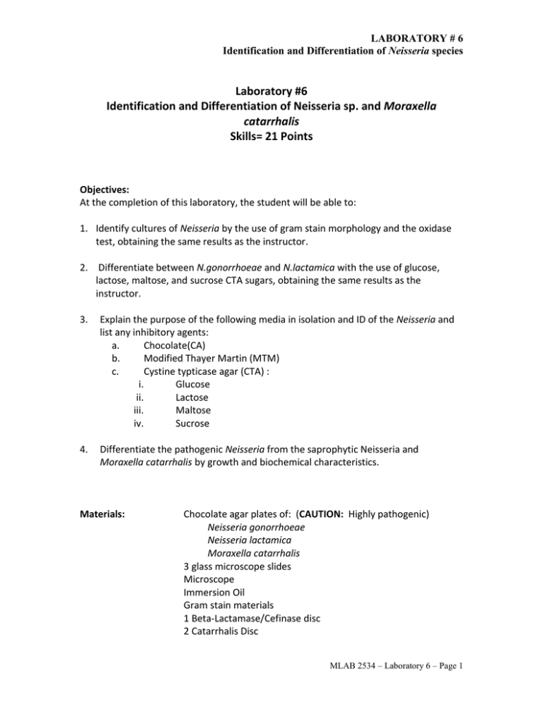
LABORATORY # 6 Identification and Differentiation of Neisseria species Laboratory #6 Identification and Differentiation of Neisseria sp. and Moraxella catarrhalis Skills= 21 Points Objectives: At the completion of this laboratory, the student will be able to: 1. Identify cultures of Neisseria by the use of gram stain morphology and the oxidase test, obtaining the same results as the instructor. 2. Differentiate between N.gonorrhoeae and N.lactamica with the use of glucose, lactose, maltose, and sucrose CTA sugars, obtaining the same results as the instructor. 3. Explain the purpose of the following media in isolation and ID of the Neisseria and list any inhibitory agents: a. Chocolate(CA) b. Modified Thayer Martin (MTM) c. Cystine typticase agar (CTA) : i. Glucose ii. Lactose iii. Maltose iv. Sucrose 4. Differentiate the pathogenic Neisseria from the saprophytic Neisseria and Moraxella catarrhalis by growth and biochemical characteristics. Materials: Chocolate agar plates of: (CAUTION: Highly pathogenic) Neisseria gonorrhoeae Neisseria lactamica Moraxella catarrhalis 3 glass microscope slides Microscope Immersion Oil Gram stain materials 1 Beta-Lactamase/Cefinase disc 2 Catarrhalis Disc MLAB 2534 – Laboratory 6 – Page 1 LABORATORY # 6 Identification and Differentiation of Neisseria species Applicator sticks Oxidase test materials CTA sugars: 3-Glucose 3-Lactose 3-Maltose 1-Sucrose (Demo only) References: 1. Mahon and Manuselis, Textbook of Diagnostic Microbiology, Fourth Edition, Chapter 1 2. Remel Catarrhalis Test Disc product insert 3. BD Oxidase Reagent product insert Principles: The genus Neisseria consists of oxidase-positive, gram-negative cocci that often appear as kidney bean-shaped diplococci on Gram stain. Most species are normal flora of the mucosa of the respiratory, alimentary, and genital tracts. Certain species such as Neisseria gonorrhoeae are recognized as true pathogens. Others such as Neisseria meningitidis, which are inhabitants of the oral and upper respiratory flora, may occasionally cause clinical disease. All species grow on blood agar except for N. gonorrhoeae, which requires an enriched medium such as chocolate or Thayer-Martin agar. Chocolate agar contains blood or hemoglobin that has been heated to 80oC until the media turns chocolate brown. This procedure makes nutrients within the RBCs accessible to the microorganism. MTM is a chocolate agar that contain antibiotics such as vancomycin( inhibits gram positives), colistin( inhibits gram negatives), nystatin( inhibits fungus) and trimethoprim(inhibits Proteus). Thayer Martin or TM has a chocolate agar base and includes vancomycin, colistin, and nystatin. Thayer Martin does not include trimethoprim. Enhanced CO2 (2% to 8%) is required for optimum incubation atmosphere. This is referred to a capnophilic environment. N. gonorrhoeae is a strict pathogen when isolated from any body site. Common sources include the urethra, cervix, and anal canal. Other infections may involve the joints, blood and conjunctiva. Transport media should be used if the specimen collected is not inoculated immediately to appropriate culture media. Colonies of N. gonorrhoeae on chocolate agar appear small, translucent, grayish, convex and shiny with entire margins. Suspected colonies are tested for oxidase and a smear is made for Gram stain. The Gram stain can be used to presumptively identify N. gonorrhoeae when intracellular gramnegative diplococcic are observed from male urethral specimens. The observance of gram-negative diplococcic from female genital specimens is not diagnostic due to other MLAB 2534 – Laboratory 6 – Page 2 LABORATORY # 6 Identification and Differentiation of Neisseria species gram-negative diplococcic which are normal flora. Definitive identification is based on carbohydrate utilization or other rapid identification techniques such as latex agglutination. Neisseria lactamica is a commensal that resides on the mucous membranes of the upper respiratory system. They may resemble pathogens or cause disease in compromised patients. N.lactamica can be confused with Neisseria meningitidis due to similar growth patterns on selective media. Moraxella catarrhalis is a member of the oral flora that has been recognized as an important agent of lower respiratory infections. Morphologically, these organisms resemble the Neisseria species. Definitive identification is based on carbohydrate utilization. The following chart shows the carbohydrate utilization reactions of Neisseria species. Species Tests/Results CTA Carbohydrates Maltose Lactose - Oxidase + Glucose + + + + - - N. lactamica + + + + - M. catarrhalis + - - - - N. gonorrhoeae N. meningitidis Sucrose - Procedure: 1. Cultural and Cellular Morphology a. Working in pairs, describe the colony morphology of isolated colonies of N. gonorrhoeae, N.lactamica and Moraxella catarrhalis and record on the report form. b. Prepare smears of each isolate. You will need a total of three (3) smears, putting two (2) smears on each slide. c. Gram stain each organism and record results on report form. 2. Cytochrome Oxidase Cytochrome oxidase is a bacterial enzyme that acts as the last link in the chain of aerobic respiration by transferring electrons (hydrogen) to oxygen, with the formation of water. The cytochrome system is possessed by aerobic or facultatively anaerobic organisms so that the oxidase test is important in identifying those organisms that either lack the enzyme or are obligate anaerobes. MLAB 2534 – Laboratory 6 – Page 3 LABORATORY # 6 Identification and Differentiation of Neisseria species The cytochrome oxidase test utilizes certain reagent dyes, such as p-phenylenediamine dihydrochloride, that substitute for oxygen as artificial electron acceptors. In the reduced state the dye is colorless; however, in the presence of cytochrome oxidase and atmospheric oxygen, p-phenylenediamine is oxidized, forming indophenol blue. The oxidase test must be performed on media without dye, since dye can interfere with color production. a. Hold the reagent dropper upright and point the tip away from yourself. Grasp with thumb and forefinger and squeeze gently to crush ampule inside the dropper. Tap bottom on tabletop a few times. Then, invert to dispense drop-by-drop. b. Add a few drops of oxidase test reagent to a strip of filter paper. NOTE: Do not place the filter paper saturated with oxidase reagent directly on the bench top because the bleach used to clean the countertops can cause a false positive reaction. c. Streak a loopful of N. gonorrhoeae on the reagent-saturated paper using either a loop or a wooden applicator stick. Observe reaction. d. Repeat steps a-c using N. lactamica and Moraxella catarrhalis. Interpretation: A positive reaction is indicated by a violet to purple color within 10-30 seconds. Negative reactions remain colorless or turn a light pink/ light purple after 30 seconds. Record results as either positive” or “negative” in report form. Note: delayed reactions should be ignored. 3. Carbohydrate Utilization with CTA Sugars Cystine trypticase agar (CTA) supports the growth of many fastidious organisms, including species of Neisseria and Moraxella. Various carbohydrates can be added to the CTA base for the purpose of testing for carbohydrate utilizations. Phenol red indicator is included in the medium and changes from red to yellow in the presence of acid, which is a waste product of carbohydrate utilization. a. Label tubes of CTA glucose, lactose, and maltose for each organism (N. gonorrhoeae, N. lactamica and Moraxella catarrhalis). b. Gather several colonies from the chocolate plate with an inoculating loop and inoculate the upper portion of the medium heavily. See picture below. MLAB 2534 – Laboratory 6 – Page 4 LABORATORY # 6 Identification and Differentiation of Neisseria species c. Fasten the caps of the tubes loosely and incubate at least 24 hours in a non-CO2 incubator. d. Record results as positive or negative on the report form. Interpretation: The development of a yellow band in the upper portion of the medium indicates production of acid and is interpreted as a positive test for utilization of the carbohydrate present. N.gonorrhoeae utilizes only glucose but N.lactamica utilizes glucose, lactose, and maltose with the production of acid. M.catarrhalis utilizes no sugars. 4. Beta-Lactamase/Cefinase β-Lactamases are enzymes that selectively destroy β-lactam molecules by attacking the β-lactam component of the molecule. Production of β-Lactamase is a significant mechanism contributing to β-lactam resistance in certain organisms, such as H. influenzae, N. gonorrhoeae and Moraxella catarrhalis, among others. Simple β-Lactamase testing is performed in the clinical laboratory to identify βLactamase production in these organisms, and a positive reaction means that the βlactam agents commonly used to treat infections caused by them (primarily ampicillin, amoxicillin, and penicillin) would be ineffective. Procedure: 1. For this lab, we will test only Neisseria gonorrhea. 2. Place the Cefinase disc on a clean microscope slide. 3. Moisten disc with one drop of DI water. 4. With an applicator stick, remove several well-isolated colonies and smear onto disc surface. 5. Observe for a color change. Interpretation: A positive reaction will show a yellow to red color change on the area when the culture was applied. A negative result will show no color change on MLAB 2534 – Laboratory 6 – Page 5 LABORATORY # 6 Identification and Differentiation of Neisseria species the disc. A positive result will usually develop within 5 minutes. Record results as either “positive” or negative” in result form. 5. Catarrhalis Test Disc Moraxella catarrhalis is a significant pathogen when isolated from respiratory cultures. The rapid ID of this organism is important, since most strains produce βlactamase and are resistant to penicillin and ampicillin. In this procedure, the enzyme butyrate, releases indoxyl from indoxyl butyrate and spontaneously forms indigo in the presence of oxygen. Procedure: a. Test only oxidase positive, gram negative diplococci. For this lab, we will test Neisseria gonorrhea and Moraxella catarrhalis. b. Using one (1) clean glass slide, take forceps and place disc on the left side of slide. c. Rub several colonies across the disc using a wooden applicator stick. d. Observe for a color change. e. Repeat steps b-d, placing a new disc on the right side of glass slide. Interpretation: A positive test is the development of a blue-green color within 2 minutes. A negative test lacks any color development within the 2 minutes. Record as either “positive” or “negative” in report form. Quality Control Quality control ensures that the information generated by the laboratory is accurate, reliable, and reproducible. This is accomplished by assessing the quality of the specimens, monitoring the performance of test procedures, reagents, media, and personnel. For the reagents used in this lab, QC should be performed weekly and on each new lot or shipment of the product using ATCC control organisms. Results should be reported in the QC log. Components of kit tests should not be interchanged with those from another kit. MLAB 2534 – Laboratory 6 – Page 6 LABORATORY # 6 Identification and Differentiation of Neisseria species Name:_____________ Date:______________ Points= 21 Lab #6: Id and Differentiation of Neisseria and Moraxella Species Report Form (1 pt. each) Test/Procedure Colony Morphology Gram stain Oxidase Glucose Lactose Maltose Β-lactamase Catarrhalis Disc Neisseria gonorrhoeae Neisseria lactamica Moraxella catarrhalis MLAB 2534 – Laboratory 6 – Page 7 LABORATORY # 6 Identification and Differentiation of Neisseria species Name:_______________ Date:________________ Lab #6: Identification and Differentiation of Neisseria sp and Moraxella Study Questions Points= 7 1. What is the difference between Thayer-Martin and Modified Thayer-Martin? What organism does this media select for? (2 pts) 2. What bacterial enzyme is being detected with the oxidase reagent? (1 pt) 3. Explain why cystine tryticase agar medium turns from red to yellow if the carbohydrate is utilized. (1 pt) 4. What diseases does Neisseria gonorrhoeae cause? (1 pt) 5. What are the optimal culture conditions for most gram-negative cocci and gramnegative pleomorphic coccobacilli? (1 pt) 6. Intracellular GNDC seen on a urethral gram stain can presumptively be identified as: (1 pt) a. Neisseria gonorrhoeae b. Neisseria meningitides c. Neisseria lactamica d. Moraxella catarrhalis MLAB 2534 – Laboratory 6 – Page 8
