Table 2 CT (ADULT)
advertisement
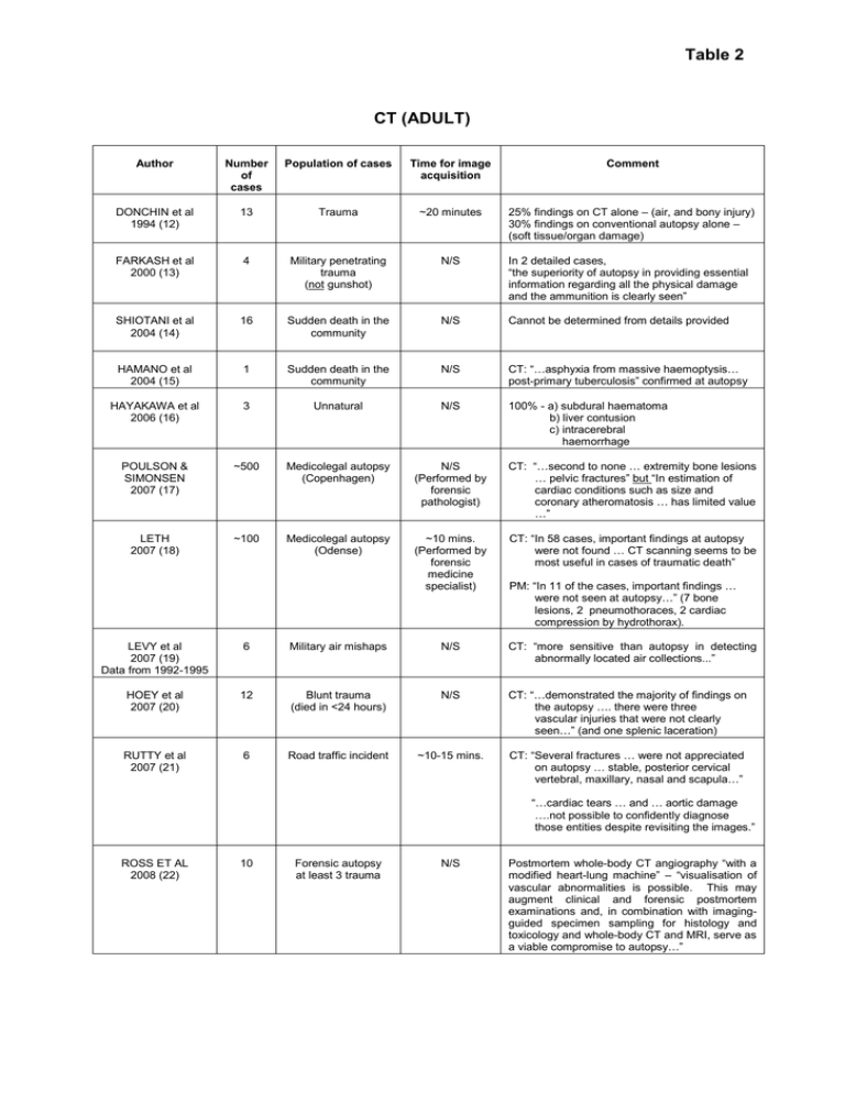
Table 2 CT (ADULT) Author Number of cases Population of cases Time for image acquisition Comment DONCHIN et al 1994 (12) 13 Trauma ~20 minutes 25% findings on CT alone – (air, and bony injury) 30% findings on conventional autopsy alone – (soft tissue/organ damage) FARKASH et al 2000 (13) 4 Military penetrating trauma (not gunshot) N/S In 2 detailed cases, “the superiority of autopsy in providing essential information regarding all the physical damage and the ammunition is clearly seen” SHIOTANI et al 2004 (14) 16 Sudden death in the community N/S Cannot be determined from details provided HAMANO et al 2004 (15) 1 Sudden death in the community N/S CT: “…asphyxia from massive haemoptysis… post-primary tuberculosis” confirmed at autopsy HAYAKAWA et al 2006 (16) 3 Unnatural N/S 100% - a) subdural haematoma b) liver contusion c) intracerebral haemorrhage POULSON & SIMONSEN 2007 (17) ~500 Medicolegal autopsy (Copenhagen) N/S (Performed by forensic pathologist) CT: “…second to none … extremity bone lesions … pelvic fractures” but “In estimation of cardiac conditions such as size and coronary atheromatosis … has limited value …” LETH 2007 (18) ~100 Medicolegal autopsy (Odense) ~10 mins. (Performed by forensic medicine specialist) CT: “In 58 cases, important findings at autopsy were not found … CT scanning seems to be most useful in cases of traumatic death” PM: “In 11 of the cases, important findings … were not seen at autopsy…” (7 bone lesions, 2 pneumothoraces, 2 cardiac compression by hydrothorax). LEVY et al 2007 (19) Data from 1992-1995 6 Military air mishaps N/S CT: “more sensitive than autopsy in detecting abnormally located air collections...” HOEY et al 2007 (20) 12 Blunt trauma (died in <24 hours) N/S CT: “…demonstrated the majority of findings on the autopsy …. there were three vascular injuries that were not clearly seen…” (and one splenic laceration) RUTTY et al 2007 (21) 6 Road traffic incident ~10-15 mins. CT: “Several fractures … were not appreciated on autopsy … stable, posterior cervical vertebral, maxillary, nasal and scapula…” “…cardiac tears … and … aortic damage ….not possible to confidently diagnose those entities despite revisiting the images.” ROSS ET AL 2008 (22) 10 Forensic autopsy at least 3 trauma N/S Postmortem whole-body CT angiography “with a modified heart-lung machine” – “visualisation of vascular abnormalities is possible. This may augment clinical and forensic postmortem examinations and, in combination with imagingguided specimen sampling for histology and toxicology and whole-body CT and MRI, serve as a viable compromise to autopsy…”
