Lab 5: Exploring the Effects of Density on Fern Development
advertisement
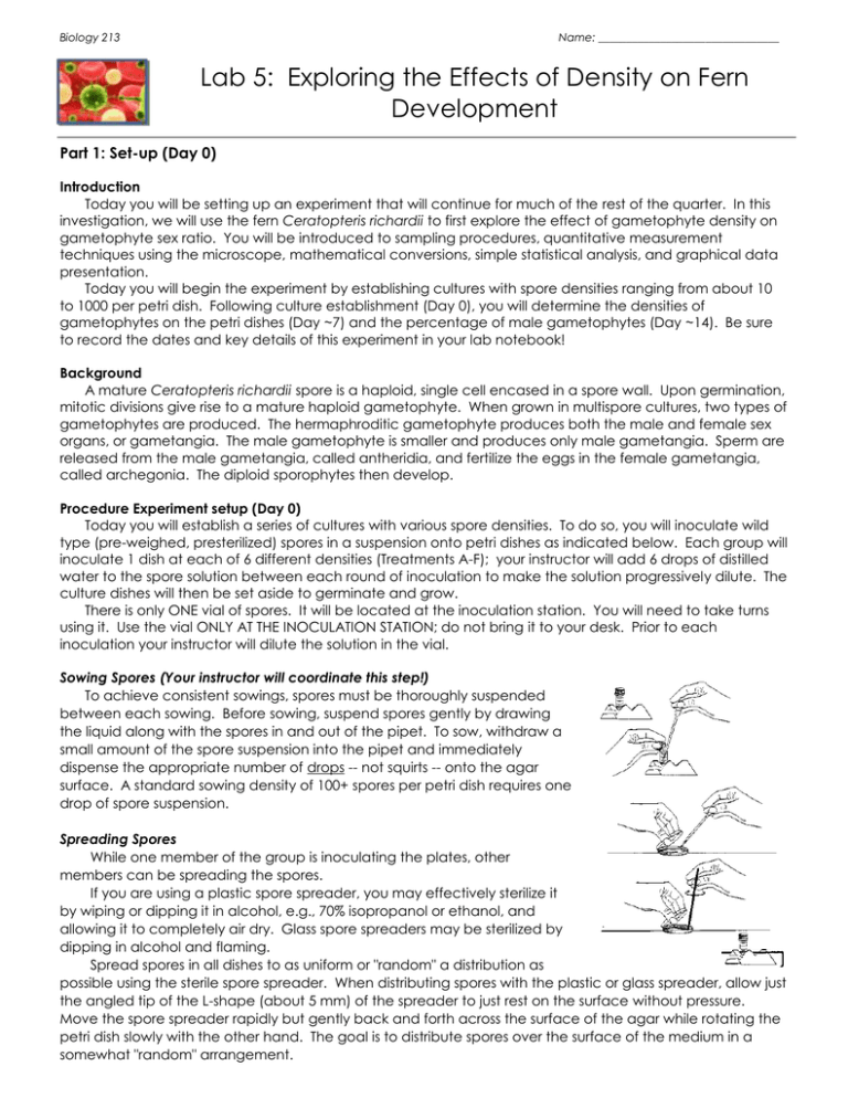
Biology 213 Name: ________________________________ Lab 5: Exploring the Effects of Density on Fern Development Part 1: Set-up (Day 0) Introduction Today you will be setting up an experiment that will continue for much of the rest of the quarter. In this investigation, we will use the fern Ceratopteris richardii to first explore the effect of gametophyte density on gametophyte sex ratio. You will be introduced to sampling procedures, quantitative measurement techniques using the microscope, mathematical conversions, simple statistical analysis, and graphical data presentation. Today you will begin the experiment by establishing cultures with spore densities ranging from about 10 to 1000 per petri dish. Following culture establishment (Day 0), you will determine the densities of gametophytes on the petri dishes (Day ~7) and the percentage of male gametophytes (Day ~14). Be sure to record the dates and key details of this experiment in your lab notebook! Background A mature Ceratopteris richardii spore is a haploid, single cell encased in a spore wall. Upon germination, mitotic divisions give rise to a mature haploid gametophyte. When grown in multispore cultures, two types of gametophytes are produced. The hermaphroditic gametophyte produces both the male and female sex organs, or gametangia. The male gametophyte is smaller and produces only male gametangia. Sperm are released from the male gametangia, called antheridia, and fertilize the eggs in the female gametangia, called archegonia. The diploid sporophytes then develop. Procedure Experiment setup (Day 0) Today you will establish a series of cultures with various spore densities. To do so, you will inoculate wild type (pre-weighed, presterilized) spores in a suspension onto petri dishes as indicated below. Each group will inoculate 1 dish at each of 6 different densities (Treatments A-F); your instructor will add 6 drops of distilled water to the spore solution between each round of inoculation to make the solution progressively dilute. The culture dishes will then be set aside to germinate and grow. There is only ONE vial of spores. It will be located at the inoculation station. You will need to take turns using it. Use the vial ONLY AT THE INOCULATION STATION; do not bring it to your desk. Prior to each inoculation your instructor will dilute the solution in the vial. Sowing Spores (Your instructor will coordinate this step!) To achieve consistent sowings, spores must be thoroughly suspended between each sowing. Before sowing, suspend spores gently by drawing the liquid along with the spores in and out of the pipet. To sow, withdraw a small amount of the spore suspension into the pipet and immediately dispense the appropriate number of drops -- not squirts -- onto the agar surface. A standard sowing density of 100+ spores per petri dish requires one drop of spore suspension. Spreading Spores While one member of the group is inoculating the plates, other members can be spreading the spores. If you are using a plastic spore spreader, you may effectively sterilize it by wiping or dipping it in alcohol, e.g., 70% isopropanol or ethanol, and allowing it to completely air dry. Glass spore spreaders may be sterilized by dipping in alcohol and flaming. Spread spores in all dishes to as uniform or "random" a distribution as possible using the sterile spore spreader. When distributing spores with the plastic or glass spreader, allow just the angled tip of the L-shape (about 5 mm) of the spreader to just rest on the surface without pressure. Move the spore spreader rapidly but gently back and forth across the surface of the agar while rotating the petri dish slowly with the other hand. The goal is to distribute spores over the surface of the medium in a somewhat "random" arrangement. Biology 213 It is important to carefully spread spores so that later observations of developed gametophytes are not obscured by clumping. The excess water from the inoculation volume will absorb into the agar over the next 24-48 hours. Spores from dishes D, E, and F may be sufficiently sparse such that only minimal spreading is required. Procedure 1. Work in groups of 4 or as assigned by your instructor. 2. Obtain 6 culture dishes. Label all dishes with your Group Name, Today's Date, Time Your Class Meets, and Treatment Code as follows: A, B, C, D, E, and F. 3. For steps 4-6 below, be sure that spores are well suspended before sowing. 4. Sow 1 drop of suspension onto your A dish, using the technique described above. It is important that only a single uniform drop -- not a squirt -- of spore suspension is sown onto a dish. 5. Return any unused solution back to the spore vial. 6. After all groups have inoculated their A plate and the Instructor has diluted the solution, sow 1 drop of spore suspension onto your B dish. Again, return any unused solution back to the spore vial. 7. Repeat for plates C-F in order, waiting for the Instructor to dilute the solution before each inoculation. 8. Check dishes E and F for sufficient spores; i.e., dish E should have about 30 spores and dish F should have about 10 spores. Adjust number if necessary, with spores remaining in vial. 9. Sterilize your spreader. 10. Spread the spores, following the directions described above. 11. Put all dishes into a Culture Dome, which your instructor will place under lights. Part 2: Determining the Effect of Density on Fern Development (Day 14+) Introduction Recall that in this investigation, we are using the fern, Ceratopteris richardii, to first explore the effect of gametophyte density on gametophyte sex ratio. Today you will be collecting data on your developing fern to determine the percentage of male and hermaphroditic gametophytes. After collecting data, you may then permit the gametophytes to mate. You will write up your results as key sections of a scientific paper. Background Recall that when grown in multispore cultures, two types of gametophytes are produced. The hermaphroditic gametophyte (far right) produces both male and female sex organs, or gametangia. The male gametophyte is smaller (near right) and produces only male gametangia. Sperm are released from the male gametangia, called antheridia (singular = antheridium), and fertilize the eggs in the female gametangia, called archegonia (singular = archegonium). This produces the zygote that becomes the large, diploid sporophyte. You can readily observe the developing gametophytes at low magnification using a dissecting microscope. Using transmitted illumination from below works best. Because some microscopes' stages become quite warm after extended use of transmitted illumination, PLEASE turn the base light off and remove the culture plates from the stage when they are not being observed. If cultures are to be observed with the petri dish lids in place, heat may cause condensation to form on the inside of the lid, obstructing observation. If spare lids from clean unused dishes are available, these can be used to replace the fogged lids. If spare lids are not available, you can remove the condensation by carefully wiping it off with a clean, preferably sterile, tissue. If lids do not need to be kept in place during observation, condensation will not be a problem. However, without the lid in place, gametophytes may begin to show signs of drying after extended observation. For this reason, it is important to replace the lid on the cultures when they are not being observed. For greater detail, you can also use a compound microscope (40-400X) to observe wet mounts of a few gametophytes. Select and remove gametophytes from the culture medium with a sterile probe or toothpick. modified 4/22/99 from material from Carolina Biological Supply Co. Procedure: Data Collection and Analysis (Day 14+) Data Collection 1. Obtain a sampling grid, your culture dishes, and a dissecting microscope. 2. Sketch one of each type of gametophyte in your lab notebook, labeling the location of antheridia and archegonia, the old spore coat, the notch meristem, and the rhizoids. 3. Determine the percentage of male and hermaphroditic gametophytes on each dish sown. Use the sampling grid to keep track of which gametophytes have been counted. Note: It may be necessary to use a sterile toothpick or dissecting needle to spread out clusters of gametophytes to allow for accurate counting. Record your results in an organized table in your lab notebook! a. For low density dishes (D-F), count and categorize all of the gametophytes on the dish. Enter data into the appropriate column in Table 1. Calculate the percentage of each sexual type by dividing the "Number of Male or Hermaphroditic Gametophytes" by the "Total Number of Gametophytes" b. For high density dishes (A-C), a sampling procedure may be used to estimate the actual percentage of the sexual types. Use the sampling grid and count the numbers of male and female gametophytes in 5 randomly selected grids. Count all of the gametophytes in each grid before proceeding to the next grid. Enter the "Number of Male Gametophytes," the "Number of Hermaphroditic Gametophytes," and the "Total Number of Gametophytes” in your notebook. Take care to include the area counted as we need to know the overall density (#spores/cm 2). Calculate the percentage of each sexual type by dividing the number of male or hermaphroditic gametophytes by the total number of gametophytes. One member of each group should record your results in the class database. 4. Keep your culture dishes for the next procedure (see below). Thought Questions 1. Why should spores be spread to a uniform or random distribution? 2. From an evolutionary and/or breeding system perspective, why would such a relationship exist between gametophyte population density and sex expression (i.e., what would the advantage be for such a relationship)? HINT: How do changing proportions of males influence the kinds of mating that can occur? 3. The relationship between gametophyte population density and sex expression provides information about how the sex expression response occurs. a. Do the data indicate that any gametophyte can develop into a male? Why or why not? b. Suggest a mechanism by which the sex expression response occurs. Use the data to explain why you suggested the mechanism you did. Part 3: Fertilization (Day 14+) Background Fertilization, or the union of sperm and egg resulting in the formation of a zygote, occurs after a sperm successfully makes its way down the archegonial neck. The archegonium is a flask-shaped organ embedded in the region behind the notch meristem. The neck of the archegonium protrudes from the surface of the gametophyte and has a central column of cells (neck canal cells) surrounded by the neck cells proper. Procedure: Fertilization of Gametophytes 1. To each petri dish, add sterile distilled water to completely cover the surface, that is about 3 mL for a 60mm petri dish. 2. After about 2-5 min, mature antheridia will release the sperm cells into the water. Initially the sperm do not swim but become motile within a short time (seconds). Place a dish (a high density one is best -why?) under the microscope (try the dissection microscope first), and observe the sperm swimming in the water and swarming toward archegonia. (The rapid movement of sperm can make observation difficult unless they are caught on something or slow down.) 3. Allow the water to stand for 30 min, pour off the water, and allow the dishes to drain upside down for 30 min on paper towels. 4. Return dishes (with their lids back on) to the culture dome, and your instructor will place it under lights. Thought Questions 1. What does water do? What limits does this place on where ferns can live? 2. How can the sperm cells find their way to archegonia and thus to the egg cell? Biology 213 Post Lab: 1. Write a good hypothesis to describe this experiment. 2. Prepare two graphs of the data. On the first, illustrate the relationship between density and sex ratio from your group’s plates. On the second, illustrate the same relationship for the class data. As always, be sure to give your graphs a descriptive title, and provide both labels and units for each axis. 3. Prepare a 1-2 paragraph discussion of the data. Your discussion should explain the data you present in your graphs, and address one or more of the “thought questions” posed in part 2 above! modified 4/22/99 from material from Carolina Biological Supply Co.
