ardea2008.doc
advertisement
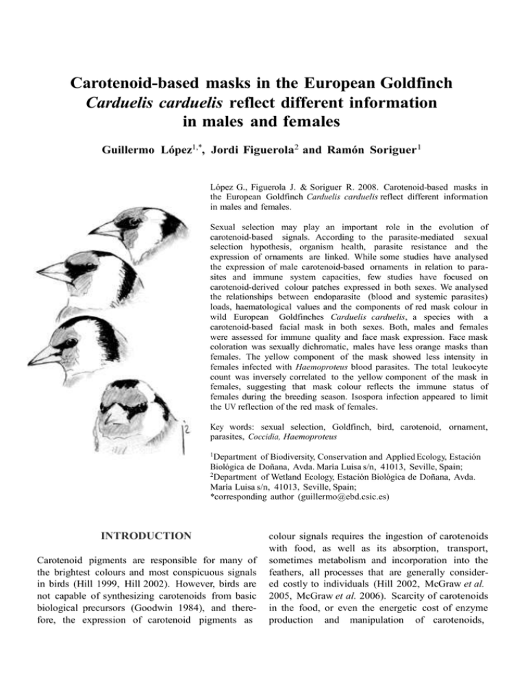
Carotenoid-based masks in the European Goldfinch Carduelis carduelis reflect different information in males and females Guillermo López1,*, Jordi Figuerola2 and Ramón Soriguer1 López G., Figuerola J. & Soriguer R. 2008. Carotenoid-based masks in the European Goldfinch Carduelis carduelis reflect different information in males and females. Sexual selection may play an important role in the evolution of carotenoid-based signals. According to the parasite-mediated sexual selection hypothesis, organism health, parasite resistance and the expression of ornaments are linked. While some studies have analysed the expression of male carotenoid-based ornaments in relation to parasites and immune system capacities, few studies have focused on carotenoid-derived colour patches expressed in both sexes. We analysed the relationships between endoparasite (blood and systemic parasites) loads, haematological values and the components of red mask colour in wild European Goldfinches Carduelis carduelis, a species with a carotenoid-based facial mask in both sexes. Both, males and females were assessed for immune quality and face mask expression. Face mask coloration was sexually dichromatic, males have less orange masks than females. The yellow component of the mask showed less intensity in females infected with Haemoproteus blood parasites. The total leukocyte count was inversely correlated to the yellow component of the mask in females, suggesting that mask colour reflects the immune status of females during the breeding season. Isospora infection appeared to limit the UV reflection of the red mask of females. Key words: sexual selection, Goldfinch, bird, carotenoid, ornament, parasites, Coccidia, Haemoproteus 1Department of Biodiversity, Conservation and Applied Ecology, Estación Biológica de Doñana, Avda. María Luisa s/n, 41013, Seville, Spain; 2Department of Wetland Ecology, Estación Biológica de Doñana, Avda. María Luisa s/n, 41013, Seville, Spain; *corresponding author (guillermo@ebd.csic.es) INTRODUCTION Carotenoid pigments are responsible for many of the brightest colours and most conspicuous signals in birds (Hill 1999, Hill 2002). However, birds are not capable of synthesizing carotenoids from basic biological precursors (Goodwin 1984), and therefore, the expression of carotenoid pigments as colour signals requires the ingestion of carotenoids with food, as well as its absorption, transport, sometimes metabolism and incorporation into the feathers, all processes that are generally considered costly to individuals (Hill 2002, McGraw et al. 2005, McGraw et al. 2006). Scarcity of carotenoids in the food, or even the energetic cost of enzyme production and manipulation of carotenoids, 234 ARDEA 96(2), 2008 remain unclear. Carotenoids are also involved in metabolic pathways related to host immunity and a trade-off between the ornamental and health functions of carotenoids has been proposed (Hõrak et al. 2004a, Møller et al. 2004). According to this hypothesis, signals based on carotenoid pigments are costly and therefore act as honest indicators of an individual’s quality. The features that each carotenoid-based ornament reflect have been mainly studied in males of very dimorphic species. Carotenoid-based ornaments have been shown to be related to body condition (McGraw et al. 2002, Saks et al. 2003, Jawor & Breitwisch 2004, Jawor et al. 2004), sexual selection (Collias et al. 1979, Hill 1991, Drachman 1997) and even parasite loads (Figuerola et al. 2003, Hill et al. 2004, Hõrak et al. 2004). In the case of parasites, studies have reported variously negative (Thompson et al. 1997, Brawner et al. 2000, Figuerola et al. 2003, Hõrak et al. 2004b), positive (Burley et al. 1991, VanHoort & Dawson 2005) and non-significant (Seutin 1994) relationships between parasites and the expression of a carotenoid-based ornament. The potential causes of these contradictory results could stem from varying methods of study (e.g. correlational vs. experimental, ranges or doses of experimental treatments used), different impacts on parasites on their hosts and environmentaldependent effects of parasites on host condition (Figuerola et al. 2003). Work published up to now has generally focused on a single type of parasite, above all blood parasites, and analysis of the relative and combined impact of different types of parasites on ornament expression is still lacking. Haematozoan parasites are blood-cell parasite protozoa transmitted by blood-sucking arthropods that are quite prevalent in wild passerines (Deviche et al. 2001). Coccidian protozoa are widespread intestinal epithelium parasites with a direct biological cycle; transmission results from the ingestion of oocysts liberated in the faeces of an infected individual. Passerines are mainly infected by genus Isospora (Hill 2002, Hõrak et al. 2006). Both haematozoan and coccidian parasites affect their hosts in a condition-dependent way (Merino et al. 2000): they have little impact when resources are abundant (Weatherhead & Bennett 1992, Friend & Franson 2001), but affect negatively the host when resources are scarce (Ots & Hõrak 1998, Ilmonen et al. 1999). The mechanisms leading Coccidia to limit the expression of carotenoid-based ornaments may work in at least two different direct ways. First, they may reduce the absorption of carotenoids through the intestine (Tyczkowski et al. 1991, Allen 1992) and, second, they may reduce the release of high-density lipoproteins (Allen 1987), which are responsible for the transport of carotenoids in the bloodstream to the tissues. Moreover, a third indirect mechanism related to body condition – to which both carotenoid ornamentation and immunity have shown to be linked – may also be at work (see Smith et al. 2007). The mechanisms through which Haematozoa may limit the expression of carotenoid-based ornaments are still unknown. Most of the relationships described up to now between parasites and the expression of ornaments have been made focusing on conspicuous male ornaments, but little attention have been traditionally paid to such a relation in female ornaments. Roulin (2001), for instance, conducted an experimental study by comparing the degree of female Barn Owl Tyto alba ornamentation (eumelanin-based spottiness) with parasite resistance in their offspring raised by foster parents. He found that in females ornamentation positively reflects parasite resistance ability. Some observational studies have also demonstrated that the expression of ornaments is negatively related to the parasite load in female birds (Hõrak et al. 2001, Piersma et al. 2001). The aim of this study was to explore the relationship between the colour of the carotenoidbased red mask of European Goldfinches, and a number of indices of condition (haematological parameters and different parasite loads) in both sexes, paying attention to possible inter-sex differences. The study was carried out with free-living birds during the breeding season, just after mate choice had occurred, when individuals were going through the costly task of raising young, because López et al.: GOLDFINCH RED MASK IMPLICATIONS under these conditions we expected the effect of parasites on their hosts to be maximal. To our knowledge, this is the first study done analysing the relationship between plumage coloration in both sexes and several groups of parasites at a time. METHODS The European Goldfinch is a 12-cm long, seed-eating finch that has a unique colour pattern on its head. The front of the face has a conspicuous crimson patch, which is known to be composed of four carotenoid pigments (Stradi et al. 1995): a) ε,εcarotene-3,3'-dione, b) 3-hydroxy-ε,ε-carotene-3'one, c) 4,4'-dihydroxy-ε,ε-carotene-3,3'-dione (isoastaxanthin), and d) 4-hydroxy-ε,ε-carotene-3,3'dione. Although ε,ε-carotene-3,3'-dione and 3-hydroxyε,ε-carotene-3'-one are very common yellow pigments in cardueline finches, isoastaxanthin and 4hydroxy-ε,ε-carotene-3,3'-dione have not yet been found in any other species studied to date. These two pigments provide the red colour in the mask together with the keratin bond arrangement the pigments have in the feathers (Stradi et al. 1995). Although both sexes are superficially similar (Cramp & Perrins 1994), small differences in the size of the patch exist, being larger on average in males (Svensson 1996). To our knowledge, differences between the sexes in mask colour have never been investigated. Fieldwork In the springs of 2004 and 2005, we trapped 13 adult female and 44 adult male goldfinches in a tree nursery in the Spanish city of Seville (37°23' N, 5°57'W) where these finches are common resident breeders. Birds were captured between sunrise and sunset in 20 twelve-metre long mist-nets. Individuals were marked with numbered aluminium rings. Sex was determined by the presence of a brood patch (only present in females) or a cloacal protuberance (only present in males), and by the colour of the lesser wing-coverts (see 235 Svensson 1996). We also measured body mass (to the nearest 0.1 g) and wing length (maximum chord). Birds were kept individually in clean ringing bags for 20 minutes to collect faecal samples. Faeces were immediately placed in individually marked vials containing 5% formol, and the time of collection was recorded for each sample. To control the mass of faecal samples, we avoided taking the urine-based part of the excretion and only collected the intestinal-based portion. We drew 0.1 ml of blood from the jugular vein using 29 G sterile insulin syringes and prepared smears on a microscopy slide as per Bennett (1970), which were air-dried, fixed and stained using Diff-Quick solution. To confirm field sexing an analysis of the cellular fraction of a drop of blood was performed. Sex was determined from blood cell DNA via a polymerase chain reaction (PCR) amplification of the CHD genes (Ellegren 1996, Griffiths et al. 1998). After blood extraction, we took two colour measurements of the frontal area of the red mask in the 57 trapped birds using a MINOLTA CM2600d spectrometer, which measures the characteristics of reflected light by illuminating the feather surface under standard light conditions. We obtained the reflectance curve of the mask, that is, the light reflection from the UVA (360 nm) to the end of the visible spectrum (740 nm), measured at 10 nm intervals (39 intervals). The UVA reflection is visible to birds and has important implications in sexual selection in some passerine species (Saetre 1994, Siitari et al. 2002, Pearn et al. 2003). Although 700 nm has been shown as a maximum wavelength for avian visual sensitivity, we included 700–740 nm interval within the analysis because birds indeed present variability in visual spectrum among different species (Bowmaker et al. 1997) and, to our knowledge, this spectrum has never been studied in the Eurasian Goldfinch. Blood smear analysis For each blood smear, we estimated the total leukocyte count (TLC) by counting the number of leukocytes on twenty 400x light microscope fields of similar density and multiplying this value by ARDEA 96(2), 2008 100 (Wiskott 2002). The differential leukocyte count was made by identifying (according to Campbell 1995) the cellular type (heterophils, eosinophils, basophiles, lymphocytes or monocytes) of 100 leukocytes at 1000x magnification. The heterophil-lymphocyte ratio (H/L) was calculated as the percentage of heterophils divided by the percentage of lymphocytes. A total of 15 000 erythrocytes were scanned for blood parasites at low (400x) and high (oil 1000x) magnification (Godfrey et al. 1987) and in infected individuals, the blood parasite load was estimated as the percentage of infected erythrocytes. Prevalence was calculated on the basis of the percentage of infected individuals. Only Haemoproteus spp. (prevalence: 23.7%) and Plasmodium spp. (7.9%) were found in the 38 samples analysed. The repeatability of all variables was estimated by counting twice the smears of ten individuals and calculated as the intra-class correlation (Lessells & Boag 1987). Repeatabilities were high for TLC (95%), H/L (90%), and blood parasite infection (92%). PC2 0,8 PC1 factor loadings 236 0,4 PC3 0,0 PC4 –0,4 –0,8 400 500 600 700 wavelength (nm) Figure 1. Factor loading of the four principal components calculated from light reflectance at 10 nm intervals between 360 and 740 nm. PC1 represents the blue-violet component, PC2 the red one, PC3 includes the yellows, and PC4 represents the UVA reflection. Coprology In the laboratory, faecal samples were passed through a double lint filter and mixed to obtain a homogeneous dilution, which was then analysed for coccidian oocysts and other endoparasite eggs using a McMaster chamber. This method only provides an estimation of the real parasite load, although it has been described as the only possible non-invasive method of research on the intestinal parasite of wild animals (Watve & Sukumar 1995). Samples were scored as positive, when coccidian oocysts were observed, and negative, when not. Only protozoan coccidia were found in the sample. Based on size and the number of sporocysts, the oocysts were identified as Isospora-like (Baker et al. 1972, Grulet et al. 1982). Repeatability of coccidian infection estimated from samples of 10 individuals scored twice was very high (97%), giving confidence in the accuracy of oocyst counts. ferent wavelength intervals. Four relevant components (Eigenvalues > 1) were obtained that together summarised 99.97% of variance (Fig. 1). The first component (PC1) summarised reflection in the visible spectrum, mainly between 400 and 530 nanometers (within violets and blues). PC2 was more positive for individuals with more reflection at 650–740 nanometers (reds) and less reflection at lower wave lengths. PC3 was negatively related to reflection in the 550–590 nanometers intervals (yellows), so this component represents the yellow carotenoids reflection. PC4 was mainly influenced by reflection in the non-visible-tohumans portion of the spectra, between 360 and 390 nanometers (UVA radiation). The repeatability of the colour measurements was calculated as the intra-class correlation of the principal components from ten individuals measured twice (Lessells & Boag 1987). The repeatability was very high for all components (PC1: 98%; PC2: 96%; PC3: 97%; PC4: 96%) because of the great accuracy of the spectrometer method (see Figuerola et al. 1999). Colour characteristics Reflectance curves were analysed by a Principal Component Analysis of reflectances at the 39 dif- Statistical analyses TLC was log-transformed to fit a normal distribution. H/L did not fit normality by any common 237 López et al.: GOLDFINCH RED MASK IMPLICATIONS RESULTS The coloration of the red mask in the European Goldfinch was sexually dichromatic. Sexes differed in reflectance along the whole visible spectrum (PC1, PC2 and PC3), especially within the yellow region (PC3), but not in the UV (PC4) (Table 1). 20 males 16 females reflectance transformation so ranked values were used in the analyses (Conover & Iman 1981). We analysed sexual dimorphism in colouration (PC1, PC2, PC3 and PC4), haematological values (TLC and H/L), and parasite (Haemoproteus and Isospora) load with ANOVAs including sex as a factor. We analysed the effects of Haemoproteus and Isospora infections (presence/absence) as factors, and the effects of TLC and ranked H/L on PC1, PC2, PC3, PC4 as dependent variables in two MANOVAs. Due to circadian rhythms affecting coccidian prevalence in passerines (López et al. 2007), morning/ afternoon factor was included in the model including Isospora infection. All the two-way interactions among covariates and sex and morning/ afternoon factor were included in the models, and stepwise backwards selection procedure was followed until all the independent variables remaining in the model increased significantly the fit of the model. 12 8 4 0 400 500 600 700 wavelength (nm) Figure 2. Mean reflection curves of the red mask of the Goldfinch along the UV and the whole visible spectrum by sex. Reflectance curves showed that males were on average much more red and less yellow than females (Fig. 2). No differences in haematological values or parasite infection were found between sexes (Table 1). None of these variables were related to PC1 or PC2 (Tables 2 and 3). Haemoproteus infection, TLC, and their interaction with sex were related to PC3 (Tables 2 and 3, Fig. 3A and B). Isospora infection, its interaction with sex, and the interaction between sex and Haemoproteus infection were related to PC4 (Tables 2 and 3, Fig. 3C). Table 1. Mean, SE and sample size for males and females for the colour, parasites and haematological variables analysed. Differences between sexes were tested by one-way ANOVA. Males PC1 PC2 PC3 PC4 Isospora Haemoproteus TLC H/L Females mean SE n mean SE n F –0.174 0.178 0.248 0.016 1.300 0.030 3.771 0.856 0.173 0.172 0.132 0.156 0.231 0.013 0.03 0.040 33 33 33 33 37 31 31 31 0.573 –0.588 –0.820 –0.051 1.020 0.002 8.838 0.704 0.261 0.260 0.407 0.426 0.282 0.002 0.080 0.033 10 10 10 10 12 7 7 7 4.64 4.93 10.80 0.03 2.37 0.92 0.87 3.25 P 0.05 0.03 <0.01 0.86 0.13 0.35 0.36 0.08 238 ARDEA 96(2), 2008 Table 2. Results of stepwise backwards selection procedure MANOVA analysing parasite infection over colour components of the red mask of the Eurasian Goldfinch. Table 3. Results of stepwise backwards selection procedure MANOVA analysing haematological values over colour components of the red mask of the Eurasian Goldfinch. Source Source Dependent variable F1, 32 P Dependent variable Df F P Sex PC1 PC2 PC3 PC4 7.21 3.09 0.03 32.91 0.014 0.092 0.872 <0.001 Sex PC1 PC2 PC3 PC4 32 32 32 32 0.81 0.58 10.38 8.24 0.551 0.783 0.002 0.498 Morning/afternooon PC1 PC2 PC3 PC4 0.18 1.64 0.45 1.81 0.675 0.214 0.508 0.192 Log (TLC+1) PC1 PC2 PC3 PC4 32 32 32 32 0.49 0.01 6.36 9.69 0.616 0.201 0.017 0.807 Haemoproteus PC1 PC2 PC3 PC4 0.47 0.88 9.80 1.72 0.499 0.358 0.005 0.204 Sex x Log (TLC+1) PC1 PC2 PC3 PC4 32 32 32 32 0.83 0.63 11.84 8.30 0.633 0.741 0.001 0.546 Isospora PC1 PC2 PC3 PC4 1.85 2.64 1.37 20.38 0.187 0.118 0.254 <0.001 Ranked H/L PC1 PC2 PC3 PC4 31 31 31 31 0.21 1.28 2.46 0.02 0.651 0.266 0.127 0.885 Sex x Haemoproteus PC1 PC2 PC3 PC4 0.60 0.26 8.91 4.89 0.446 0.614 0.007 0.037 Sex x Isospora PC1 PC2 PC3 PC4 2.00 1.38 0.26 28.54 0.171 0.253 0.616 <0.001 DISCUSSION Colour dichromatism has not been reported before in the masks of the European Goldfinch. Our results show that hues differ between sexes: males reflect reds more strongly than females, but reflect yellows and oranges with lower intensity than females. The carotenoids expressed in the mask, are qualitatively the same in both sexes (Stradi 1995). McGraw et al. (2002) also found that male American Goldfinches Carduelis tristis artificially- fed with ad libitum canthaxantin were more colourful than females, due to a higher carotenoid concentration in the feathers. The larger accumulation of red pigments in males than in females seems thus to be the most plausible option for explaining colour differences in European Goldfinches. Testosterone, by means of its capacity to upregulate lipoprotein status, has been proposed as the responsible agent for such differences in the American Goldfinch (McGraw et al. 2006), but there are also studies showing opposite outcomes in House Finches Carpodacus mexicanus (Stoehr & Hill 2001). Diet differences between sexes, health variations or the effect of other hormones should not be discarded to explain this sexual dichromatism. Even a differential selection of ornaments between sexes could also be an underlying factor regarding the dichromatism. Unfortunately, no information is available on any of these aspects in the Eurasian goldfinch. López et al.: GOLDFINCH RED MASK IMPLICATIONS 2 A 1 PC3 0 –1 –2 –3 males females –4 3.4 3.6 3.8 4.0 4.2 log (TLC+1) 1 0.8 B mean PC4 mean PC3 0 –1 –2 –3 –4 C 0.6 0.4 0.2 0.0 –0.2 males females negative positive Haemoproteus infection –0.4 negative positive Isospora infection Figure 3. A) Scatter graph of log-transformed TLC over the PC3 values, by sex. B) Mean PC3 in relation to Haemoproteus infection state by sex. C) Mean PC4 in relation to Isospora infection state by sex. When interpreting our results relating coloration to health it is important to consider that the study was carried out in the spring. European Goldfinches moult their masks in late summer (Jenni & Winkler 1994), around six months before reproduction takes place. The health status of individuals is expected to change during this time, although the signals involved in sexual selection act in the early spring at the time of mate choice (Cramp et al. 1994). The red of the mask is not fully developed at the time of the moult and is only completed during the spring due to the abrasion of melanin derived feather tips (Svensson 1996). We sampled birds at the time of the year when the expression of the ornament is at its 239 fullest, just when the indicator function of a sexual selection signal should be at work. We found a relationship between presence of different parasites and different colour components of red masks. However, these effects were sex-dependent and only significant for females. Female European Goldfinches infected with Haemoproteus blood parasites and those with higher TLC values were more orange, that is with a higher yellow component, than those uninfected or with lower values. A higher intensity in yellow component may be due to 1) a decrease in the intensity of red pigments, or 2) an increase in the intensity of yellow ones. Because red pigments are predominant in the red mask, we think that the first option is more plausible than the second one. In this way, the red of the mask may reflect Haemoproteus parasitemia or infection resistance in females. Also, female European Goldfinches with higher TLC values were more orange (with a higher yellow component) than those with lower values. Since high TLC values are linked to chronic or acute infections (Campbell 1995), the red of the mask may reflect immune levels or infection resistance in females. In this way, the most infected females would have less red ornaments, a finding that suggests that red masks act as an honest indicator of general infection in female European Goldfinches. This relationship is not significant in males, probably due to the different reproductive role of each sex, since egg-laying and incubation, a very expensive process, is carried out only by females (Cramp et al. 1994). UV reflection was higher in Isospora non-infected females than in infected ones in our study. This result seems to indicate that UV reflection acts as an honest indicator of Isospora infection in females. Finally, our results suggest that double-infected animals (with Haemoproteus and Isospora) reflect violets with a higher intensity that those non-infected. This fact could be due to the lack of red and yellow pigments (carotenoids) that those individuals have in their feathers. Although H/L has shown to be related with some aspects of stress and condition in passerines (Ots et al. 1998, Groombridge et al. 2004), it was not related to any colour variables in our study. 240 ARDEA 96(2), 2008 How is it possible that breeding roles had an effect on the relationship between coloration and parasites if plumage was developed several months before breeding? We suggest that under the stress derived from breeding activities females in worst condition or with a less active immune system are less able to control already present infections and/or exclude new infections when exposed to pathogens. A similar process was experimentally demonstrated to work in male Greenfinches Carduelis chloris experimentally infected with Sindbis virus (Lindström & Lundstrom 2000). In conclusion, our study shows that (1) sexual dichromatism exists in the colour of the mask of the European Goldfinch, and (2) the red colour of the mask reflects different signals in both sexes and may be a reliable indicator of parasite infection during the breeding season, at least in females. ACKNOWLEDGEMENTS The Spanish Health Ministry via its Thematic Research Net ‘EVITAR’ funded our research. Alberto Álvarez, Alicia Cortés, Ángel Mejía, Ara Villegas, Beatriz Fernández, Carmen Gutiérrez, Chari Terceño, Cristina Sánchez, Elena Fierro, Enrique Sánchez, Esteban Serrano, Francisco Miranda, Grego Toral, Inma Cancio, Joaquín Díaz, José Antonio Sánchez, Mari Carmen Roque, Olga Jiménez, Pedro Sáez, Rafael Reina, and Samuel del Río helped with fieldwork. Beatriz Sánchez helped with fieldwork and provided moral support. Cuqui Rius helped with the design, statistical models, and focusing. Francisco Jamardo, Manuel Sánchez, Manuel Vázquez, Miguel Carrero, and Oscar González helped with the bird ringing and taking of samples. REFERENCES Allen P. 1987. Effect of Eimeria acervulina infection on chick (Gallus domesticus) high density lipoprotein composition. Comp. Biochem. Physiol. 87: 313–319. Allen P. 1992. Long segmented filamentous organisms in broiler chickens: Possible relationship to reduced serum carotenoids. Poultry Sci. 71: 1615–1625. Baker J.R., Bennett G.F., Clark G.W. & Laird M. 1972. Avian blood coccidians. Adv. Parasitol. 10: 1–30. Bennett G.F. 1970. Simple technique for making avian blood smears. Can. J. Zool. 50: 353–356. Bowmaker J.K., Heath L.A., Wilkie S.E. & Hunt D.M. 1997. Visual pigments and oil droplets from six classes of photoreceptor in the retinas of birds. Vision Res. 37: 2183–2194. Brawner W.R., Hill G.E. & Sundermann C.A. 2000. Effects of coccidial and mycoplasmal infections on carotenoid-based plumage pigmentation in male House Finches. Auk 117: 952–963. Burley N., Tidemann S.C. & Halupka K. 1991. Bill colour and parasite levels in zebra finches. Oxford University Press, Oxford. Campbell T.W. 1995. Avian Hematology and Cytology. Iowa. Collias E., Collias N., Jacobs C., McAlary F. & Fujimoto J. 1979. Experimental evidence for facilitation of pair formation by bright color in weaverbirds. Condor 81: 91–93. Conover W.J. & Iman R.L. 1981. Rank transformations as a bridge between parametric and nonparametric statistics. Am. Stat. 35: 124–129. Cramp S. & Perrins C.M. 1994. Handbook of the birds of Europe, the Middle East and North Africa. The birds of the Western Palearctic. Oxford University Press, Oxford. Deviche P., Greiner E.C. & Manteca X. 2001. Interspecific variability of prevalence in blood parasites of adult passerine birds during the breeding season in Alaska. J. Wildl. Dis. 37: 28–35. Drachman J. 1997. Sexual selection in the Linnet. Dep. Ecology and Genetics. University of Aarhus. Ellegren H. 1996. First gene on the avian W chromosome (CHD) provides a tag for universal sexing of nonratite birds. Proc. R. Soc. Lond. B 263: 1635–1641. Figuerola J., Senar J.C.& Pascual J. 1999. The use of a colorimeter in field studies of blue tit Parus caeruleus coloration. Ardea 87: 269–275. Friend M. & Franson J.C. 2001. Field manual of wildlife diseases: General field procedures and diseases of birds. Biological Resources Division. U.S. Geological Survey. Godfrey R.J., Fedynich A. & Pence D. 1987. Quantification of hematozoa in blood smears. J. Wildl. Dis. 23: 558–565. Goodwin T.W. 1984. The biochemistry of carotenoids. Chapman & Hall, New York. Griffiths R., Double M.C., Orr K. & Dawson R.J.G. 1998. A DNA test to sex most birds. Mol. Ecol. 7: 1071–1075. Groombridge J.J., Massey J.G., Bruch J.C., Brosius C.N., Okada M.M. & Sparklin B. 2004. Evaluating stresslevels in a Hawaiian honeycreeper, Paroreomyza montana, following translocation using different container designs. J. Field Ornithol. 75: 183–187. López et al.: GOLDFINCH RED MASK IMPLICATIONS Grulet O., Landau I. & Baccam D. 1982. Isospora from the domestic sparrow; multiplicity of species. Ann. Parasitol. Hum. Comp. 57: 209–235. Hill G.E. 1991. Plumage coloration is a sexually selected indicator of male quality. Nature 350: 337– 339. Hill G.E. 1999. Mate choice, male quality, and carotenoid-based plumage coloration. In: Adams N. & Slowtow R. (eds). Proc 22 Int. Ornithol. Congr. University of Natal, Durban. Hill G.E. 2002. A red bird in a brown bag: The function and evolution of colorful plumage in the House Finch. Oxford University Press, Oxford. Hill G.E., Farmer K.L. & Beck M.L. 2004. The effects of mycoplasmosis on carotenoid plumage coloration in male house finches. J. Exp. Biol. 207: 2095–2099. Hõrak P., Saks L., Karu U. & Ots I. 2006. Host resistance and parasite virulence in greenfinch coccidiosis. J. Evol. Biol. 19: 277–288. Hõrak P., Saks L., Karu U., Ots I., Surai P.F. & McGraw K.J. 2004a. How coccidian parasites affect health and appearance of greenfinches. J. Anim. Ecol. 73: 935–947. Hõrak P., Surai P.F., Ots I. & Møller A.P. 2004b. Fat soluble antioxidants in brood-rearing great tits Parus major: relations to health and appearance. J. Avian Biol. 35: 63–70. Hõrak P., Ots I., Vellau H., Spottiswoode C. & Møller A.P. 2001. Carotenoid-based plumage coloration reflects hemoparasite infection and local survival in breeding great tits. Oecologia 126: 166–173. Ilmonen P., Hakkarainen H., Koivunen V., Korppimäki E., Mullie A. & Shutler D. 1999. Parental effort and blood parasitism in Tengmalm’s owl: effects of natural and experimental variation in food abundance. Oikos 86: 79–86. Jawor J.M., Gray N., Beall S.M. & Breitwisch R. 2004. Multiple ornaments correlate with aspects of condition and behaviour in female northern cardinals, Cardinalis cardinalis. Anim. Behav. 67: 875–882. Jawor J.M. & Breitwisch R. 2004. Multiple ornaments in male northern cardinals, Cardinalis cardinalis, as indicators of condition. Ethol. 110: 113–126. Jenni L. & Winkler R. 1994. Moult and ageing of European passerines. Academic Press, London. Lessells C.M. & Boag P.T. 1987. Unrepeatable repeatabilities – A common mistake. Auk 104: 116–121. López G., Figuerola J. & Soriguer R. 2007. Time of day, age, and feeding habits influence coccidian oocyst shedding in wild passerines. Int. J. Parasitol. 37: 559–564. McGraw K.J., Correa S.M. & Adkins-Regan E. 2006. Testosterone upregulates lipoprotein status to control sexual attractiveness in a colorful songbird. Behav. Ecol. Sociobiol. 60: 117–122. 241 McGraw K.J., Hill G.E. & Parker R.S. 2005. The physiological costs of being colourful: nutritional control of carotenoid utilization in the American goldfinch (Carduelis tristis). Anim. Behav. 69: 653–660. McGraw K.J., Mackillop E.A., Dale J. & Hauber M.E. 2002. Different colors reveal different information: how nutritional stress affects the expression of melanin- and structurally based ornamental coloration. J. Exp. Biol. 205: 3747–3755. Merino S., Moreno J., Sanz J.J. & Arriero E. 2000. Are avian blood parasites pathogenic in the wild? A medication experiment in blue tits (Parus caeruleus). Proc. R. Soc. Lond. B 267: 2507–2510. Møller A. P., Martin-Vivaldi M. & Soler J.J. 2004. Parasitism, host immune defence and dispersal. J. Evol. Biol. 17: 603–612. Ots I. & Hõrak P. 1998. Health impact of blood parasites in breeding great tits. Oecologia 116: 441–448. Ots I., Murumägi A. & Hõrak P. 1998. Haematological health state indices of reproducing Great Tits: methodology and sources of natural variation. Funct. Ecol. 12: 700–707. Pearn S.M., Bennett T.D. & Cuthill I. 2003.The role of ultraviolet-A reflectance and ultraviolet-A-induced fluorescence in Budgerigar mate choice. Ethol. 109: 961–970. Piersma T.T., Mendes L.L., Hennekens J.J., Ratiarison S.S., Groenewold S.S. & Jukema J.J. 2001. Breeding plumage honestly signals likelihood of tapeworm infestation in females of a long-distance migrating shorebird, the bar-tailed godwit. Zoology 104: 41–48. Roulin A. 2001. Female-and male-specific signals of quality in the barn owl. J. Evol. Biol. 14: 255–266. Saetre G-P., Dale S. & Slagsvold T. 1994. Female pied flycatchers prefer brightly coloured males. Anim. Behav. 48: 1407–1416. Saks L., Ots I. & Hõrak P. 2003. Carotenoid-based plumage coloration of male greenfinches reflects health and immunocompetence. Oecologia 134: 301–307. Seutin G. 1994. Plumage redness in redpoll finches does not reflect hemoparasitic infection. Oikos 70: 280–286. Siitari H., Honkavaara J., Huhta E. & Viitala J. 2002. Ultraviolet reflection and female mate choice in the pied flycatcher, Ficedula hypoleuca. Anim. Behav. 63: 97–102. Smith H.G., Råberg L., Ohlsson T., Granbom M. & Hasselquist D. 2007. Carotenoid and protein supplementation have differential effects on pheasant ornamentation and immunity. J. Evol. Biol. 20: 310–319. Stoehr A.M. & Hill G.E. 2001. The effects of elevated testosterone on plumage hue in male House Finches. J. Avian Biol. 32: 153–158. 242 ARDEA 96(2), 2008 Stradi R., Celentano G., Rossi E., Rovati G. & Pastore M. 1995. Carotenoids in bird plumage - I. The carotenoid pattern in a series of Paleartic Carduelinae. Comp. Biochem. Physiol. 110: 131–143. Svensson L. 1996. Guía para la identificación de los passeriformes europeos. SEO, Madrid. Thompson C.W., Hillgarth N., Leu M. & McClure H.E. 1997. High parasite load in House Finches (Carpodacus mexicanus) is correlated with reduced expression of a sexually selected trait. Am. Nat. 149: 270–294. Tyczkowski J., Hamilton P. & Ruff M. 1991. Altered metabolism of carotenoids during pale-bird syndrome in chickens infected with Eimeria acervulina. Poultry Sci. 70: 2074–2081. VanHoort H. & Dawson R.D. 2005. Carotenoid ornamentation of adult male Common Redpolls predicts probability of dying in a salmonellosis outbreak. Funct. Ecol. 19: 822–827. Watve M.G. & Sukumar R. 1995. Parasite abundance and diversity in mammals: correlates with host ecology. Proc. Nat. Acad. Sci. 92: 8945–8949. Wiskott M. 2002. Vergleich verschiedener Methoden zur Leukozytenzählung bei Vögeln. Diss. Med. Vet. Wien. Weatherhead P.J. & Bennett G.F. 1992. Ecology of parasitism of brown-headed cowbirds by haematozoa. Can. J. Zool. 70: 1–7. SAMENVATTING Verondersteld kan worden dat de kleurenpracht van het verenkleed bij vogels afhangt van de gezondheidstoestand van het individu. Of dat zo is werd onderzocht aan de hand van de markante koptekening van Putters Carduelis carduelis. Bij vrouwtjes – niet bij mannetjes – werden duidelijke verbanden gevonden tussen de kleur van het rood op de kop en bloedwaarden en de aanwezigheid van parasieten in het lichaam. Individuen die geïnfecteerd waren met de bloedparasiet Haemoproteus hadden een minder intensief gekleurde kop (vooral in het gele deel van het spectrum). Daarnaast was de UV-reflectie van de rode koptekening minder wanneer de vogels geïnfecteerd waren door de coccidiose veroorzakende protozoë Isospora. Bovendien bleek er een verband te bestaan tussen de kleuring van de kop en de dichtheid aan witte bloedlichaampjes, hetgeen een aanwijzing vormt dat de kopkleur een indicatie is voor de aktiviteit van het immuunsysteem. (BIT) Corresponding editor: B. Irene Tieleman Received 26 January 2008; accepted 31 July 2008
