D24BT8_Cryo Electron Microscopy.ppt
advertisement

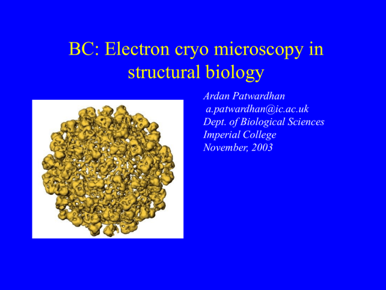
BC: Electron cryo microscopy in structural biology Ardan Patwardhan a.patwardhan@ic.ac.uk Dept. of Biological Sciences Imperial College November, 2003 Specimen contrast Amplitude Contrast Phase Contrast Phase Contrast • • • • Is not directly observable Converted to amplitude contrast by defocusing specimen Limited to study of thin specimens (<1000Å) Same technique used in light microscopy to study unstained specimens • Why not use stain? - May affect macromolecular structure Cryo specimen preparation • Preserve native environment • High vacuum need frozen specimens! • Snap freezing for amorphous ice phase, not crystalline ice phase Cryo EM “grid” Supporting carbon film Metal grid Ice holes An ice hole • Particles are randomly positioned and orientated EM images • 2D projections of 3D objects • Similar to x-ray images EM images are very noisy!! • Beam damage limits exposure • At our disposal: Thousands of randomly oriented macromolecular images with very poor signal to noise ratio • Image processing techniques used to combine thousands of 2D images into a 3D reconstruction of the particle Particle Picking • Objective: identify particles in micrograph and cut out patches containing one particle each • Can be done automatically, in some cases, especially if the molecule possesses icosahedral symmetry • Most cases still done manually - tedious, difficult and boring • Need to collect between 1000 and 10000 particles to get going (the more the better) Translational Alignment • Requires reference image(s) to align to Rotational Alignment • Requires reference image(s) to align to Classification • Combine like views to improve signal to noise Chicken and egg problem • The class “sum” images can be used as references for alignment • The quality of the classification depends on how well aligned the data is • In general, steps of alignment and classification have to be repeated several times Angular reconstitution • Determine angles of projections relative to each other in 3D • Find common line projections to determine relative angles Slice through 3D Reprojection • 3D density map can be used to generate projections that can be used to realign the raw images • Process may have to be repeated several times Pros and cons • Excellent tool for difference studies • Resolution not yet as good as for x-ray crystallography and NMR Examples: Ribosome References • M. van Heel, B. Gowen, R. Matadeen, E. Orlova, R. Finn, T. Pape, D. Cohen, H. Stark, R. Schmidt, M. Schatz and A. Patwardhan: Single-particle electron cryo-microscopy: towards atomic resolution. Quarterly Review of Biophysics 33(4), 307 - 369(2000) Credits Biological Sciences • Prof. Marin Van Heel • Dr. Tillman Pape • Dr. Elena Orlova • Alexis Rohou • David Carpentier • Martin Bommer • Richard Hall • Dr. Pampa Ray Division of Medicine • Dr. Edward P. Morris • Danielle Paul


