Parasitology 6th lecture
advertisement
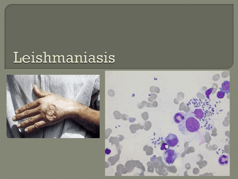
Leishmaniasis is a disease caused by protozoan parasites that belong to the genus Leishmania and is transmitted by the bite of certain species of sand fly. Most forms of the disease are transmissible only from animals (zoonosis), but some can be spread between humans. Human infection is caused by about 21 of 30 species that infect mammals. Cutaneous leishmaniasis is the most common form of leishmaniasis. Visceral leishmaniasis is a severe form in which the parasites have migrated to the vital organs. The most important species of Leishmania is L. donovani causes visceral leishmaniasis (Kala-azar, black disease, dumdum fever). Amastigote (leishmanial form) is oval and measures 2-5 microns by 1 - 3 microns, whereas the leptomonad (Leishmania promastigotes) measures 14 - 20 microns by 1.5 - 4 microns, trypanosomes. a similar size to Leishmania promastigotes. This is the form that lives in the sandfly and is injected into humans or animals. Notice the flagella. Spindle-shaped cells with a single flagellum that endows them with motility. This form of the parasite can be easily cultured in tissue culture medium Leishmania amastigote. This is the form that lives in the human. Notice the lack of flagella. The amastigotes are smaller, oval-shaped cells that have only a residual flagellum and lack motility. Leishmaniasis can be transmitted in many tropical and sub-tropical countries, and is found in parts of about 88 countries. Approximately 350 million people live in these areas. The settings in which leishmaniasis is found range from rainforests in Central and South America to deserts in West Asia and the Middle East. It affects as many as 12 million people worldwide, with 1.5–2 million new cases each year. The visceral form of leishmaniasis has an estimated incidence of 500,000 new cases and 60,000 deaths each year. More than 90 percent of the world's cases of visceral leishmaniasis are in India, Bangladesh, Nepal, Sudan, and Brazil. The organism is transmitted by the bite of several species of bloodfeeding sand flies (Phlebotomus) which carry the promastigote in the anterior gut and pharynx. The parasites gain access to mononuclear phagocytes where they transform into amastigotes and divide until the infected cell ruptures. The released organisms infect other cells. The sandfly acquires the organisms during the blood meal; the amastigotes transform into flagellate promastigotes and multiply in the gut until the anterior gut and pharynx are packed. Dogs and rodents are common reservoirs. Phlebotomus (sandfly) The symptoms of leishmaniasis are skin sores which erupt weeks to months after the person affected is bitten by sand flies. Other consequences, which can become manifest anywhere from a few months to years after infection, include fever, damage to the spleen and liver, and anaemia. In the medical field, leishmaniasis is one of the famous causes of a markedly enlarged spleen, which may become larger even than the liver. Leishmaniasis may be divided into the following types: Cutaneous leishmaniasis Mucocutaneous Visceral leishmaniasis leishmaniasis Cutaneous leishmaniasis – the most common form which causes a sore at the bite site, which heals in a few months to a year, leaving an unpleasant looking scar. Mucocutaneous leishmaniasis – commences with skin ulcers which spread causing tissue damage to (particularly) nose and mouth. Visceral leishmaniasis – the most serious form and potentially fatal if untreated. Cutaneous leishmaniasis ulcer Visceral leishmaniasis or "Kala-azar" is caused by infection with Leishmania donovani. Parasiteinfected cells can be found in spleen, liver, lymph glands, intestinal mucosa and bone marrow. This can result in a very enlarged liver and spleen. Leishmaniasis is diagnosed in the haematology laboratory by direct visualization of the amastigotes (Leishman-Donovan bodies). Buffy-coat preparations of peripheral blood or aspirates from marrow, spleen, lymph nodes or skin lesions should be spread on a slide to make a thin smear, and stained with Leishman's or Giemsa's stain (pH 7.2) for 20 minutes. Amastigotes are seen with monocytes or, less commonly in neutrophil in peripheral blood and in macrophages in aspirates. Sodium stibogluconate (Pentostam) is the drug of choice. Pentamidine isethionate is used as an alternative. Control measures involve vector control and avoidance. Immunization has not been effective. Amphotericin (AmBisome) is now the treatment of choice; its failure in some cases to treat visceral leishmaniasis (Leishmania donovani) has been reported in Sudan, but this may be related to host factors such as coinfection with HIV or tuberculosis rather than parasite resistance. Miltefosine (Impavido) is a new drug for visceral and cutaneous leishmaniasis
