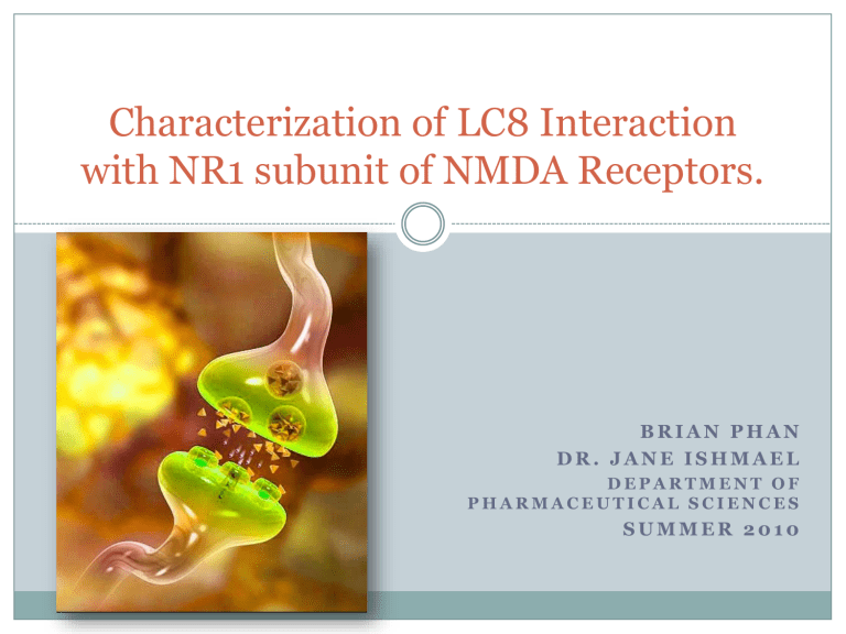
Characterization of LC8 Interaction with NR1 subunit of NMDA Receptors.

Characterization of LC8 Interaction with NR1 subunit of NMDA Receptors.
B R I A N P H A N
D R . J A N E I S H M A E L
D E P A R T M E N T O F
P H A R M A C E U T I C A L S C I E N C E S
S U M M E R 2 0 1 0
NMDA-type Glutamate Receptors
N-methyl-D-Aspartate receptors
(NMDAR)
Glutamate receptors important for synaptic plasticity and memory function.
Tetramer made of 2 different subunits (NR1 and NR2)
Ligand and voltage-gated ion channel that facilitates movement of calcium across neural cell membranes.
LC8
LC8
Conserved and dimeric cargo binding subunit of dynein motor complex.
Increases ordered structure of various proteins.
LC8 can be found in postsynaptic sites in neurons.
Post-synaptic sites
Glutamate is the major excitatory amino acid neurotransmitter in mammalian CNS.
NMDAR regulates neurotransmission at postsynaptic sites.
Protein-protein interactions facilitate assembly of NMDA receptors.
Significance
Understanding in the functional organization at pre- and post-synaptic sites.
Insight into cellular mechanisms underlying neurological disorders.
Importance in drug discovery.
NR1 Structure
NR1-1a
Techniques
• Use HEK293 cells as model system to transiently express NR1 and LC8.
• Co-immunoprecipitation reaction to identify protein complexes in cells.
Working Hypothesis
Direct binding of LC8 onto the C-terminus of the
NR1-1a subunit will facilitate NMDAR assembly and transport to the neural synapse.
NH
2
Tetramerization with NR2 dimer?
C0 C1
… KDTST …
C2 COOH
LC8
Dimerization with another NR1.
Preliminary Results
NR1-4a (NR1 without LC8 motif) still shows that
NR1 and LC8 are still associated together in a complex.
NH
2 C0
…
X
…
C2’
LC8
COOH
Immunoblot Analysis
Transient expression of NMDAR1 splice variants and FLAGtagged LC8 in
HEK293 cells using
Calcium Phosphate transfection.
150
100
75
NR1-1a NR1-4a
Western Blot analysis to identify expressed proteins X hr. later
15
10
FLAG-LC8
Co-Immunoprecipitation
1. Recognize FLAG-LC8 with antibody (m2 alpha-FLAG).
2. Use protein G-sepharose beads to recognize and bind to the antibody-FLAG-LC8 complex.
3. Elute FLAG-LC8 from lysate and determine if it is (or is not) associated with NR1.
Preliminary Results
Hypothesis
Another LC8 binding motif common in both
NR1-1a and NR1-4a exists on the NR1 C-tail that interacts with LC8 to order and form the
NR1 homodimer.
Methods
Remove C1/C2 from NR1-1a (1-863)
NH
2
C0 C1 C2 COOH
NH
2
C0 COOH
Does C0 contain another LC8 binding motif?
Methods
Remove entire C-terminus from NR1-1a (1-834)
NH
2
C0 C1 C2 COOH
NH
2
COOH
Is the LC8 binding domain within the C-terminus?
Methods
Mouse Anti-Glutamate Receptor Monoclonal
Antibody (Epitope 660-811)
Used to detect NR1 which migrates to ~120kDa on
SDS/PAGE.
NH
2 C-tail COOH
Construction of Eukaryotic Expression Vectors
Design new primers and use PCR to amplify NR1 mutant inserts.
HindIII
XBa1
Ligate new NR1 insert into a cloning vector.
Construct construction (cont.)
Transform ligated insert into bacterial cells (XL-
Blue10).
Select single colonies
Mini-prep to extract DNA.
DNA digestion to confirm successful ligation and transformation.
Transfect new plasmid into
HEK293 cells.
5.4 kb
2.5 kb
Transient expression in HEK293 cells
New DNA transfected into HEK293 cells.
Success in producing NR1 (1-863) construct and expression in HEK293.
Results of co-immunoprecipitation study
• FLAG-LC8 appears to interact with NR1 (1-863) in
HEK 293 cells
• Results need to be repeated for verification with NR1-1a without FLAG-LC8 as a control.
Future Work
Sequence pCDNA3/ NR1 (1-863) DNA to verify that mutations were not introduced by PCR.
Continue constructing NR1 (1-834) and determine if an LC8 binding site exists on C-tail.
Biophysical approaches to further structurally characterize LC8-NR1 complex.
New Skills and Techniques
Producing and testing new DNA constructs.
Transient transfection into eukaryotic cells.
Cell culture and maintaining a cell line.
Co-immunoprecipitation.
Acknowledgements
Dr. Jane Ishmael
Andrew Hau
Xiao Liu
HHMI
OSU Dept. of Pharmaceutical Sciences


