AbstractID: 2900 Title: Dose calculation and quantitative PTV expansion using... MRI images
advertisement
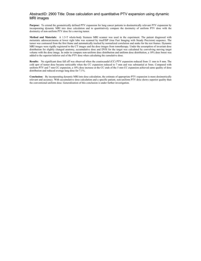
AbstractID: 2900 Title: Dose calculation and quantitative PTV expansion using dynamic MRI images Purpose: To extend the geometrically defined PTV expansion for lung cancer patients to dosimetrically relevant PTV expansion by incorporating dynamic MRI into dose calculation and to quantitatively compare the dosimetry of uniform PTV dose with the dosimetry of non-uniform PTV dose for a moving tumor. Method and Materials: A 1.5-T whole-body Siemens MRI scanner was used in the experiment. The patient diagnosed with metastatic adenocarcinoma at lower right lobe was scanned by trueFISP (true Fast Imaging with Steady Precision) sequence. The tumor was contoured from the first frame and automatically tracked by normalized correlation and snake for the rest frames. Dynamic MRI images were rigidly registered to the CT images and the dose images from tomotherapy. Under the assumption of invariant dose distribution for slightly changed anatomy, accumulative dose and DVH for the target was calculated by convolving moving target volume with the dose image. In order to compare non-uniform does distribution and uniform dose distribution, a 10% does boost was added to the superior/inferior end of the PTV dose when calculating the cumulative dose. Results: No significant dose fall off was observed when the craniocaudal (CC) PTV expansion reduced from 11 mm to 8 mm. The cold spot of tumor dose became noticeable when the CC expansion reduced to 7 mm and was substantial at 5mm. Compared with uniform PTV and 7 mm CC expansion, a 10% dose increase at the CC ends of the 5 mm CC expansion achieved same quality of dose distribution and reduced average lung dose for 7.1%. Conclusion: By incorporating dynamic MRI into dose calculation, the estimate of appropriate PTV expansion is more dosimetrically relevant and accuracy. With accumulative dose calculation and a specific patient, non-uniform PTV dose shows superior quality than the conventional uniform dose. Generalization of this conclusion is under further investigation.

