AbstractID: 4882 Title: Soft-Tissue Detectability Limits in Cone-Beam CT: 2AFC... Human Observer Performance in Relation to Contrast, Spatial Resolution, and...
advertisement
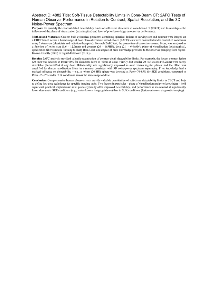
AbstractID: 4882 Title: Soft-Tissue Detectability Limits in Cone-Beam CT: 2AFC Tests of Human Observer Performance in Relation to Contrast, Spatial Resolution, and the 3D Noise-Power Spectrum Purpose: To quantify the contrast-detail detectability limits of soft-tissue structures in cone-beam CT (CBCT) and to investigate the influence of the plane of visualization (axial/sagittal) and level of prior knowledge on observer performance. Method and Materials: Custom-built cylindrical phantoms containing spherical lesions of varying size and contrast were imaged on a CBCT bench across a broad range of dose. Two-alternative forced choice (2AFC) tests were conducted under controlled conditions using 7 observers (physicists and radiation therapists). For each 2AFC test, the proportion of correct responses, Pcorr, was analyzed as a function of lesion size (1.6 – 12.7mm) and contrast (20 – 165HU), dose (2.1 – 6.4mGy), plane of visualization (axial/sagittal), apodization filter (smooth Hanning to sharp Ram-Lak), and degree of prior knowledge provided to the observer (ranging from SignalKnown-Exactly (SKE) to Signal-Unknown (SUK)). Results: 2AFC analysis provided valuable quantitation of contrast-detail detectability limits. For example, the lowest contrast lesion (20 HU) was detected at Pcorr>70% for diameters down to ~6mm at doses >2mGy, but smaller 20 HU lesions (<3.2mm) were barely detectable (Pcorr<60%) at any dose. Detectability was significantly improved in axial versus sagittal planes, and the effect was amplified by sharper apodization filters in a manner consistent with 3D noise-power spectrum asymmetry. Prior knowledge had a marked influence on detectability – e.g., a ~6mm (20 HU) sphere was detected at Pcorr~70-85% for SKE conditions, compared to Pcorr~55-65% under SUK conditions across the same range of dose. Conclusion: Comprehensive human observer tests provide valuable quantitation of soft-tissue detectability limits in CBCT and help to define low-dose techniques for specific imaging tasks. Two factors in particular – plane of visualization and prior knowledge – hold significant practical implications: axial planes typically offer improved detectability, and performance is maintained at significantly lower dose under SKE conditions (e.g., lesion-known image guidance) than in SUK conditions (lesion-unknown diagnostic imaging).

