AbstractID: 5527 Title: Spatial resolution-matched comparison between fan-beam and
advertisement

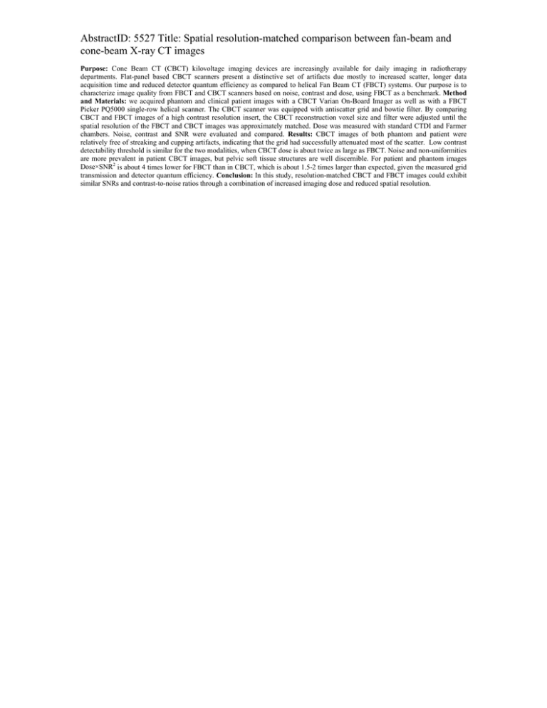
AbstractID: 5527 Title: Spatial resolution-matched comparison between fan-beam and cone-beam X-ray CT images Purpose: Cone Beam CT (CBCT) kilovoltage imaging devices are increasingly available for daily imaging in radiotherapy departments. Flat-panel based CBCT scanners present a distinctive set of artifacts due mostly to increased scatter, longer data acquisition time and reduced detector quantum efficiency as compared to helical Fan Beam CT (FBCT) systems. Our purpose is to characterize image quality from FBCT and CBCT scanners based on noise, contrast and dose, using FBCT as a benchmark. Method and Materials: we acquired phantom and clinical patient images with a CBCT Varian On-Board Imager as well as with a FBCT Picker PQ5000 single-row helical scanner. The CBCT scanner was equipped with antiscatter grid and bowtie filter. By comparing CBCT and FBCT images of a high contrast resolution insert, the CBCT reconstruction voxel size and filter were adjusted until the spatial resolution of the FBCT and CBCT images was approximately matched. Dose was measured with standard CTDI and Farmer chambers. Noise, contrast and SNR were evaluated and compared. Results: CBCT images of both phantom and patient were relatively free of streaking and cupping artifacts, indicating that the grid had successfully attenuated most of the scatter. Low contrast detectability threshold is similar for the two modalities, when CBCT dose is about twice as large as FBCT. Noise and non-uniformities are more prevalent in patient CBCT images, but pelvic soft tissue structures are well discernible. For patient and phantom images Dose×SNR2 is about 4 times lower for FBCT than in CBCT, which is about 1.5-2 times larger than expected, given the measured grid transmission and detector quantum efficiency. Conclusion: In this study, resolution-matched CBCT and FBCT images could exhibit similar SNRs and contrast-to-noise ratios through a combination of increased imaging dose and reduced spatial resolution.

