AbstractID: 3527 Title: Clinical Experience with a 3-D Ultrasound Prostate... System
advertisement

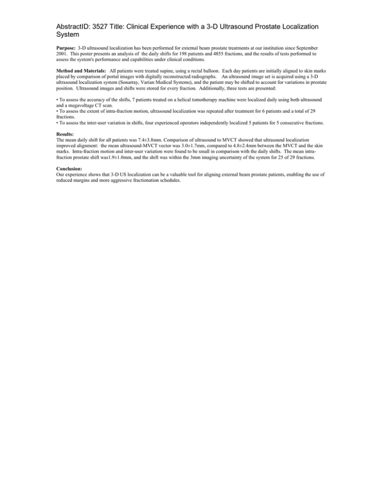
AbstractID: 3527 Title: Clinical Experience with a 3-D Ultrasound Prostate Localization System Purpose: 3-D ultrasound localization has been performed for external beam prostate treatments at our institution since September 2001. This poster presents an analysis of the daily shifts for 198 patients and 4855 fractions, and the results of tests performed to assess the system's performance and capabilities under clinical conditions. Method and Materials: All patients were treated supine, using a rectal balloon. Each day patients are initially aligned to skin marks placed by comparison of portal images with digitally reconstructed radiographs. An ultrasound image set is acquired using a 3-D ultrasound localization system (Sonarray, Varian Medical Systems), and the patient may be shifted to account for variations in prostate position. Ultrasound images and shifts were stored for every fraction. Additionally, three tests are presented: • To assess the accuracy of the shifts, 7 patients treated on a helical tomotherapy machine were localized daily using both ultrasound and a megavoltage CT scan. • To assess the extent of intra-fraction motion, ultrasound localization was repeated after treatment for 6 patients and a total of 29 fractions. • To assess the inter-user variation in shifts, four experienced operators independently localized 5 patients for 5 consecutive fractions. Results: The mean daily shift for all patients was 7.4±3.8mm. Comparison of ultrasound to MVCT showed that ultrasound localization improved alignment: the mean ultrasound-MVCT vector was 3.0±1.7mm, compared to 4.8±2.4mm between the MVCT and the skin marks. Intra-fraction motion and inter-user variation were found to be small in comparison with the daily shifts. The mean intrafraction prostate shift was1.9±1.0mm, and the shift was within the 3mm imaging uncertainty of the system for 25 of 29 fractions. Conclusion: Our experience shows that 3-D US localization can be a valuable tool for aligning external beam prostate patients, enabling the use of reduced margins and more aggressive fractionation schedules.


