AbstractID: 2616 Title: Daily localization of post-prostatectomy patients with combined
advertisement
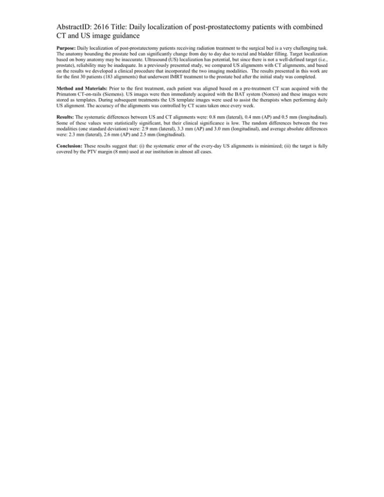
AbstractID: 2616 Title: Daily localization of post-prostatectomy patients with combined CT and US image guidance Purpose: Daily localization of post-prostatectomy patients receiving radiation treatment to the surgical bed is a very challenging task. The anatomy bounding the prostate bed can significantly change from day to day due to rectal and bladder filling. Target localization based on bony anatomy may be inaccurate. Ultrasound (US) localization has potential, but since there is not a well-defined target (i.e., prostate), reliability may be inadequate. In a previously presented study, we compared US alignments with CT alignments, and based on the results we developed a clinical procedure that incorporated the two imaging modalities. The results presented in this work are for the first 30 patients (183 alignments) that underwent IMRT treatment to the prostate bed after the initial study was completed. Method and Materials: Prior to the first treatment, each patient was aligned based on a pre-treatment CT scan acquired with the Primatom CT-on-rails (Siemens). US images were then immediately acquired with the BAT system (Nomos) and these images were stored as templates. During subsequent treatments the US template images were used to assist the therapists when performing daily US alignment. The accuracy of the alignments was controlled by CT scans taken once every week. Results: The systematic differences between US and CT alignments were: 0.8 mm (lateral), 0.4 mm (AP) and 0.5 mm (longitudinal). Some of these values were statistically significant, but their clinical significance is low. The random differences between the two modalities (one standard deviation) were: 2.9 mm (lateral), 3.3 mm (AP) and 3.0 mm (longitudinal), and average absolute differences were: 2.3 mm (lateral), 2.6 mm (AP) and 2.5 mm (longitudinal). Conclusion: These results suggest that: (i) the systematic error of the every-day US alignments is minimized; (ii) the target is fully covered by the PTV margin (8 mm) used at our institution in almost all cases.
