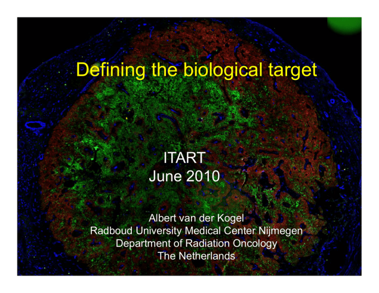
Defining the biological target ITART June 2010
advertisement

Defining the biological target ITART June 2010 Albert van der Kogel Radboud University Medical Center Nijmegen Department of Radiation Oncology The Netherlands The primary biological targets for RT: yp &p proliferation hypoxia SBRT? HYPOXIA VASCULARITY PROLIFERATION A classic resistance mechanism in radiotherapy: yp hypoxia HYPOXIA VASCULARITY PROLIFERATION Hypoxia is not a static condition, and generally changes in response to irradiation and many chemotherapeutic and biological agents Immunohistochemical imaging of hypoxia and proliferation Biopsy of head/neck squamous cell carcinoma blood vessels proliferating cells (IdUrd +) Hypoxia yp (pimonidazole +) necrosis Hypoxia imaging from PET hypoxic voxel to microregional distribution biopsy pO2 gradient! Hypoxia is not binary, and identical hypoxic voxels may represent different biological characteristics h t i ti 18F-misonidazole autoradiography vs. pimonidazole IHC: correlation related to tumor structure Gray-value images after immunohistochemical staining of pimonidazole (left) and 18FFMISO autoradiography (right) of SCCNij3 (A), SCCNij153 (B), and SCCNij86 (C) xenografted human squamous cell carcinomas of head and neck. Pimonidazole and 18F-FMISO correlate for SCCNij3 and SCNij153 but not for SCCNij86. SCCNij86 Troost et al, Eur J Nucl Med 2008 Different types of hypoxia in same tumor IIntermittent/acute i / hypoxia Intermittent/acute hypoxia Chronic/diffusion limited h limited hypoxia i Chronic/diffusion hypoxia heterogeneity in hypoxia (R1 tumor) heterogeneity in hypoxia (R1 tumor) Effect of irradiation on hypoxia 0.25 hypoxic yp fraction 0.2 0 hours ours 26 hoursh 0.15 0.1 0.05 0 11 days 0 40 80 120 160 200 240 necrosis Bussink et al., Rad Res. 2000 10 HF 12 % HF 4 % HF 28 % Temporal changes in hypoxia and proliferation after irradiation of seg-1 esophageal s.c.c. (15 Gy SD) day 6 day 2 d unirradiated control green: hypoxic cells red: proliferating cells (KI-67) white: blood vessels Blue: perfusion (Hoechst ) day 10 Overall treatment time-related loss of effect: p proliferation HYPOXIA VASCULARITY PROLIFERATION Larynx carcinoma xenografts showing different proliferation patterns Hypoxia (pimonidazole) Blood vessels (CD31 a.o.) Perfused vessels (Hoechst dye) Proliferating cells (BrdU) Proliferation & hypoxia in human s.c.c. xenograft control unirradiated Hypoxic fraction 10% Ki67 PIMO Vessels T Kruser et al, Clin Canc Res 2010 Proliferation & hypoxia in human s.c.c. xenograft after 8 X 3 Gy/4 weeks Proliferation: ↑↑ Hypoxia: H i ↓↓ (10% ➙1.7%) Ki67 PIMO Vessels Proliferation & hypoxia after 8 X 3 Gy/4 weeks + VEGFR2 inhibitor Proliferation: ↑ Hypoxic fraction: 6% Ki67 PIMO Vessels T Kruser et al, Clin Canc Res 2010 FLT-PET: weak correlation with immunohistochemical IdUrd labeling p<0.0001 Multiple FLT-PET in oropharyngeal tumors 10 patients with oropharyngeal tumours undergoing (chemo)radiation 3 FLT-PET-CT scans with i.v. contrast agent 2nd PET‐CT 3rd PET‐CT 8 fractions 18 fractions Start of treatment 1st PET‐CT ‐ 5 days Troost et al, 2009 & 2010 CT vs FLT‐PET : repeated measurements • The mean gross tumor volume on CT g (GTVCT) was 12.7 cm3 prior to treatment, 11.1 cm3 in the second and 5.0 cm3 in the fourth week of treatment • GTVCT decreased significantly after 18 fractions (p < 0.001) • GTVFLT/PET did not change significantly Troost et al, JNM 2010 Individual differences in reduction of FLT/PET signal Troost et al, 2010 Boosting of highly proliferative subvolume • Definition of subvolume using 80% of maximum SUV (GTV80%) in first first and second second FLT‐PET scan • Creation of radiation treatment plan using IMRT with g simultaneous integrated boost Troost et al, 2010 conclusions • Both hypoxia and proliferation are highly dynamic and heterogenously distributed in tumors • Hypoxia is not a binary event, and the most hypoxic cells that dominate a hypoxic yp voxel may y be biologically the least relevant • Clearly dose-painting or adaptive RT needs repeated imaging before f and (early) ( ) during therapy • Later into therapy, extensive cell death and vascular injury will mask the extent of clinically relevant hypoxia and proliferation Acknowledgements Nijmegen Jan Bussink Esther Troost H Hans P Peters t Hans Kaanders Maastricht Madison Brad B dW Wouters t Kasper Rouschop Ludwig Dubois Philippe pp Lambin Deric D i Wh Wheeler l Robert Jeraj Soren Bentzen Paul Harari the Nijmegen radiobiology team Jan Bussink Hans Kaanders Esther Troost Saskia Rademakers Bianca Hoeben Hanneke Stegeman Paul Span Monique Nijkamp Anika Nagelkerke Marloes Wennemers Wenny Peeters Jasper Lok Hans Peters Paul Rijken