Introduction visualization of surgical clips in electronic portal images, was monitored... portal images. The setup errors and organ motion were measured...
advertisement

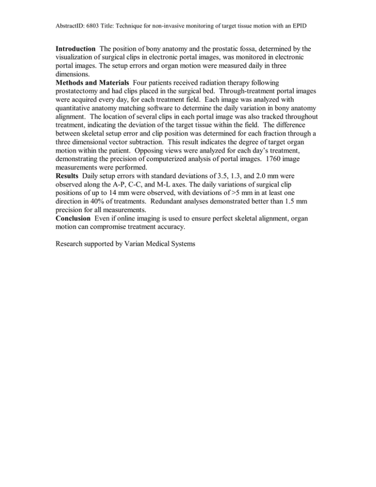
AbstractID: 6803 Title: Technique for non-invasive monitoring of target tissue motion with an EPID Introduction The position of bony anatomy and the prostatic fossa, determined by the visualization of surgical clips in electronic portal images, was monitored in electronic portal images. The setup errors and organ motion were measured daily in three dimensions. Methods and Materials Four patients received radiation therapy following prostatectomy and had clips placed in the surgical bed. Through-treatment portal images were acquired every day, for each treatment field. Each image was analyzed with quantitative anatomy matching software to determine the daily variation in bony anatomy alignment. The location of several clips in each portal image was also tracked throughout treatment, indicating the deviation of the target tissue within the field. The difference between skeletal setup error and clip position was determined for each fraction through a three dimensional vector subtraction. This result indicates the degree of target organ motion within the patient. Opposing views were analyzed for each day’s treatment, demonstrating the precision of computerized analysis of portal images. 1760 image measurements were performed. Results Daily setup errors with standard deviations of 3.5, 1.3, and 2.0 mm were observed along the A-P, C-C, and M-L axes. The daily variations of surgical clip positions of up to 14 mm were observed, with deviations of >5 mm in at least one direction in 40% of treatments. Redundant analyses demonstrated better than 1.5 mm precision for all measurements. Conclusion Even if online imaging is used to ensure perfect skeletal alignment, organ motion can compromise treatment accuracy. Research supported by Varian Medical Systems


