Simulating Lunar Surface Missions:
advertisement

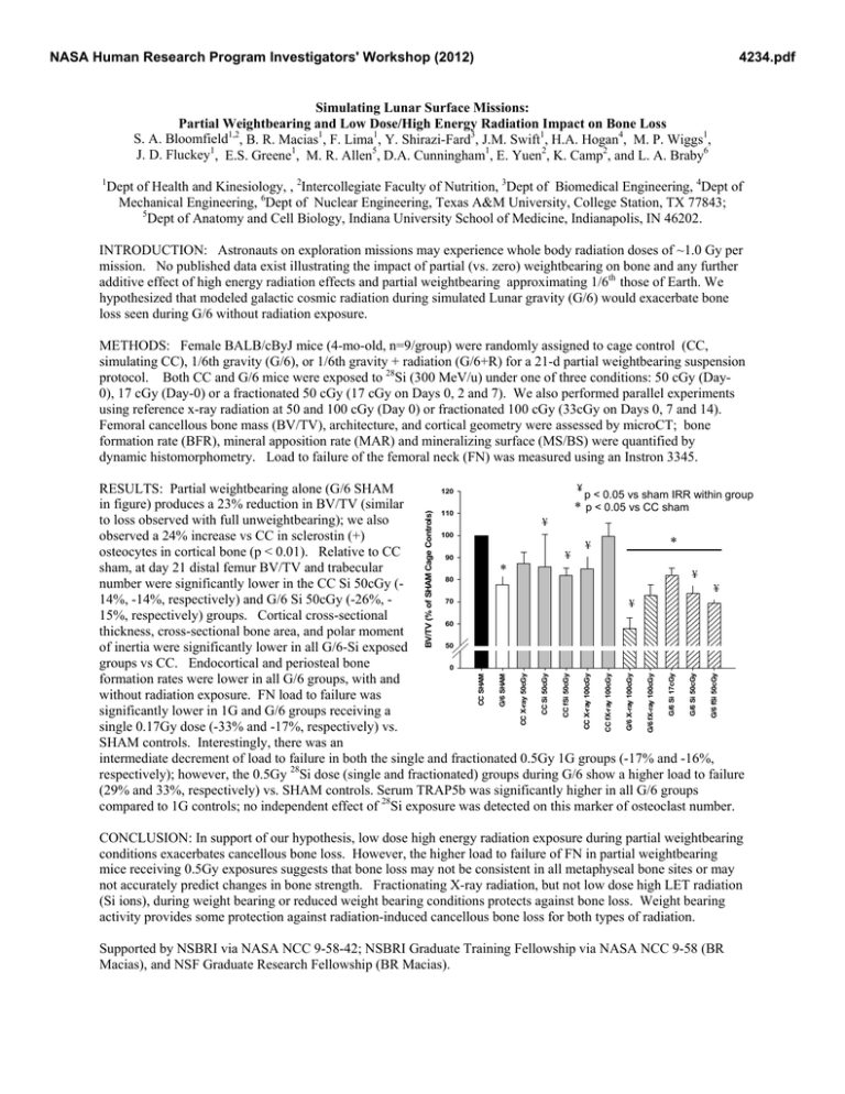
NASA Human Research Program Investigators' Workshop (2012) 4234.pdf Simulating Lunar Surface Missions: Partial Weightbearing and Low Dose/High Energy Radiation Impact on Bone Loss S. A. Bloomfield1,2, B. R. Macias1, F. Lima1, Y. Shirazi-Fard3, J.M. Swift1, H.A. Hogan4, M. P. Wiggs1, J. D. Fluckey1, E.S. Greene1, M. R. Allen5, D.A. Cunningham1, E. Yuen2, K. Camp2, and L. A. Braby6 1 Dept of Health and Kinesiology, , 2Intercollegiate Faculty of Nutrition, 3Dept of Biomedical Engineering, 4Dept of Mechanical Engineering, 6Dept of Nuclear Engineering, Texas A&M University, College Station, TX 77843; 5 Dept of Anatomy and Cell Biology, Indiana University School of Medicine, Indianapolis, IN 46202. INTRODUCTION: Astronauts on exploration missions may experience whole body radiation doses of ~1.0 Gy per mission. No published data exist illustrating the impact of partial (vs. zero) weightbearing on bone and any further additive effect of high energy radiation effects and partial weightbearing approximating 1/6th those of Earth. We hypothesized that modeled galactic cosmic radiation during simulated Lunar gravity (G/6) would exacerbate bone loss seen during G/6 without radiation exposure. METHODS: Female BALB/cByJ mice (4-mo-old, n=9/group) were randomly assigned to cage control (CC, simulating CC), 1/6th gravity (G/6), or 1/6th gravity + radiation (G/6+R) for a 21-d partial weightbearing suspension protocol. Both CC and G/6 mice were exposed to 28Si (300 MeV/u) under one of three conditions: 50 cGy (Day0), 17 cGy (Day-0) or a fractionated 50 cGy (17 cGy on Days 0, 2 and 7). We also performed parallel experiments using reference x-ray radiation at 50 and 100 cGy (Day 0) or fractionated 100 cGy (33cGy on Days 0, 7 and 14). Femoral cancellous bone mass (BV/TV), architecture, and cortical geometry were assessed by microCT; bone formation rate (BFR), mineral apposition rate (MAR) and mineralizing surface (MS/BS) were quantified by dynamic histomorphometry. Load to failure of the femoral neck (FN) was measured using an Instron 3345. G/6 fSi 50cGy G/6 Si 50cGy G/6 Si 17cGy G/6 fX-ray 100cGy G/6 X-ray 100cGy CC fX-ray 100cGy CC X-ray 100cGy CC Si 50cGy CC fSi 50cGy CC X-ray 50cGy G/6 SHAM CC SHAM BV/TV (% of SHAM Cage Controls) ¥ RESULTS: Partial weightbearing alone (G/6 SHAM 120 p < 0.05 vs sham IRR within group in figure) produces a 23% reduction in BV/TV (similar * p < 0.05 vs CC sham 110 to loss observed with full unweightbearing); we also ¥ 100 observed a 24% increase vs CC in sclerostin (+) * ¥ osteocytes in cortical bone (p < 0.01). Relative to CC ¥ 90 sham, at day 21 distal femur BV/TV and trabecular * ¥ 80 number were significantly lower in the CC Si 50cGy (¥ 14%, -14%, respectively) and G/6 Si 50cGy (-26%, 70 ¥ 15%, respectively) groups. Cortical cross-sectional 60 thickness, cross-sectional bone area, and polar moment 50 of inertia were significantly lower in all G/6-Si exposed groups vs CC. Endocortical and periosteal bone 0 formation rates were lower in all G/6 groups, with and without radiation exposure. FN load to failure was significantly lower in 1G and G/6 groups receiving a single 0.17Gy dose (-33% and -17%, respectively) vs. SHAM controls. Interestingly, there was an intermediate decrement of load to failure in both the single and fractionated 0.5Gy 1G groups (-17% and -16%, respectively); however, the 0.5Gy 28Si dose (single and fractionated) groups during G/6 show a higher load to failure (29% and 33%, respectively) vs. SHAM controls. Serum TRAP5b was significantly higher in all G/6 groups compared to 1G controls; no independent effect of 28Si exposure was detected on this marker of osteoclast number. CONCLUSION: In support of our hypothesis, low dose high energy radiation exposure during partial weightbearing conditions exacerbates cancellous bone loss. However, the higher load to failure of FN in partial weightbearing mice receiving 0.5Gy exposures suggests that bone loss may not be consistent in all metaphyseal bone sites or may not accurately predict changes in bone strength. Fractionating X-ray radiation, but not low dose high LET radiation (Si ions), during weight bearing or reduced weight bearing conditions protects against bone loss. Weight bearing activity provides some protection against radiation-induced cancellous bone loss for both types of radiation. Supported by NSBRI via NASA NCC 9-58-42; NSBRI Graduate Training Fellowship via NASA NCC 9-58 (BR Macias), and NSF Graduate Research Fellowship (BR Macias).


