Why Didn’t It Live? Hypothermia and Starvation in Michigan Lambs
advertisement

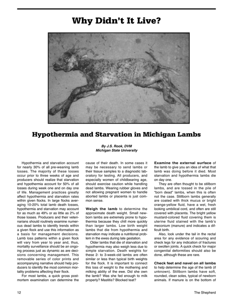
Why Didn’t It Live? Hypothermia and Starvation in Michigan Lambs By J.S. Rook, DVM Michigan State University Hypothermia and starvation account for nearly 30% of all pre-weaning lamb losses. The majority of these losses occur prior to three weeks of age and producers should realize that starvation and hypothermia account for 50% of all losses during week one and on day one of life. Management practices greatly affect hypothermia and starvation rates within given flocks. In large flocks averaging 10-20% total lamb death losses, hypothermia and starvation may account for as much as 49% or as little as 2% of those losses. Producers and their veterinarians should routinely examine numerous dead lambs to identify trends within a given flock and use this information as a basis for management decisions. Lamb loss patterns within a given flock will vary from year to year and, thus, mortality surveillance should be an ongoing process just as dynamic as are decisions concerning management. This removable series of color prints and accompanying narrative should help producers to identify the most common mortality problems affecting their flock. For most lambs, a quick gross postmortem examination can determine the 12 cause of their death. In some cases it may be necessary to send lambs or their tissue samples to a diagnostic laboratory for testing. All producers, and especially women of childbearing age, should exercise caution while handling dead lambs. Wearing rubber gloves and not allowing pregnant women to handle aborted lambs or placenta is just common sense. Weigh the lamb to determine the approximate death weight. Small newborn lambs are extremely prone to hypothermia because they chill more quickly than larger lambs. Low birth weight lambs that die from hypothermia and starvation may indicate a nutritional problem in the ewes during late gestation. Older lambs that die of starvation and hypothermia may also weigh less due to simple starvation. Death weights for these 2- to 3-week-old lambs are often similar or less than typical birth weights for the flock. It is important to connect this loss of weight to the mothering and milking ability of the ewe. Did she own the lamb? Was she fed enough to milk properly? Mastitis? Blocked teat? Examine the external surface of the lamb to give you an idea of what that lamb was doing before it died. Most starvation and hypothermia lambs die on day one. They are often thought to be stillborn lambs, and are tossed in the pile of “born dead” lambs, when this is often not the case. Stillborn lambs generally are coated with thick mucus or bright orange-yellow fluid, have a wet, fresh looking umbilical cord, and often are still covered with placenta. The bright yellow mustard-colored fluid covering them is uterine fluid stained with the lamb’s meconium (manure) and indicates a difficult birth. Also, look under the tail in the rectal area for any evidence of scouring and check legs for any indication of fractures or swollen joints. A quick check for major congenital deformities should also be done, although these are rare. Check feet and navel on all lambs to help determine the age of the lamb (if unknown). Stillborn lambs have soft, rounded, clean soles, typical of newborn animals. If manure is on the bottom of The Shepherd the feet and the soles are worn, the lamb was strong enough to stand and was looking for nutrition and warmth. This is commonly the case with newborn lambs that producers just find dead shortly after birth. These lambs were born alive and had a chance to survive. The umbilical cord also begins to dry and shrivel shortly after birth and usually falls off between 3 to 10 days of age. This will also help document age if the time of birth is unknown. Routinely necropsy lambs no matter how sure you are of the cause of death since routine examination allows you to recognize normal structures and findings. You will not be prone to miss things if your exam is done the same way each time. Lay the dead lamb on its right side with the head to your left and feet towards you. Grab the front upper leg in your left hand and lift up (away from the rib cage) cutting the skin under the armpit, thus reflecting the front limb away from you exposing the entire rib cage. Look for signs of bruising over the ribs as fractured ribs are a common result of trauma. Also look for signs of pale discoloration of the muscle which might indicate vitamin E and selenium problems or hemorrhage. Do not mistake injection stains (yellow LA200 or white penicillin) under the elbow for abscesses as this is a common site for injections. Normal newborn lambs have a layer of tan fat that follows the junction of the ribs to the cartilage of the sternum and the junction of the ribs with the spine. This fat is readily apparent in stillborn lambs but totally absent in starvation animals. After necropsying starvation and hypothermia lambs, the comparison is obvious – look for it. Next make a long curved incision from front to rear through the cartilage junction of the ribs and sternum and continue the cut up across the abdomen to the area just in front of the hip. Lift up on the ribs and push them away from you (you may actually break the ribs at their connection to the spine) allowing all the internal organs of the chest and abdomen to be viewed like an oyster on the half-shell. Trim any skin or muscle in your way. Finally, make a cut through the back of the muscles of the ham area to check for uneven, pale muscle discoloration characteristic in this area for whitemuscle disease. November 2013 With the lamb opened in a routine manner, the diagnosis of hypothermia and starvation and other common diseases can now be easily made while asking yourself several questions. 1. ARE THE LUNGS NORMAL AND INFLATED? This is the first important question to answer, especially in lambs dying from hypothermia and starvation during day one. If the lamb is stillborn, the lungs will be an even dark purple coloration throughout – much the same color and feel as the liver. Normal aerated lungs, however, will be spongy to the touch and an even colored pink. Variations in color and feel with dark purple firm areas toward the bottom and front of the lungs with normal feeling spongy pink areas toward the top and back might indicate pneumonia or some other cause of death. Starvation and hypothermia lambs should have normal lungs unless some compounding problem exists. Pneumonia is often found secondary to starvation problems so don’t be surprised to observe both problems in the same lamb. Hypothermia lambs that die very shortly after birth may also have a small amount of clear amber-colored fluid in the chest. 2. IS THERE FAT IN THE HEART GROOVES? While looking at the lungs, cut through the heart sac and expose the bare heart muscle. Normal stillborn lambs have a good supply of tan fat in the grooves on the external surface of the heart. This fat is lacking in starvation lambs that have metabolized it for energy. 3. IS THERE MILK IN THE STOMACH AND INTESTINES? Normal newborn lambs have a very large true stomach filled with a clear mucus and small rudimentary forestomachs. The absence of milk in the abomasum or true stomach is common in hypothermia and starvation lambs. Normally, when a lamb nurses, the milk bypasses the forestomachs and goes directly to the abomasum where it forms cottage cheese-like curds during digestion. Weak lambs tube fed by producers often have fluid milk (lacking curds) present in the forestomachs and abomasum (since suckling did not take place) and sometimes the extreme upper portion of the small intestine. Milk in the forestomachs, lack of curdling and absence of milk in the lower intestinal tract indi- cate tube feeding as a last resort. Do not rule out starvation simply by the presence or absence of milk. The presence of large amounts of silage, hay or grass in the stomach of very young lambs may also support starvation. 4. IS KIDNEY FAT PRESENT IN NORMAL AMOUNT AND COLOR? Kidney fat color and consistency is probably the most remarkable change occurring in starvation and hypothermia lambs. As you examine stillborn lambs, look at the large amount of light brown fat Mother Nature deposits around the kidney. This fat acts as a source of energy for newborn lambs until they receive adequate nutrition. As starvation ensues the fat changes from a light tan color to a dark purple gelatinous material (about the color of black cherry jello) as it is gradually metabolized for energy. Kidneys in older starvation lambs are often totally devoid of any fat. In extremely cold weather, when the starving lamb requires lots of calories for heat production, this change in color may occur over the 3-12 hours preceding death. In warmer weather, starvation may occur over several days and result in kidneys that are totally devoid of fat. If you look closely, a small gland known as the adrenal gland is also more obvious in older starvation lambs. The adrenal is normally small and hidden under the renal fat just in front of and to the inside of the kidney. The adrenal enlarges to produce more cortisone-like products in response to the stress of starvation and becomes more obvious due to the lack of fat. Hypothermia and starvation lambs dying very early in life will not have obvious adrenal enlargement due to the acuteness of their condition. Older starvation lambs may. Remember to check kidney fat on all dead lambs to appreciate normal amounts and color, and avoid missing starvation-related infectious diseases. Conclusion Remember that 50% of all Michigan lambs that were born alive and died during week one of life died from hypothermia and starvation. Approaching the nutritional, environmental and management factors contributing to starvation and hypothermia after lambing has begun is simply plugging the dike. Think about your operation and where changes could be made for next year. Continued on page 26 13 Why Didn’t It Live? From page 13 Figure 1: Typical stillborn lamb positioned on its right side and ready to be necropsied. Notice the yellow meconium staining and soft rounded hooves commonly seen in stillborn lambs. Figure 2: Cut the skin under the front leg and fold the cut leg over the back of the lamb. Next cut the cartilage junction of the ribs and sternum and continue this cut through the skin and muscle up the flank to the point of the hip. Figure 3: Stillborn lamb. After cutting through the rib cartilage, lift the ribs and push them away from you, breaking the ribs at their attachment to the spine. This should open the chest and belly cavity for easy viewing. Notice the dark purple non-inflated lungs typical of a lamb that never breathed. Also, notice the straw-colored mucus normally found in the newborn’s stomach and the large deposits of tan-colored fat surrounding the kidney. Inflated lungs of a lamb that had breathed would be reddish-pink in color and spongy feeling compared to these dark purple lungs that are of similar consistency to raw liver. Figure 4: Notice the normal amount and color of the kidney fat In a newborn lamb. This tan fat serves as an energy source during the first few days at life. Compare this to the pictures of starvation and hypothermia lambs. Figures 6 & 7: Notice the normal amounts of tan fat covering the ribs and chest muscles of a newborn lamb (Figure 6). Compare this to the lack of fat cover in a 5-day old starvation lamb (Figure 7). Figure 8: This picture illustrates the progressive fat loss and color changes commonly observed in hypothermia and starvation lambs. In cold weather; progression from the normal fat color and consistency observed in the upper left kidney to the black cherry jello color of the lower left or upper middle kidney may only span several hours. Warm weather starvation lambs surviving several days may totally deplete the kidney at fat as observed in the kidney in the lower center row or on the far right. Figure 5: Non-inflated lungs of a stillborn lamb. Areas of hemorrhage (dark purple spots) on the lung surface are commonly observed. 26 The Shepherd Figure 9: All the components of hypothermia and starvation are present. Notice the sharp, manure-stained hooves indicating that this lamb was up and walking. Notice the inflated lungs, empty stomach and intestines and the typical color change in the kidney fat. Figures 12 & 13: Lambs dying from pneumonia usually will have a sharp line of demarcation between normal (pink and spongy) and diseased lung tissue. Figure 10: Trauma lambs often show blood loss into the chest or abdominal cavity. This is normally due to fractured ribs or ruptured livers. Notice the clotted blood around the liver of this traumatized lamb. Figure 11: Many lambs die from more than one cause. Notice the fractured ribs and punctured lungs typical of a traumatized lamb. The owner thought that this lamb had been crushed. However, further examination revealed a starvation kidney and empty stomach. Starvation was the primary cause of death. Starvation underlies many trauma and pneumonia deaths. November 2013 The dark reddish-purple, firm diseased area is usually located to the front and bottom of the lungs with the normal area usually positioned to the top and back. This is obvious in both Figures 12 and 13. Figure 12 shows the sudden severe pneumonia that often occurs in 1- to 2-day~old lambs. Figure 13 shows a more chronic condition with round, yellow abscesses distributed throughout the diseased portions of the lung. Figures 14, 15 & 16: Abortion causes are often difficult to document. Figure 14 illustrates the circular, doughnutshaped areas on the liver sometimes noted with vibrionic abortion. Figures 15 and 16 are commonly seen with toxoplasmosis abortion. Figure 15 illustrates a typical set of twins aborted from toxoplasmosis. Notice the mummified fetus and more normal looking twin. Figure 16 illustrates the white, granular appearance to the buttons of the placenta commonly seen with toxoplasmosis abortions. The four-page Why Didn’t It Live? by J.S. Rook, DVM, Michigan State University, is available as a PDF document that can be emailed to producers for further reprinting and distribution. Email requests to: editor@theshepherdmagazine.com. 27


