SBRT (I): Clinical and Radiobiological Considerations, part 2 Educational Objectives
AAPM 2008 Annual Meeting, Houston, TX
Therapy Continuing Education Course
Monday, July 28, 2008
SBRT (I): Clinical and Radiobiological
Considerations, part 2
Brian D. Kavanagh, MD, MPH
Department of Radiation Oncology
University of Colorado
Comprehensive Cancer Center
SBRT (I): Clinical and Radiobiological
Considerations, part 2
Educational Objectives
1.
Review and understand the major issues related to the use SBRT for tumors in the spine or paraspinous region, including key clinical observations
• Covered by Dr Chang
2.
Review and understand the common clinically observed normal tissue responses to SBRT and the inferences to be drawn regarding the practical radiobiology of high dose per fraction therapy
3.
Explore and highlight the major issues related to clinical application of SBRT for tumors in the lung and liver, including key clinical observations
Clinical observation
SBRT (I): Clinical and Radiobiological
Considerations, part 2 covering objectives 2 and 3 using a
“ping-pong question” outline
Segue from Dr Chang:
MSKCC spinal SBRT outcomes
Practical radiobiological correlate
SBRT as focal anti-angiogenic?
Is there a clinical dose-response?
Should we abandon the LQ model?
What do other clinical observations imply?
Any normal tissue lessons?
SBRT for NSCLC outcomes summary
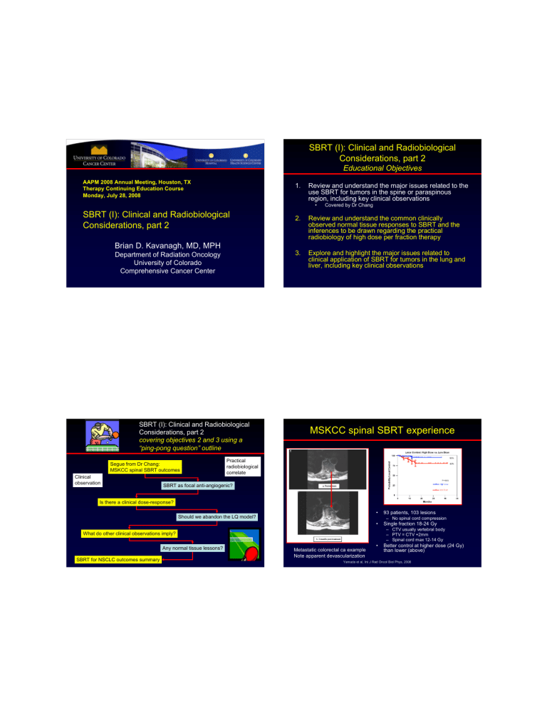
MSKCC spinal SBRT experience
Metastatic colorectal ca example
Note apparent devascularization
• 93 patients, 103 lesions
– No spinal cord compression
• Single fraction 18-24 Gy
– CTV usually vertebral body
– PTV = CTV +2mm
– Spinal cord max 12-14 Gy
• Better control at higher dose (24 Gy) than lower (above)
Yamada et al, Int J Rad Oncol Biol Phys, 2008
MSKCC argument why SBRT works well:
Radiation as potent, focused anti-angiogenic agent
• Fibrosarcoma and melanoma models
• Growth delay after RT influenced by apoptotic capacity
• Dose-dependence of percent apoptosis in endothelial cells
Garcia-Barros et al, Science, 2003 Threshold?
Apoptosisincompetent
Apoptosiscompetent
SBRT dose-response observation:
McCammon R et al, in press, IJROBP 2008
60
50
40
30
20
10
Liver SBRTPhase I dose escalation study
Lung SBRT Phase I dose escalation study
0
1998 1999 2000 2001 2002 2003
Treatment Date
2004 2005 2006
• On MVA, dose only significant predictor of local control after 3-fraction
SBRT
• Borderline significant: tumor volume
• 141 pts, 246 lesions
• 3 yr local control 89% for highest dose cohort
But think about it…
• Median dose in middle cohort was 42 Gy in 3 fractions
• LQ-based BED
10Gy
• 42(1+14/10)= 100.8 Gy
10
• equivalent to approx 84 Gy in 2 Gy fractions
• …and we still only got 60% local control?
– disappointing
The problem of modeling SBRT doses using LQ formalism—deviation at high dose
Park et al, IJROBP 70(3): 847–852, 2008
UTSW Proposed solution:
The “Universal Survival Curve”
• Essentially a step function
– Below the transition dose,
D
T
, the LQ model is used
– Above D
T
, the multi-target model is used
• Backed by some experimental evidence
Park et al, IJROBP 70(3): 847–852, 2008
Derived from the USC:
Single fraction equivalent dose (SFED)
“BED” reserved for conventionally fractionated RT modeling here
Missing from the UT-SW USC:
Marples-Joiner Low dose hypersensitivity
• First described 1993,
Marples and Joiner, for CHO cells
• Modeled with an
“Induced Repair” formula
– Requires 2 separate alpha values and a conversion dose parameter
Marples B, Collis SJ. IJROBP. 2008 Apr 1;70(5):1310-8.
“Toward a Unified Survival Curve”
• I like the USC and SFED and applaud
Park and colleagues, but maybe there is still room for improvement
• First, the high-dose region of the curve approaches a straight line on a log-linear graph: x
→ ∞
, S x
+
δ
=
S x e
−
K omega
•
δ
Kavanagh BD, Newman F, Int J Rad Oncol Biol Phys, 2008
“Unified Survival Curve”, continued
• A “shoulder” caused by dose-related increase in the exponent may be easily incorporated:
S
= e
−
K omega
• x
•
(
1
− e
−
K omegaGROWT H
•
) x
“Unified Survival Curve”, continued
• But let’s not forget low-dose hypersensitivity, which involves a decaying exponent:
S
= e
−
K psi
• x
• e
(
−
K psiDECAY
• x
)
Kavanagh BD, Newman F, Int J Rad Oncol Biol Phys, 2008 Kavanagh BD, Newman F, Int J Rad Oncol Biol Phys, 2008
S
=
e
−
K psi
• x
•
( e
−
K psiDECAY
• x
)
−
K omega
• x
•
(
1
− e
−
K omegaGROWT H
• x
)
And then put it all together, adding in some hypersensitivity data points to the Park data and re-fit the total curve:
At least concordant with the “Universal Survival Curve”:
SBRT regimens converted to 2 Gy/fxn equivalent doses using that formula
Here is where we have 90% control
Here is where we have
~60% control
Park et al, IJROBP 70(3): 847–852, 2008
Kavanagh BD, Newman F, Int J Rad Oncol Biol Phys, 2008
Liver SBRT trials
Fractionation Median FU Author
Herfarth et al.
Number of lesions
55 1 x 14-26 Gy 6 mo
Actuarial
Local Control
18mo 67%
Hoyer et al.
141* 3 x 15 Gy 4.3 yrs 2yr 79%
Milano et al.
293** 10 x 5 Gy 41mo*** 2yr 67%
Mendez
Romero et al.
U of Colorado
(unpublished)
45
49
3 x 12.5 Gy#
3 x 20 Gy
13 mo
16 mo
* Total number of CRC metastases; 44 liver metastases
** Total number of lesions treated; 45% of patients had hepatic metastases treated
*** In surviving patients
#Different fractionation (3 x 10Gy or 5 x 5Gy) used for patients with HCC or lesions 4 cm
2yr 82%
2yr 92%
SBRT for liver mets
1
0.8
0.6
0.4
0.2
0 y = 0.0098x + 0.4083
R
2
= 0.7177
10 20 30
SFED, Gy
40 50 60
SBRT normal tissue effects in liver and lung
Strategies for setting normal liver dose constraints
• NTCP-based
– Eg, PMH experience
• I defer to Dr Dawson on this one!
• Critical volume model
– At least 700 cc normal liver received < 15 Gy cumulative
2500
2000
1500
1000
500
0
0
This difference must be > 700
10 20 dose
30 40 50
Sample case:
54 Gy cohort
(18 Gy x 3 fxns)
71F T1N2cM0 nasopharynx ca
– s/p initial cisplatin and 70
Gy, salvage dissection and brachytherapy for neck recurrence, and later weekly gemcitabine and cisplatin for biopsy-proven liver met
• Transient minor response
– 6 mos later, Phase I SBRT study enrollment
Case study: pre-op SBRT for liver metastasis
– Pre-op chemoRT for rectal cancer
– SBRT for solitary liver met
– At time of APR, resection of treated liver and new, previously unknown other liver met
Typical post-SBRT normal liver image a few mos after SBRT
III II I
Necrosis
Hepatocyte/endothelial damage, central v occlusion
“red cell lakes”
Fibrosis/degenerating tumor
Olsen et al, in press, IJROBP
Reed GB and Cox AJ. The Human Liver after Radiation Injury. Am J Pathol 1966; 48:597-611
Also common: transient normal liver volume reduction
And another example
Liver V30 and Mean dose versus percent volume change
Example case:
T1N0 medically inoperable lung cancer
• 52-year-old male, h/o emphysema, on 3-5 L/min supplemental O
2
– 70 pack-yr smoker
• CT in 2003 revealed LUL lesion
(?)
– Repeat CT 3 mos later showed enlargement of the left upper lobe nodule, up to 1.5 x 2 cm
• Biopsy: adenocarcinoma
• FEV1 0.9 L (28% of predicted)
Olsen et al, in press, IJROBP
Treatment: 60 Gy/3 fractions
Characteristic radiographic findings
baseline
4 mos, CR
8 mos, subtle fibrosis
12 mos, mature fibrosis
18 mos, NED
Timmerman et al, J Clin Oncol, 2006
Plausible radiobiological anatomy large proximal airways serial architecture
Caveats interpreting the proximal airway observations
• Note that location could have had an effect on dose calculation
– with possibly higher doses given to central tumors as a result of the lack of heterogeneity correction?
• Overlooked (?) is the fact that tumor volume was also a significant predictor of toxicity (p = 0.017)
– GTV>10 cc had 8-fold higher risk of high-grade toxicity
• The grade 5 toxicities assigned by the data monitoring as possibly related to SBRT are as follows:
– Four cases of pneumonia
• Note that pts with medically inoperable NSCLC are susceptible to this event, regardless
– One patient died from a pericardial effusion
– One death occurred in a patient who had proven local recurrence and subsequently had massive bleeding—scored as treatment-related toxicity instead of local recurrence!!!
Chest Wall Volume Receiving more than 30 Gy predicts risk of severe pain and/or rib fracture after lung SBRT
Dunlap et al, oral presentation, ASTRO collaboration between University of Virginia and University of Colorado absolute V30 v. risk of severe pain or fracture
0.7
0.6
0.5
0.4
0.3
0.2
0.1
0
0 of 5 pts
V30<10cc
2 of 4 pts
V30 10-40 cc
6 of 13 pts
5 of 8 pts
V30 41-120cc V30 >120cc
Severe pain or rib fracture is a function of volume of chest wall receiving more than 30 Gy in 3 fxns
Spectrum of potential indications for SBRT
• Intensified treatment to a primary cancer
– Stage I lung cancer
• Best studied to date
– Primary HCC
– Pancreas cancer
– Prostate cancer
• Favorable due to low alpha/beta ratio
• Treatment of selected spinal/paraspinal lesions
• Palliation for challenging sites of recurrence
– Retroperitoneal
– Previously irradiated volumes
• Adjuvant systemic cytoreductive therapy
– “Radical” treatment for isolated liver, lung, and other mets
Prospective Trials of SBRT for Stage I NSCLC*
Institution
Indiana University
N
4
7
SBRT dose and fractionation
24-66 Gy/3 fractions
30 Gy/1 fraction
37.5 Gy/3-5
60 Gy/3 fractions
Results
Phase I study; maximum tolerated dose (MTD) not reached for T1 lesions;
MTD 66 Gy for T2 lesions
2 yr local control 95% Indiana University
Aarhus University
Kyoto University
Air Force General Hospital,
Beijing
University of Marburg
Technical University,
Munich
Oncology Group (RTOG)
0236
6
8
3
3
7
0
4
3
4
5
4
0
7
0
60-66 Gy/3
45 Gy/3 fractions
48 Gy/4 fractions
50 Gy/10 fractions
*probably an incomplete list!
2 yr local control 85%
2 yr local control 95%
1 yr local control 95%
3 yr local control 80%
3 yr local control 87%
1 yr local control 98%
Overall Survival after SBRT for early stage NSCLC vs conventional
100
1988-2001 SEER database overall survival after RT for early stage NSCLC in versus reported survivals from prospective and retrospective SBRT series
75
50
25
0
0
SEER average results, stage I and II
SEER stage I patients, 1988-2001
SEER summary data, stage I
SEER stage II patients, 1988-2001
SEER summary data, stage II prospective, Timmerman N = 70 prospective, Koto N = 31 prospective, Nagata stage Ia N = 20 prospective, Nagata stage Ib N = 25
Onishi retrospective, medically inoperable
N = 158
Uematsu retrospective N = 50
Wulf retrospective N = 20
Scorsetti retrospective N = 43
Baumann retrospective N = 138
Zimmerman retrospective N = 30
Fritz retrospective N = 33
1 2 3 follow-up, yrs
4 5
SEER reference: Wisnivesky J et al. Radiation Therapy for the Treatment of Unresected Stage I-II Non-small Cell
Lung Cancer. CHEST 2005; 128:1461–1467.
