Pediatric Radiation Therapy: Applications of Proton Beams for Pediatric Treatment Disclosure
advertisement
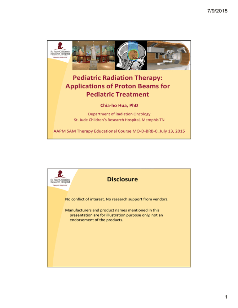
7/9/2015 Pediatric Radiation Therapy: Applications of Proton Beams for Pediatric Treatment Chia-ho Hua, PhD Department of Radiation Oncology St. Jude Children’s Research Hospital, Memphis TN AAPM SAM Therapy Educational Course MO-D-BRB-0, July 13, 2015 Disclosure No conflict of interest. No research support from vendors. Manufacturers and product names mentioned in this presentation are for illustration purpose only, not an endorsement of the products. 1 7/9/2015 Outline 1. Pediatric proton therapy: patterns of care 2. Proton dosimetric advantages and predictions of radiation necrosis and second cancer risk 3. Challenges in pediatric proton therapy 4. Proton techniques for pediatric CSI 5. Proton techniques for pediatric Hodgkin lymphoma 6. Controversy on brainstem necrosis in children 7. Bowel gas, metal artifact, beam hardening 8. Summary Pediatric Proton Therapy: Patterns of Care 13,500 children/adolescents diagnosed with cancer each year in US (~10,000 excluding leukemias) (COG data 2014). ~3000 require RT as part of frontline management (Siegel 2012 CA). # of proton patients in US ↑from 613 to 722 (from 2011 to2013). Survey on 11 operating proton centers in 2013 (Chang & Indelicato 2013 IJPT) 99% with curable intent Medulloblastoma is the most common, followed by ependymoma , low grade glioma, rhabdomyosarcoma, craniopharyngioma, and Ewing’s sarcoma. Majority were enrolled on single/multi-institution registry studies or therapeutic trials Multi-room centers in the past but single room facilities have arrived Majority with passively scattered beams due to limited access to scanning beams and large spot sizes. IMPT with spot scanning of smaller spots has been delivered in new centers. 2 7/9/2015 Pediatric Proton Therapy: Dosimetric Advantages in Critical Organs Rhabdomyosarcoma in mediastinum Tomotherapy RapidArc IMPT Fogliata et al, Radiotherapy and Oncology 2009:4:2 Pediatric Proton Therapy: Dosimetric Advantages in Critical Organs Fogliata et al, Radiotherapy and Oncology 2009:4:2 3 7/9/2015 Pediatric Proton Therapy: Necrosis Risk Prediction VMAT PSPT Confomity Index (IMPT vs. PSPT vs. VMAT) IMPT Brain necrosis risk (PSPT vs. VMAT) Brain necrosis risk (IMPT vs. VMAT) Freund et al, Cancers 2015:7:617-630 Pediatric Proton Therapy: Second Cancer Risk Prediction Excess absolute risk of proton vs. photon Moteabbed et al, PMB 2014:59:2883-2899 4 7/9/2015 Pediatric Proton Therapy: Challenges Biology and clinical Limited knowledge on in-vivo biological effect. Uncertain RBE effect at distal edge Concerns about brain and brainstem necrosis in treatment of posterior fossa tumors Limited data on clinical outcomes and normal tissue tolerance. Demonstrate clinical significance. Physics and technical Range uncertainty (e.g. requiring margin of 3.5% ×tumor depth) Larger spot sizes at lower energies (conformity of shallow target in small children) Limited options for beam angle (avoid going through bowel gas and high heterogeneous tissues) Motion interplay effects with proton scanning (mitigation strategies were proposed) Workflow and application Longer wait for beam ready after patient setup (motion while beam switching from room to room) Longer delivery time (dose rate, layer switching, longer scanning with larger volume, SBRT-type?) Is proton (especially scanning beams) better for SIB or reirradiation? Fiscal challenges (referral, more staff and room time, affordability, financial burden on centers) Proton Craniospinal Irradiation for Children Dose reduction in mandible, parotid gland, thyroid gland, lung, kidney, heart, ovary, uterine, and other non-target intracranial structures (St Clair 2004 IJROBP, Lee 2005 IJROBP, Howell 2012 IJROBP). IMPT achieves better OAR sparing than passive scattered beams while maintaining cribriform plate coverage (Dinh 2013 RO). 36Gy(RBE) prescribed CSI dose, Passive Scatter vs. IMPT Dinh et al, Radiat Oncol 2013:8:289 Models predict lower risk of second cancer, lower rate of pneumonitis, cardiac failure, xerostomia, blindness, hypothyroidism, and ototoxicity (Mirabell 2002 IJROBP, Newhauser 2009 PMB, Thaddei 2010 PMB, Brodin 2011 Acta Oncol, Zhang 2013 PMB). 23.4 Gy(RBE) CSI to 4 y.o. predicted life time risk of second cancer is 24.6% for passive scatter proton CSI risk for photon CSI is 5.6 times higher (Zhang 2013 PMB) 5 7/9/2015 Proton Craniospinal Irradiation for Children Current clinical techniques: Supine position is common. Many centers require all fields are set up and filmed prior to treatment of the first field. More common to treat with scattered beams but will change with the advent of scanning beams. Two posterior oblique beams for whole brain are common for lens sparing (Cochran 2008 IJROBP, Mahajan 2014 IJPT). Single PA spot scanning beam for uniform dose to the whole brain is feasible. Use one or more PA beams to cover spinal targets. Compensator use for passive scattered beams increased heterogeneity within the brain (Jin 2011 JACMP, Dinh 2013 RO). Many do not use compensators for whole brain. Clinical outcomes No published data yet on long term effects of proton CSI Acute toxicity is mild – 40% experienced nausea requiring antiemetic for nausea prophylaxis and most patients experienced some degree of alopecia and dry skin (Mahajan 2014 IJPT). Proton CSI setup Indiana University Setup In house short and long CSI carbon fiber boards Indexed, homogeneous, torso-length No sharp thickness changes Buchsbaum et al, Med Dosim 2013:38:70-76 MDACC Setup Neutral head position and straight cervical spine/back 10cm thick styrofoam to elevate patient to prevent the posterior oblique whole brain fields from intersecting the couch edges. Mass General Hospital Setup Giebeler et al, Radiat Oncol 2013:8:32 Prone head holder with chin and forehead pads Anterior face mask Commercial BOS (base of skull) couch inserts www.civco.com Min et al, Radiat Oncol 2014:9:220 Allow aperture to get close to patient to minimize penumbra No flat base so more freedom to choose beam angles www.qfix.com 6 7/9/2015 Proton CSI: Whole Brain Techniques MGH patient treatment (Cochran 2008 IJROBP) Posterior oblique beams (20° in the posterior direction) spare lens more than opposed laterals for passive scattered beams. MDACC IMPT paper study (Stoker 2014 IJROBP) 2 cranial fields-mirrored anterior oblique beams, angled 75° laterally with S-I rotation to prevent the ipsilateral eye from eclipsing the target. Cochran et al, Int J Radiat Oncol Biol Phys 2008:70:1336-1342 PSI and Scripps patient treatment (Timmermann 2007 Strahlenther Stoker et al, Int J Radiat Oncol Biol Phys 2014:90:637-644 Onkol, Chang PTCOG meeting 2015) A single PA beam of spot scanning for whole brain and spinal axis. Allow for a precise individual conformation of dose to the frontal subarachnoid space (Timmermann 2007 Strahlenther Onkol). Timmermann et al, Strahlenther Onkol 2007:12:685-688 Proton CSI: Low Gradients Across Spine Field Junction to Remove Junction Changes U Penn patient treatment (Lin 2014 IJROBP) No junction change. 5-8 cm overlap region between fields 4 equally spaced “gradient volumes” optimized to achieve low dose-gradient junctions Scripps patient treatment (Chang 2015 PTCOG meeting) Two isocenters for entire CSI and two fields overlap 5-6 cm Overlaps in high thoracic region to avoid thyroid & esophagusLin et al, Int J Radiat Oncol Biol Phys 2014:90:71-78 Commercial IMPT TPS to create 2%/mm smooth dose gradients MDACC paper study (Stoker 2014 IJROBP): 10-cm overlap region between fields Target divided along the cranio-caudal axis into 4 to 10 equally sized tapering segments 3 staged IMPT optimization OAR sparing as good or better than passive scattered plans Stoker et al, Int J Radiat Oncol Biol Phys 2014:90:637-644 7 7/9/2015 Pediatric Proton CSI without Junction Changes Via Robust Optimization Robust optimized IMPT plan can achieve a low dose gradient in overlapped junctions, is less sensitive to junction mismatch, and may eliminate the need for junction shifts. 10 cm overlap is needed to achieve max 5% dose variations applying a 3mm shift. Conventional MFO optimization applying Robust optimization applying 3mm intra-fractional junction shift 3mm intra-fractional junction shift Courtesy of Xiaodong Zhang. Liao et al. AAPM 2014 meeting TH-C-BRD-12 Pediatric Proton CSI: Vertebral Body Inclusion (Symmetric Bone Growth Vs. Bone Marrow Sparing) Common practice is to include the entire vertebral body for irradiation for younger children (prepubertal, not yet reaching the skeletal maturity, often <15 y.o.) to prevent differential growth of the spine (Krejcarek 2007 IJROBP, Giebeler 2013 Radiat Oncol, Lin 2014 IJROBP). But spare esophagus and thyroid. For older children (postpubertal), spare the vertebral body and the bone marrow inside. Allow for better tolerance of chemotherapy. Typically only the spinal canal is included with a few mm extension into the vertebral bodies to account for distal range uncertainty (Krejcarek 2007 IJROBP, Giebeler 2013 Radiat Oncol). May decide based on evidence of wrist epiphyseal closure on plain film (McMullen 2013 Pract Radiat Oncol) Giebeler et al. Radiat Oncol 2013:8:32 8 7/9/2015 Pediatric Proton CSI: Vertebral Body Inclusion (Bone Tolerance Dose) The exact proton tolerance for pediatric growing bone is yet to be determined. For photon, 20 Gy tolerance in children < 6 y.o. and 35 Gy for older children (scoliosis, kyphosis, bony hypoplasia). Recommended a homogeneous dose profile within the vertebral bodies in younger children (Dorr 2013 Strahlenther Onkol). Dorr et al, Strahlenther Onkol 2013:189:529-534 Lower CSI dose (18-23.4Gy) creates a dilemma regarding vertebral body coverage. St Jude photon data showed lumbar spine (L1-L5) was more affected by radiation than cervical or thoracic spine. Radiation insult to the more rapidly growing posterior components of the lumbar spine could contribute to greater lumbar lordosis (Hartley 2008 IJROBP). Source: http://ww.spinalstenosis.org Proton Therapy for Pediatric Brain Tumors craniopharyngioma Ependymoma medulloblastoma Low grade glioma Commonly – medullo/PNET, ependymoma, craniopharyngioma, and low grade glioma. RT late effects – vision (chiasm, lens, optic nerve), hearing (cochlea, auditory nerve), endocrine (hypothalamus, pituitary), neurocognition (brain, medial temporal lobe). IMPT with MFO produces better target conformity and OAR sparing than SFUD (SFO) and passively scattered plans (Yeung 2014 Pediatr Blood Cancer) For IMPT, smaller spot sizes result in better plan quality. But pediatric brain tumors, typically 5-10cm deep, require lower beam energies which have larger spot sizes. The use of range shifter to treat <4cm deep tumors further degrade the spot sizes. Min et al, Radiat Oncol, 2014:9:220 Shih et al, Cancer, 2015: 121:1712-1719 Safai et al, Transl Cancer Res, 2012:1:196-206 9 7/9/2015 Proton Therapy for Pediatric Brain Tumors Common planning rules - Avoid beams passing bony anatomy that could drastically change WEPL with a small rotation setup error, e.g. sinus cavities - Avoid partially clipping couch corners or small high density setup devices - Avoid stopping all distal edges within OAR - Be aware of device inhomogeneity and stability over time (e.g. head cushion, head rest) Wroe et al, Technol Cancer Res Treat, 2014:13:217-226 Be aware of skin dose for single proton beam (permanent alopecia reported with concurrent chemo) Be aware of anatomy and tumor changes during proton course – steroid use, tumor growth, early response, cyst changes, CSF shunting. Repeat MRI/CT may be needed for surveillance and replanning. Beltran et al, Int J Radiat Oncol Biol Phys, 2012:82:e281e287 Therapeutic Trends for Pediatric Hodgkin Lymphoma Conventional to contemporary targeting Late toxicities of pediatric Hodgkin treatment continue to emerge as patients survive longer (heart disease, second cancers). (review paper by Hodgson 2011 Hematology) 2 most recent thrusts within the RT community (Hoppe 2014 IJROBP). - treat a minimal target volume, the “involved node” or “involved site” as defined by volumetric and PET imaging - modify radiation doses based on chemotherapy response (responseadapted) Merchant, Semin Radiat Oncol, 2013:23:97-108 15 patients Proton therapy is expected to further reduce the integral dose and late effects. Hoppe et al, Int J Radiat Oncol Biol Phys, 2014;89;1053-1059 10 7/9/2015 Proton Techniques for Pediatric Hodgkin Lymphoma Hoppe et al, IJROBP, 2014:89:1053-1059 Plastaras et al, Semin Oncol, 2014:41:807-819 Holtzman, Acta Oncologica, 2013:52:592-594 Andolino et al, IJROBP, 2014:81:e667-e671 Unless pre-chemo FDG PET can be performed in RT position, usually have to position RT patients to match pre-chemo imaging position for better image registration. 4DCT is typically performed to assess motion. Breath hold may be used to reduce heart and lung doses. UFPTI OAR priorities (after mean lung dose<18Gy): Heart > Lungs > Breasts (woman only) > esophagus (Hoppe 2014 IJROBP) Cardiac radiation exposure of ≥15Gy increased the relative hazard of congestive heart failure, myocardial infarction, pericardial disease, and valvular abnormalities by 2-6 fold compared to non-irradiated survivors (Mulrooney 2009 BMJ). Pediatric Hodgkin Lymphoma: Proceed With Caution Appropriate margins to account for range uncertainty and going through heterogeneous tissues? Distal edges in critical organs. Uncertain increased RBE effect? Robustness evaluation or robust optimization for range and setup uncertainties Accuracy of proton dose calculation in thorax? CT image artifacts in thorax and shoulder regions Interplay effect significant from respiratory motion and pencil beam scanning? Volumetric image guidance is not available in many proton centers Patient selection for proton therapy depends on disease location and extent? For more discussions, see the following publications Lohr et al, Strahlenther Onkol, 2014:190:864-871 Hodgson & Dong, Leuk & Lymphoma, 2014:51:1397-1398 11 7/9/2015 Controversy on Brainstem Necrosis from Proton Therapy Unanticipated complication of brainstem necrosis developed in pediatric patients receiving proton therapy. - 43% post-PT MRI changes in brain/brainstem of ependymoma patients (MDACC, Gunther 2015 IJROBP) 3.8% incidence for >50.4 CGE to brainstem, but 10.7% for patients with posterior fossa tumors and 12.5% for <5 y.o. (UFPTI, Indelicato 2014 Sabin et al, Am J Neuroradiol, 2013:34:446-450 Acta Oncologica) Researchers suspected increased RBE at the end of range explains brainstem necrosis and proposed biological proton planning considering RBE variation. So far no evidence of association between RBE/LET distribution and brainstem toxicity or recurrence - Elevated RBE values due to increased LET at the distal end of Paganetti, Phys Med Biol, 2012:57:R99-R117 treatment fields do not clearly correlate with radiation induced brainstem injury (Giantsoudi 2015 PTCOG meeting, Giantsoudi 2014 IJROBP). - No correlation between recurrence and Monte-Carlo calculated LET distribution in medulloblastoma patients receiving proton therapy (Sethi 2014 IJROBP). Physical dose Dose weighted LET Wedenberg et al, Med Phys, 2014:41:091706 Controversy on Brainstem Necrosis from Proton Therapy Approaches to miRgate effects of ↑RBE at distal edge - Multiple fields with large angular separation - Proper angles to avoid distal ends of SOBP inside critical structures - Smear the distal fall off: split the dose for a field in half; deliver half of the dose as planned and then other half with range modified by 3mm (Buchsbaum 2014 RO) No consensus on brainstem tolerance for proton therapy. Currently err on the side of caution with brainstem. UFPTI guidelines: Dmax to brainstem ≤ 56.6 Gy D50% to brainstem ≤ 52.4 Gy For young patients with posterior fossa tumors who undergo aggressive surgery, more conservative dosiemetric guidelines should be considered. (Indelicato Buchsbaum et al, Radiat Oncol, 2014:9:2 Buchsbaum et al, Radiat Oncol, 2014:9:2 Acta 2014 Oncologica) 12 7/9/2015 Affecting Proton Range: Bowel Gas, Metal Artifact, and Beam Hardening Bowel gas Often near neuroblastoma, Wilm’s tumor, rhabdomyosarcoma, and bone sarcoma in abdomen and pelvis Vary in size and location every day Avoid shooting through bowel gas Override density within beam path on planning CT? Expect to average out? Pose a problem for whole abdominal RT Metal artifact Spinal implant, dental braces, surgical clips Apply metal artifact reduction on CT? Need to overwrite CT numbers Need to know hardware material to assign proper proton stopping power Beam hardening artifact without metal Summary Proton therapy is compelling for children and adolescents because of the promise in reducing late effects and second cancer risk. Most children are currently treated with passively scattered beams but IMPT with scanning beams of smaller spot sizes has arrived. Data on OAR tolerance and RBE effects in children are extremely limited. Planners and physicists should be careful in translating photon experience into proton (CT scan, margin design, OAR constraints, beam angle selection, setup and immobilization devices, etc). Opportunities await and abound for physicists – • • • • • technical guidance on patient selection for proton therapy safe and efficient delivery to this vulnerable patient population disease-specific treatment techniques including reirradiation uncertainty analysis and margin design sharing planning and delivery experience with the community 13 7/9/2015 Acknowledgement • • • • St. Jude CHLA MDACC Scripps Jonathan Gray, Jonathan Farr, Thomas Merchant Arthur Olch Xiaodong Zhang, Ronald Zhu, Michael Gillin Annelise Giebeler, Atmaram Pai-Panandiker, Andrew Chang, Lei Dong • UFPTI Zuofeng Li and Daniel Indelicato • Hitachi, Ltd. Power System Group and project manager Kazuo Tomida 14
