The Proton Beam Therapy Facility at PSI Talk outline
advertisement

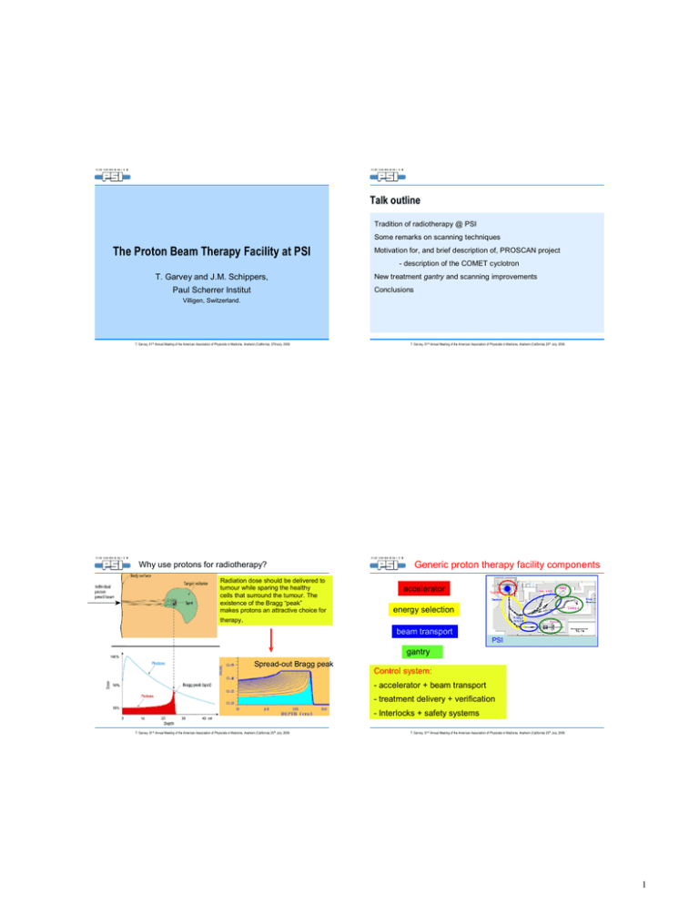
Talk outline Tradition of radiotherapy @ PSI Some remarks on scanning techniques The Proton Beam Therapy Facility at PSI Motivation for, and brief description of, PROSCAN project - description of the COMET cyclotron T. Garvey and J.M. Schippers, Paul Scherrer Institut New treatment gantry and scanning improvements Conclusions Villigen, Switzerland. T. Garvey, 51st Annual Meeting of the American Association of Physicists in Medicine, Anaheim (California), 27thJuly, 2009. Why use protons for radiotherapy? Radiation dose should be delivered to tumour while sparing the healthy cells that surround the tumour. The existence of the Bragg “peak” makes protons an attractive choice for therapy. T. Garvey, 51st Annual Meeting of the American Association of Physicists in Medicine, Anaheim (California) 25th July, 2009. Generic proton therapy facility components accelerator energy selection beam transport PSI gantry Spread-out Bragg peak Control system: - accelerator + beam transport - treatment delivery + verification - Interlocks + safety systems T. Garvey, 51st Annual Meeting of the American Association of Physicists in Medicine, Anaheim (California) 25th July, 2009. T. Garvey, 51st Annual Meeting of the American Association of Physicists in Medicine, Anaheim (California) 25th July, 2009. 1 Parasitic use of research facility for patient treatment. Classical “scattered” proton beam delivery technique Passive scattering – essentially for eye treatments. Ring cyclotron 590 MeV, 2 mA Energy modulator Injector I cyclotron eye treatment (1984) > 5000 patients treated at OPTIS-1. Shaped collimator bolus Scatter foils Gantry 1 (1996) ~ 400 patients treated. T. Garvey, 51st Annual Meeting of the American Association of Physicists in Medicine, Anaheim (California) 25th July, 2009. T. Garvey, 51st Annual Meeting of the American Association of Physicists in Medicine, Anaheim (California) 25th July, 2009. Spot scanning technique developed at PSI Better for larger, deep seated tumours. Allows a “pencil” proton beam to deliver a high dose with high precision for a precisely specified period of time. Spot scanning: step & shoot Results in better dose distribution. Beam size 7 mm FWHM 5 mm steps Compact “Gantry-1” at PSI (1996) Lucite plates set depth scanning proton pencil beam 10‘000 spots/liter (21 x 21 x 21) Dose painted only once ~1 Gy /litre/minute Table motion Transverse motions via fast scanning magnet and patient table movement. Depth variation by range shifter (lucite plates) in front of patient. T. Garvey, 51st Annual Meeting of the American Association of Physicists in Medicine, Anaheim (California) 25th July, 2009. Eros Pedroni T. Garvey, 51st Annual Meeting of the American Association of Physicists in Medicine, Anaheim (California) 25th July, 2009. 2 Gantry 1 – in use at PSI for treatment since 1996. Most compact of all existing gantry designs, φ ~ 4 m Limitations of spot-scanning technique with Gantry 1 • Beam spreading due to multiple scattering in the lucite plates. - less sharply demarcated dose distributions • Relatively slow motion of table movement • Dynamic treatment suffers from tumour or organ movement during scan - under/over dosage may occur T. Garvey, 51st Annual Meeting of the American Association of Physicists in Medicine, Anaheim (California) 25th July, 2009. Motivation for the construction of the PROSCAN Facility •Difficulties of parasitic use of Ring Cyclotron in a multi-user research environment • Ring cyclotron and spallation neutron targets require long maintenance shutdowns ~ 4 months per year during which no treatment is available. • Further development of PSI spot-scanning technology • Fast scanning techniques to deal with organ motion • Transfer of technology to industry and radiation therapy centers T. Garvey, 51st Annual Meeting of the American Association of Physicists in Medicine, Anaheim (California) 25th July, 2009. The PROSCAN facility consists of; A 250 MeV proton cyclotron – COMET (COmpact MEdical Therapy cyclotron) An energy “degrader” Beam lines equipped with diagnostics Therapy equipment previously existing gantry 1 new gantry new eye treatment facility – OPTIS-2 experimental beam line. T. Garvey, 51st Annual Meeting of the American Association of Physicists in Medicine, Anaheim (California) 25th July, 2009. T. Garvey, 51st Annual Meeting of the American Association of Physicists in Medicine, Anaheim (California) 25th July, 2009. 3 Characteristics of the COMET cyclotron Cyclotron (1930) Ernest Lawrence Magnet (1901-1958) pole coil oscillating high voltage on “Dee” electrode Ion source Vdee~ Energy gain E=Vdee T. Garvey, 51st Annual Meeting of the American Association of Physicists in Medicine, Anaheim (California) 25th July, 2009. The 250 MeV Cyclotron Based on a design from NSCL (H. Blosser). Delivered by ACCEL/Varian. RF System Frequency RF power Dee voltage 72.8 MHz (h = 2) 100 kW 80 – 130 kV Beam properties RF-Electrodes: 2 “Dees” At each electrode border: COMET is a compact 4-sector AVF isochronous cyclotron employing a superconducting magnet. Extractor: -HV Energy Extracted current (max.) Extraction efficiency Vert. emittance Horiz. emittance Momentum spread # of turns 250 MeV 800 nA 80%* < 3 mm-mrad < 5 mm-mrad ± 0.2% * Important for activation 650 issues. T. Garvey, 51st Annual Meeting of the American Association of Physicists in Medicine, Anaheim (California) 25th July, 2009. Main components of cyclotron Iron yoke: shapes field Proton source Strong ACCEL/PSI Collaboration. Ordered – April 2001 First extracted beam – April 2005 Beam to gantry – June 2006 First patient – February 2007. T. Garvey, 51st Annual Meeting of the American Association of Physicists in Medicine, Anaheim (California) 25th July, 2009. Coils (super conducting) RF: accelerates protons T. Garvey, 51st Annual Meeting of the American Association of Physicists in Medicine, Anaheim (California) 25th July, 2009. 4 Internal proton source Internal proton source cathode at -HV anode pole Chimney with slit ~5 cm pole -80 kV + Dee 1 - - - Vertical + + + deflection plates anode cathode at -HV pole T. Garvey, 51st Annual Meeting of the American Association of Physicists in Medicine, Anaheim (California) 25th July, 2009. Intensity control Max. intensity set by: proton source pole T. Garvey, 51st Annual Meeting of the American Association of Physicists in Medicine, Anaheim (California) 25th July, 2009. Central region Vertical deflection plate: - Beam on/off - Intensity modulation -V +V Deflector plate: sets requested intensity - within 50 %s - 5% accuracy T. Garvey, 51st Annual Meeting of the American Association of Physicists in Medicine, Anaheim (California) 25th July, 2009. T. Garvey, 51st Annual Meeting of the American Association of Physicists in Medicine, Anaheim (California) 25th July, 2009. 5 RF-System Image of lower “Dee’s” + + - Liner ( ) T. Garvey, 51st Annual Meeting of the American Association of Physicists in Medicine, Anaheim (California) 25th July, 2009. + - + 4x “Dee”: ~100 kV 72 MHz T. Garvey, 51st Annual Meeting of the American Association of Physicists in Medicine, Anaheim (California) 25th July, 2009. Extraction from COMET Cryogenics: closed He-system 4x1.5 W 80% Electrostatic extraction elements T. Garvey, 51st Annual Meeting of the American Association of Physicists in Medicine, Anaheim (California) 25th July, 2009. Low activation 24 h after beam off (extracted beam integral: 200 µA.h) on pole: 400 - 500 µSv/h mid plane: 250 - 400 µSv/h 10 cm unshielded degr. 500-1000 µSv/h next to shielded degrader 10-20 µSv/h T. Garvey, 51st Annual Meeting of the American Association of Physicists in Medicine, Anaheim (California) 25th July, 2009. 6 Beam-energy adjustment Degrader for fast beam energy change. Main disadvantage of cyclotron is its “fixed” energy. “Degrader” is used to alter energy of beam delivered to patient. Degrader unit Cyclotron Quad. triplet Steere r Variable thickness of material is inserted in front of beam to vary energy from 70 to 238 MeV. 5 mm range variation produced in 50 ms. T. Garvey, 51st Annual Meeting of the American Association of Physicists in Medicine, Anaheim (California) 25th July, 2009. Kicker dispersive focus; position E-error Carbon wedge degrader 238-70 MeV 5 mm ∆Range in 50 ms T. Garvey, 51st Annual Meeting of the American Association of Physicists in Medicine, Anaheim (California) 25th July, 2009. Availability 2008 (first 9 months) Collimating apertures define acceptance of beam transport system and limit emittance of degraded and “scattered” beam. Translatable carbon wedge degrader Collimator for emittance limitation 3516 hours puller puller 95% Cavity Dipole magnet and energy selection slits limit E/E to ± 0.7% T. Garvey, 51st Annual Meeting of the American Association of Physicists in Medicine, Anaheim (California) 25th July, 2009. T. Garvey, 51st Annual Meeting of the American Association of Physicists in Medicine, Anaheim (California) 25th July, 2009. 7 Fast pencil beam scanning & IMRT The problem in dynamical treatments: Organ movement Cont. scanning “TV” mode After each layer: Energy change in 80 ms Danger of underdose and/or overdose - Multiple scans of tumor - increase scan speed - intensity modulation intensity kHz-Intensity modulation PROSCAN approach: 0 T. Garvey, 51st Annual Meeting of the American Association of Physicists in Medicine, Anaheim (California) 25th July, 2009. 10 7 s for a 1 liter volume. Target repainting: 15-30 scans / 2 min. T. Garvey, 51st Annual Meeting of the American Association of Physicists in Medicine, Anaheim (California) 25th July, 2009. 3D Pencil beam scanning E=155 => rgy ne) E=150 MeV e n la ne np V oto (sca E=145 Me r P h t p de time (ms) New Gantry 2 at PSI (2009) MeV scanning magnets (x & y) for 2D // scanning No patient specific R=components in nozzle 3.5 m ~180 t L=12 m an sc ts e am gn be Ma ci l n pe Eros Pedroni T. Garvey, 51st Annual Meeting of the American Association of Physicists in Medicine, Anaheim (California) 25th July, 2009. T. Garvey, 51st Annual Meeting of the American Association of Physicists in Medicine, Anaheim (California) 25th July, 2009. 8 Gantry - 2 Gantry-1 connection: check point Beam verification by user: Measurements at Check point Connection to Gantry-1 Intensity monitor collimator T. Garvey, 51st Annual Meeting of the American Association of Physicists in Medicine, Anaheim (California) 25th July, 2009. Profile monitor Halo monitor T. Garvey, 51st Annual Meeting of the American Association of Physicists in Medicine, Anaheim (California) 25th July, 2009. DIAGNOSTICS cyclotron radial probe phase probe +SE M 1-500 nA +SE M m aterial irradiation OPTIS2 <10 nA ~2 nA range shifter CP kicker individual collim ator m odulator scatter foils range shifter degrader ~0.4 nA range shifter (single) Gantry 2 CP M achine C ontrol S yst. set-up: centering, energy ... continuous: Gantry 1 current, centering, loss --> CP ~0.4 nA inser-/fixed ionization chamber table m onitors current profile position halo loss range shifter direct/without current m easurem ent m ulti-leaf Faraday cups Faraday cups/stoppers slits collim ators dipoles/steerer/quadrupoles can enforce stability and reproducibility U ser C ontrol S ystem current --> dosim etry Pa tient S afety S ystem current, position, transm ission, dosimetry (+ non diagnostics) --> safety T. Garvey, 51st Annual Meeting of the American Association of Physicists in Medicine, Anaheim (California) 25th July, 2009. T. Garvey, 51st Annual Meeting of the American Association of Physicists in Medicine, Anaheim (California) 25th July, 2009. 9 Conclusions • The long tradition of radiotherapy at PSI is strengthened by the PROSCAN project. • The choice of a superconducting cyclotron as a dedicated accelerator has proven to be judicious. • COMET is performing in a satisfactory and reliable fashion • The installation of such a facility in a hospital environment could be envisaged. Acknowledgement: I should like to thank the PSI communications department, and my colleagues, H. Fitze, R. Dölling, J. Düppich, S. Forss and R. Köferli for providing Images. The PROSCAN project is the result of the work of many people at PSI. Thank you for your attention! T. Garvey, 51st Annual Meeting of the American Association of Physicists in Medicine, Anaheim (California) 25th July, 2009. 10
