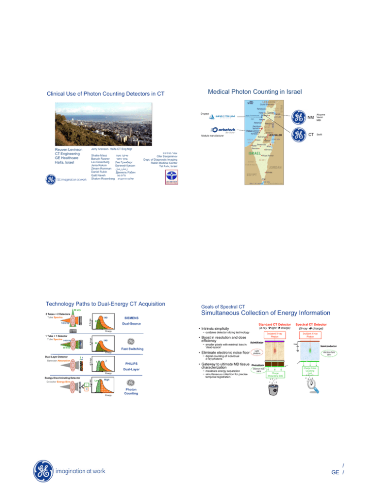
Medical Photon Counting in Israel NM CT
advertisement

Clinical Use of Photon Counting Detectors in CT Medical Photon Counting in Israel D spect NM CT Module manufacturer Swift Jerry Arenson- Haifa CT Eng Mgr Reuven Levinson CT Engineering GE Healthcare Haifa, Israel Shaike Maoz Baruch Rosner Lev Greenberg Jenia Kuksin ( ! Zimam Romman Daniel Rubin "# Galit Naveh Shalom Rosenberg ! ) *'+, Ofer Benjaminov Dept. of Diagnostic Imaging Rabin Medical Center Tel Aviv, Israel $% &' Technology Paths to Dual-Energy CT Acquisition 80 kVp Goals of Spectral CT Simultaneous Collection of Energy Information # X-rays 2 Tubes + 2 Detectors Tube Spectra 140 kVp 140 SIEMENS 80 Dual-Source # X-rays 1 Tube + 1 Detector Tube Spectra 140 kVp • Boost in resolution and dose efficiency 80 Fast Switching # X-rays Detector Absorption 1 2 PHILIPS Energy L L H # X-rays Detector Energy Bins Low light charge) Spectral CT Detector charge) (X-ray Incident X-ray Photon Scintillator Incident X-ray Photon bias Semiconductor Light photons electron-hole pairs − digital counting of individual x-ray photons Dual-Layer Energy Discriminating Detector − smaller pixels with minimal loss in ‘dead-space’ • Eliminate electronic noise floor Energy 1 2 (X-ray − outdates detector slicing technology 140 80 kVp Standard CT Detector • Intrinsic simplicity Energy Dual-Layer Detector Alcyone Ventri MBI • Gateway to ultimate MD tissue characterization − maximize energy separation − simultaneous collection for precise temporal registration Photodiode electron-hole pairs Charge Integrating DAS Charge Pulse Counting DAS High Energy Photon Counting / GE / Spectral CT: Pulse Counting Electronics direct conversion sensor common cathode discriminators pixilated anode current compensation • Incoming photon pulses stripped off flowing detector current • Pulse heights proportional to keV • Threshold discriminators trigger high or low digital counters pulse counters Digital output shaper / filter X-ray photon Optimum Imaging Performance D/A - Digital output preamplifier D/A “energy” thresholds voltage current voltage voltage HV dark/bias current CT Detector Challenges photo-current X-ray on Photon pulses ‘riding’ on photo-current and bias current time ‘zero’ electronic noise floor Precise energy separation Simultaneous energy acquisition Fully adjustable energy bins Supports multiple (>2) energy acquisition • • • • • threshold level test input + Clean Digital Signal Processing X-ray on time Photon pulses following base-line restoration X-ray on time X-ray on time • Count rates >100 Mcps/mm2 • Demanding stability requirements Narrow bi-polar pulses Digital pulses trigger counter Photon-Counting CT system: detector imaging parameters Optimal Spectral CT Performance: Paths to High-Flux X-ray Photon Counting • Smaller pixels • Today’s high-power scanners deliver >100 Mcps/mm2 count rates at the detector CT Pixel size Sub-pixelization 1mm 4x 0.5mm • Faster photon-counting DAS 1x1 mm2 Multi-slice Geometry 2D (1000x32) Flux rate (cps/mm2) 105-108 Counts/view (1 msec) 102-105 No. of bins • Future systems expected to double this requirement 20nsec shaper Channel still hasn’t reached saturation at 50 Mcps • Hybrid Counting/Integrating NO = 5 Mcps Non-paralyzable detector response and linearization calibration ameliorate pile-up issues 2 Linear Integration High Flux Readout Low-Energy Bin Photon Counting High-Energy Bin NO = 3.5 Mcps • Layered Photon-Counting Low-Energy Bin NO is when OCR=ICR/2 High-Energy Bin / GE / Swift Spectral CT Main Components • 100% simultaneous dual-energy acquisition • High-resolution direct-conversion detector array • Ultra-dose-efficient photon-counting detection • GPU-based recon and display system First Swift Phantom Scan (May 10, 2006) A very happy hour Wood Swift: The World’s First EDCT Scanner Swift 32-slice Spectral CT system Aculon Recon and Display console Teflon Iodine Air Aluminum bowtie Pixilated detector array & ASICs Plug&Play all-digital DAS 15 cm FOV 15 cm FOV Water VCT-64 gantry GPU technology First Swift Patient Scanning (May 2007) Axial Curved AVA V. Endoscopy New images in dual energy CT 2D MIP Images Conventional CT (HU) Dual Energy VR & Bone LM VR With Hard Plaque Removal 3D MIP Radial MIP monoE (mono-energetic equivalent (HU)) VNC (material density image (mg/cc)) Iodine (material density image (mg/cc)) Scan parameters: Helical, 32x0.625 mm, 140 kVp, 14 mA (eff), 1-sec rotation, pitch=0.5 / GE / Theory (dual energy) Proc, Recon and Images in dual Energy Attenuation basis functions •Basis processes: Photo-electric (w/o K-edge) & Compton Scatter Image Space Recon Non- linear processing µT(E 1)= µPE(E1)+ µComp(E1) µT(E 2)= µPE(E2)+ µComp(E2) CT IMG (E1) FBP Prep data (E1) Raw data (E1) •Basis Materials: Al, Delrin µAl(E)= xPEµPE(E)+ xComp µComp(E) Prep data (E2) Raw data (E2) µDelrin(E)= yPE µPE(E)+ yComp µComp(E) Material Decomposition 2 basis function => 2 unknowns (amount of each component) => 2 measurements @ 2 different energies m Bea har Projection Space Recon Raw data (E1) I2= exp(- L Al µ Al(E2)-LDel µDel(E2) 2-Material Basis Decomposition CT IMG (E2) FBP FBP Material density image A FBP Material density image B Linear combinations Raw data (E2) Inversion Material density images ing d en Prep data (E1, E2) I1= exp(-LAl µ Al (E1)-LDel µDel(E1) (I1, I2)= G(LAl, LDel) Linear combinations MonoE (LAl, LDel )=G-1(I1, I2) Aluminum image Aculon image Source/Detector: influence on dose efficiency 2M-PPU Cal Factor Phantom scan Al prep Ac prep Aculon (Acetal) ~50 HU Iodine [mg/ml] Calcium (CaCl2) [mg/ml] H2O Al image Ac image I 10 14cm diam. H2O BH-free B&W image I 15 I 20 I 20 I 30 Phantom legend Air Ca 80 I 10 Ca 160 H2O Ca 80 Ca 240 I 30 H2O Ca 320 B&W monoenergy image Status (vs Conventional CT) Function form Detector DQE Same; except low flux performance required for low energy beam Empirical detector data Bin energy separation NEW: does not exist in single energy EL, EH Bin flux ratios NEW: does not exist in single energy f L, f H Conventional CT σ 2= (1 / N ) DQE Dual Energy σ 2g σ 2g A B Tkaczyk et al, SPIE 2009 (7258-15) µ 2 B ( EH ) µ 2 B ( E L ) 1/ N = (µ A ( EL )µ B ( EH ) − µ A ( EH )µ B ( EL ))2 µ 2 A ( EH ) µ 2 A ( E L ) Bin energy separation f -1(L) f -1(H) DQE Bin flux ratios / GE / Energy separation/bin flux ratio Variance vs flux (photon-counting vs energy integrating) Energy Integrating Photon Counting Pile-up Electronic noise Carotid Arteriography Mono-energetic Images Mono60 Mono75 Mono100 / GE / Virtual Non-contrast Imaging Virtual Non-contrast Imaging Now you see it. Now you don’t Swift Clinical Studies: Swift Clinical Studies: VNC Performance Abdominal Imaging Energy Integrating TUE-MCI Delay-MCI Delay-VUE Spectral CT Virtual Unenhanced processing removes iodine while preserving calcium. Photon Counting 15 min delay from contrast injection images displayed with and w/o iodine. Excreted Contrast Medium No need for pre-contrast study. Pre-contrast images Calcified Structure Vs. Excreted Contrast Medium Calcified Structure / GE / Swift Clinical Studies: Full FOV Abdominal Imaging Mono 82KeV + C World’s 1st Spectral CT abdominal study Z-map* images MCI-70 keV *Color-mapping according to tissue atomic number Mono 82KeV + C Mono 82KeV + C / GE / Mono 82KeV + C VNC (+C) VNC – True Unh VNC Performance AI: Can VNC (+C) replace conventional (-C)? VCT -C VNC -C Iodine VNC (+C) Mono 82KeV + C VNC +C VNC +C Lesion Fat Muscle VNC -C 21 -87 58 VNC +C 19 -85 54 -4 31 / GE / -2 32 / GE / / GE / Adrenal Lesion Conventional CT vs Dual Energy CT (µ vs material density) VCT (-C) MCI (-C) VNC (-C) VNC (+C) MCI (+C) Subject 1 (Rt. Adrenal Les ion) 22 23 20 18 25 23 Subject 2 (Lt. Upper Lesion) -2 -2 2 1 35 Delayed MCI N/A Subject 2 (Lt. Lower Lesion) 30 22 20 19 60 N/A Subject 2 (Rt Upper Les ion) -12 -15 -14 -14 31 N/A Subject 3 (Rt. Adrenal Les ion) 3 -5 4 5 40 19 Subject 7 (Lt. Adrenal Les ion) 3 5 7 5 57 5 Subject 8 (Rt. Adrenal Les ion) -2 5 2 -5 45 14 Subject 8 (Lt. Adrenal Les ion) -3 -10 1 8 30 0 Average 5 3 5 5 40 12 Complete specificity: k-edge CT Liver Spleen Aorta Muscle Retro. Fat Gall Bladder Portal vein conv CT-C VNC -C 51.1 55.0 42.2 49.2 34.2 45.4 34.0 48.6 -101.0 -83.6 17.0 19.4 37.2 36.1 (Gd contrast) Partial Spec. No Specificity Single Dual (Photo-Electric) Gd contrast Soft tissue calcium Soft tissue No Gd contrast calcium Complete Spec. Partial Spec. Gd Contrast only Soft tissue calcium No Gd contrast Dual (Compton) Triple (kedge) Overlayed Gd contrast w/ Single Energy image / GE / Complete specificity – PET /CT Summary • Results on clinical trials show equivalent image quality for single energy scanning and potential for low-dose scanning • PC delivers a single-tube, single-detector configuration for high-quality dual energy CT imaging • PC provides a path for future k-edge imaging ' Great are the lights God created Pleasant is their radiance in all the world CT PET PET/CT - . 0 Thank you for your attention / GE /
