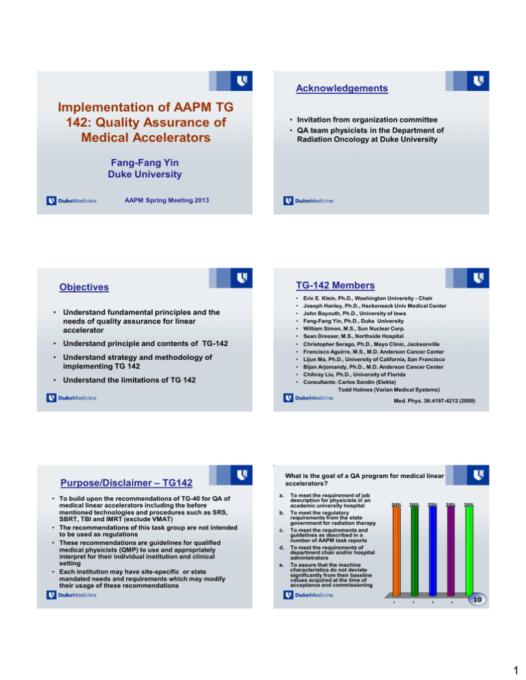
Implementation of AAPM TG 142: Quality Assurance of Medical Accelerators Acknowledgements
advertisement

Acknowledgements Implementation of AAPM TG 142: Quality Assurance of Medical Accelerators • Invitation from organization committee • QA team physicists in the Department of Radiation Oncology at Duke University Fang-Fang Yin Duke University AAPM Spring Meeting 2013 TG-142 Members Objectives • • • • • • • • • • • • • Understand fundamental principles and the needs of quality assurance for linear accelerator • Understand principle and contents of TG-142 • Understand strategy and methodology of implementing TG 142 • Understand the limitations of TG 142 Eric E. Klein, Ph.D., Washington University - Chair Joseph Hanley, Ph.D., Hackensack Univ Medical Center John Bayouth, Ph.D., University of Iowa Fang-Fang Yin, Ph.D., Duke University William Simon, M.S., Sun Nuclear Corp. Sean Dresser, M.S., Northside Hospital Christopher Serago, Ph.D., Mayo Clinic, Jacksonville Francisco Aguirre, M.S., M.D. Anderson Cancer Center Lijun Ma, Ph.D., University of California, San Francisco Bijan Arjomandy, Ph.D., M.D. Anderson Cancer Center Chihray Liu, Ph.D., University of Florida Consultants: Carlos Sandin (Elekta) Todd Holmes (Varian Medical Systems) Med. Phys. 36:4197-4212 (2009) What is the goal of a QA program for medical linear accelerators? Purpose/Disclaimer – TG142 • To build upon the recommendations of TG-40 for QA of medical linear accelerators including the before mentioned technologies and procedures such as SRS, SBRT, TBI and IMRT (exclude VMAT) • The recommendations of this task group are not intended to be used as regulations • These recommendations are guidelines for qualified medical physicists (QMP) to use and appropriately interpret for their individual institution and clinical setting • Each institution may have site-specific or state mandated needs and requirements which may modify their usage of these recommendations a. b. c. d. e. To meet the requirement of job description for physicists in an academic university hospital To meet the regulatory requirements from the state government for radiation therapy To meet the requirements and guidelines as described in a number of AAPM task reports To meet the requirements of department chair and/or hospital administrators To assure that the machine characteristics do not deviate significantly from their baseline values acquired at the time of acceptance and commissioning 20% 1 20% 20% 2 3 20% 4 20% 5 10 1 Why QA is Needed? Discussion • Answer: e • The goal for QA to ensure the quality and safety of the machines meet the criteria and guidelines obtained from ATP and commissioning • References: TG-40, TG-142 When QA is Needed? • Baseline values are entered into treatment planning systems to characterize and/or model the treatment machine, and therefore can directly affect treatment plans calculated for every patient treated on the machine • The principle of Linac QA: ICRU recommends that the dose delivered to the patient be within ±5% of the prescribed dose • Many steps involved in delivering dose to a target volume in a patient, each step must be performed with accuracy better than 5% to achieve this recommendation • The goal of a QA program for linear accelerators is to assure that the machine characteristics do not deviate significantly from their baseline values acquired at the time of acceptance and commissioning Choose the most appropriate list of components in a QA protocol for medical linear accelerators: a. b. • Machine parameters can deviate from their baseline values – – – – – – Machine malfunction Mechanical breakdown Physical accidents Component failure Major component replacement Gradual changes as a result of aging • Theses patterns of failure must be considered when establishing a periodic QA program c. d. e. Dose output, method, every day, tolerance, MD approval, Radiation Safety Officer, documentation Parameter, electrometer, frequency, tolerance, physicist, performer, daily output Parameter, what tank, frequency, sub-millimeter ruler, action, performer, computer Parameter, method, frequency, tolerance, action, performer, documentation Parameter, method, ion chamber, tolerance, performer, administrator, Therapist 20% a. Discussion • Answer: d • The process of developing a QA protocol should include several major components: the parameter to be measured, the method and tools used for the measurement, the frequency of measurement, the tolerance can be accepted for the measurement, action levels needed for the data generated, the person to perform measurement, and the method of documentation for audit. • References: TG-40, TG-142 20% 20% b. c. 20% d. 20% e. 10 General QA Considerations • Measurement parameters • Measurement methods – Phantoms – Devices – Procedures and policies • • • • • Measurement frequencies Measurement tolerances/criteria Action levels Personnel: training, efforts, finances, …. Documentation 2 Rationale for TG 142 • TG 100 task to develop QA rationales – TG 100 – A Method for Evaluating QA Needs in Radiation Therapy -- (based on “Failure Modes and Effects Analysis”) – Promotes individual department to be responsible for development of unique QA programs based on procedures and resources performed at individual institutions • TG-142 fill gap between TG-40 and TG-100 – Give performance-based recommendation – Provide process-oriented concepts and advancements in linacs since 1994 Considerations for QA Tolerances The original tolerance values in TG-40 were adapted from AAPM Report 13 which used the method of quadratic summation to set tolerances These values were intended to make it possible to achieve an overall dosimetric uncertainty of ±5% and an overall spatial uncertainty of ±5 mm These tolerances are further refined in this report and those quoted in the tables are specific to the type of treatments delivered with the treatment unit QA of Medical Accelerators Considerations for QA Frequency • Are we doing too much for QA? • The underlying principles for test frequency follow those of TG-40 and attempt to balance cost and effort • Several authors (Schultheiss, Rozenfeld, Pawlicki) have attempted to develop a systematic approach to developing QA frequencies and action levels • More recently the work being performed by Task Group 100 of the AAPM – still under evaluation Considerations for Efficiency • Challenges: – Time – Effort For an SRS system, combine WL test with daily IGRT QA • Potential solutions – Combine different tasks – Use of integrated software • Develop QA plans in Eclipse/ARIA – Some available commercial software • DoseLab • PIPSpro • RIT113 – … Table I: Daily QA • Report has 6 tables of recommendations – Linac daily (T1), Monthly (T2), Annual (T3) – Contain tests for asymmetric jaws, respiratory gating, and TBI/TSI – Dynamic/virtual/universal wedges (T4), MLC (T5), Imaging (T6) • Each table has specific recommendations based on the nature of the treatment delivered on machine – Non-IMRT, non-SRS – IMRT – SRS/SBRT • Explicit recommendations based on equipment manufacturer as a result of design characteristics of these machines 3 Table II: Monthly Table II: Monthly – Special Notes a. Dose monitoring as a function of dose rate b. Light/radiation field coincidence need only be checked monthly if light field is used for clinical setups c. Tolerance is summation of total for each width or length d. Asymmetric jaws should be checked at settings of 0.0 and 10.0. e. Lateral, longitudinal, and rotational f. Compensator based IMRT solid compensators require a quantitative value for tray position wedge or blocking tray slot set at a maximum deviation of 1.0mm from the center of the compensator tray mount and the cross hairs g. Check at collimator/gantry angle combination that places the latch toward the floor Respiratory Gating • AAPM report 91 (TG-76, Med Phys 2006) described all aspects of the management of respiratory motion in radiation therapy, including imaging, treatment planning, and delivery Respiratory Gating QA Daily protocol Duke Univ. • All respiratory techniques fundamentally require a synchronization of the radiation beam with the patient respiration • Characterization of the accelerator beam under respiratory gating conditions Monthly protocol • Recommend dynamic phantoms which simulate respiratory organ motion to test target localization and treatment delivery • Tables II and III include tests for respiratory gated accelerator operation • Daily tests were added in our institution Annually protocol Table III: Linac Annual QA - 1 Table III: Linac Annual QA - 2 4 Table IV: Dynamic/Universal/Virtual Wedges Table V: Multileaf Collimation (MLC) Multi-leaf Collimator (MLC) Sample MLC QA Test Combined to VMAT QA - to test the accuracy of dose rate and gantry speed control with P-F method Early recommendations Varian (Klein, Galvin, Losasso) Elekta (Jordan) Das (Siemens) 1998 AAPM TG-50 to address multi-leaf collimation, including extensive sections on multi-leaf collimator QA not specific for MLCs as used for IMRT Geometry accuracy: Leaf position, speed, gantry angles, etc. 1.18 Relative Dose TG-142 recommend testing (Table V) that depends on whether or not the MLC system is used for IMRT 1.20 1.16 1.14 1.12 Off y-axis : -100 mm Off y-axis : 0 mm Off y-axis : 100 mm 1.10 1.08 1.06 1.04 Dosimetry accuracy: 1.02 Abutting field, travel speed, gantry angles, dose rate, 1.00 -200 -150 -100 -50 0 50 100 150 200 Off X-Axis Position (mm) Sample MLC QA Test Sample MLC QA Test DMU/Dt Dq Dq/Dt (MU/min) (degree) (degree/s) 111 222 333 443 554 600 600 90 45 30 22.5 18 15 12.9 5.54 5.54 5.54 5.54 5.54 5.00 4.30 Ave D (%) Measurement ROIs (same MUs) Combinations of leaf speed/dose-rate to give equal dose to four strips in a RapidArc 1.1 0.5 0.0 0.1 -0.2 -0.5 -1.1 Duke University 5 Sample MLC QA Test Sample MLC QA: End-to-End Tests Combinations of leaf speed/dose-rate to give equal dose to four strips in a VMAT Dosimetry and positioning verification • From simulation to delivery for a pelvic phantom • Or a patient-specific QA • Check the data consistency acquired at different times Leaf Speed (cm/s) 0.92 0.46 1.84 2.76 Bedford et al Red J 2009 Doserate (MU/min) 138 277 544 544 Table VI: Imaging TBI/TSI • Total Body Photon irradiation (TBI) is described in detail in AAPM Report 17 (TG-29) and Total Skin Electron Therapy (TSET) in AAPM Report 23 (TG-30) • This report recommends repeating a subset of the commissioning data for TBI or TSET on an annual basis to ensure the continued proper operation of the accelerator – Should replicate commissioning test conditions i.e. Special dose rate mode for TBI/TSET treatment, Extended distance, TBI/TSET modifiers d. kV imaging refers to both 2D fluoroscopic and radiographic imaging. Functionality Modifiers’ transmission constancy TPR or PDD constancy Off-axis factor (OAF) constancy Output constancy e. Imaging dose to be reported as effective dose for measured doses per TG 75. The IGRT QA program for an imaging system attached to a linear accelerator is primarily designed to check Geometric accuracy, imaging quality, safety, and imaging dose b. Positioning and repositioning, noise, and CTDI, software accuracy c. Geometric accuracy, pixel number consistency, contrast, imaging dose d. Isocenter accuracy, Conebeam CT dose, safety, imaging dose e. Detector sag, reconstruction algorithm, resolution, and CT/CBCT dose Discussion a. 20% b. Scaling measured at SSD typically used for imaging. c. Baseline means that the measured data are consistent with or better than ATP data. • Annual TBI/TSET (Table 3) performed in the TBI/TSET mode for the clinical MU range at clinical dose rates – – – – – a. Or at a minimum when devices are to be used during treatment day. 20% 20% 20% 20% • Answer: a • IGRT QA is aimed to check the geometric accuracy-the coincidence between imaging isocenter and delivery system isocenter; the proper imaging dose, the proper image quality is maintained compared to accepted system, and operational safety such as collision detection etc. • References: TG-142, TG-104 a. b. c. d. e. 10 6 Components for IGRT QA Artifacts in kV CBCT • The goal for imaging is to improve accuracy and precision • Geometric accuracy • Cupping and streaks due to hardening and scatter (A&B) • Gas motion streak (C) • Rings in reconstructed images due – Geometric center coincidence – Positioning and repositioning to dead or intermittent pixels (D) • Streak and comets due to lag in the • Image quality flat panel detector (E) – Resolution, noise, contrast, artifacts, image fusion, etc. • Distortions (clip external contours • Safety and streaks) due to fewer than 180 degrees + fan angle projection angles (F) – Collision interlocks, warning indications, etc. • Imaging dose – 2D, 3D, 4D, fluoroscopy, etc. Artifacts in CT Imaging Crescent Artifact in CBCT Scans An apparent shift of the bow tie profile from projection to projection deriving most likely from minor mechanical instabilities, such as a tilt of the source or a shift of the focal spot W Giles et al: Crescent artifacts in cone-beam CT Med Phys 2011 Apr;38(4):2116-21. Calibration for CBCT - Coordinates Calibration of 2D System - Coordinates Isocenter calibration phantom KV Detector Video/IR Camera OBI KV Detector OBI KV tube MV Detector 1. MV Localization (0o) of BB; collimator at 0 and 90o. 2. Repeat MV localization of BB for gantry angles of 90o, 180o, and 270o. qg 3. Adjustment of BB to treatment isocenter. +1mm qg u (ExacTrac System) Recessed ExacTrac KV tube v -1mm -180 qg +180 x-ray calibration phantom Reconstruction 4. Measurement of BB location in kV radiographic coordinates (u,v) vs. q g. 5. Analysis of ‘Flex Map’ and storage for future use. 6. Use ‘Flex Map’ during routine clinical imaging. 7 Geometry – kV/MV & CBCT Combined Testing (for Iso & Positioning) Geometry – kV/MV 2D Imaging Test AP MV RLat KV Touch a pedal to get beep Touch a pedal to get beep Put a cube phantom @iso Put a cube phantom @iso Set OBI/PV at 50, 0, 0 Set OBI/PV at 50, 0, 0 S Take AP MV/RLat KV Measure the distance between BB and graticle S L R I Take CBCT Perform matching Take AP MV/RLat kV A P Shift couch: Vert +0.5cm; Lat +1.0cm; Lng +2.0cm; I Geometry – kV/MV & CBCT Combined Testing (For Iso & Positioning) Apply shift Measure the distance between BB and graticle Geometry - Imaging Fusion Software Test Daily Bladder as image In-room CT Correcting actions: Image alignment Image fusion Couch shift 6-D rotations ….. Bladder in planning CT as contour overlay Bony Structure is off Variable rectal filling observed In-room CT Prostate target is aligned with the CT image Reference CT Match BBs – Contour from CT vs CBCT IGRT QA Outcome Analysis Mechanical Accuracy Test Imaging system QA of a medical accelerator NovalisTx for IGRT per TG 142: our 1 year experience Chang et al JACMP 13 (2012) The measured discrepancies of the coincidence of CBCT imaging and treatment isocenters: 1.0 mm over 12 months Align the center of the detector – traveling distance test 8 Geometric Alignment per Gantry Rotation – 2D System S L S R S P I G270 PA A R I G0 Rt Geometric Scaling Accuracy Test Circuit-board S L A I G90 AP P I G180 Lt Image Quality: 2D Imaging Image Quality: CBCT System CTP528 – Spatial resolution Image quality Geometric CTP404 distortion HU linearity Spatial linearity Slice thickness MVD CTP515 – Low contrast resolution Image for QA analysis CTP486 – HU uniformity & noise CT number check for CBCT kV Beam Quality/Dose – Fluoroscopy kV Beam Quality/Dose - Radiography Setup : As diagramed, R/F High X-ray detector is inverted on the table with the aluminum plate placed 4.5 inches above it. Isocenter lies at the center of the high dose detector. The longest dimension of the detector is aligned along with H-F laser or cross-hair. X-ray tube with Titanium filter is placed at PA position with ABS on : • Unfors Xi: The long axis of the detector should be perpendicular to the anode-cathod axis of the tube • Detector center at isocenter or at surface Chang et al JACMP 13 (2012) # Fluoro mode 1 2 3 LD ABC HD ABC LD No ABC, @ max kV/mA with Large focal spot HD No ABC, @ max kV/mA, with Large focal spot 4 Blades XxY 26.4 x 19.8 26.4 x 19.8 26.6x20 kVp 26.6x20 140 77 77 140 Console setting mA mGy/ R/ min min 12 45.58 5.09 12 44.95 5.13 6.0 82.3 11.9 161.5 kVp 76.0 76.1 134.3 138.6 Baseline R/ HVL min 4.62 3 4.63 3 7.65 5.1 14.3 5.17 9 Sample QA for an Integrated System Imaging Dose: CBCT • • • • • • • Detector at the center of CT dose phantom • The center of phantom at the isocenter Duke Center for SRS/SBRT (Novalis Tx) • Detectors QA Consideration for QA Phantoms • • • QA for delivery system QA for imaging system QA for planning system QA for immobilization system QA for patient specific plan (IMRT/RapidArc) QA for record & verifying system QA for match software QA for gating system QA for 6D couch movement …… QA Considerations for QA Devices How to select: Imaging Daily QAphantom phantom Block Tray Dosimetry phantom CT phantom IMRT phantom 4D motion phantom Tissue phantom • Purpose • Multiple purposes • Accuracy • Ease of use • Simplicity • Size and weight • Quality • Cost • ….. Films Electrometers and cables Detectors Analysis software IMRT QAscanner device Beam data • Maintenance Sample QA Protocols and Documents at Duke University Hospital • • • • • • Daily QA Monthly QA Annually QA Gating QA - monthly Gating QA – annually Imaging QA - annually How to • Acceptance testing • Functionality • Calibration • Maintenance • ….. QA Considerations for the Process • QA will not be done automatically • QA will not automatically and correctly done • We know human makes mistakes, even you have policies and procedures in place • QA policies and procedures should be in place before machine use and be updated periodically • Policy for monitoring QA program • Mechanism for auditing QA documents • Education/training and re-education/re-training • …… 10 Sample of QA Process Error Sample of QA Process Error Event of Daily Output Check Setup • Physicist A decided to perform a monthly output check after patient treatments for the day were complete (follow the guideline). • In the evening, Physicist A assembled the monthly output check in SSD setup rather than the designed SAD setup. • The measurements showed that the photon beam outputs were 8% low, and the electron beam outputs were 2%–4% low. SSD Setup SAD Setup • 2 physicists/2 linacs, each for one linac and backup for the other linac. This primary/backup arrangement was switched once a year. • Each physicist independently designed his own monthly output check, one using an SSD setup and the other an SAD setup. • One day when Physicist A was on-site alone, the therapists reported >3% daily output based on diode measurements on his backup linac. Sample of QA Process Error • So what can we learn from this description? – Education: two different QA procedures for the two linacs (importance of standardized procedures) – Communication: not clearly understood setups by both physicists – Results of lack of education for Physicist A: • the linac worked (outputs for each modality/energy are controlled by separate boards, making it highly unlikely for all of them to suddenly be 2%–8% low) • the daily QA measurement worked (knowing that the diode response changes over time due to radiation damage, probably causing the observed underdose). Summary TG 142 provides an effective guidelines for quality assurance of medical linear accelerators. Implementation of TG 142 requires a team efforts from different expertise to support all QA activities and develop necessary policies and procedures. Institution-specific baseline and absolute reference values for all QA measurements should be established and also be evaluated for proper use and appropriateness of the particular QA test • After attempting to contact Physicist B without success, Physicist A decided to increase the machine outputs based on his measurements. • The next morning, the two physicists discussed this issue. On hearing of such a large adjustment of all energies and modalities, Physicist B investigated further, and discovered the setup discrepancy. • The outputs were immediately corrected, but unfortunately six patients had already received 8% higher doses that day. Sample of QA Process Error • Results of lack of training for Physicist A – in output adjustment (not performing an independent check of output after adjustment with the daily QA device – not minimizing the risk of such a large change by adjusting by 50% of the measured difference pending further investigation) • Results of lack of communication by Physicist A – failing to contact other physicists at nearby affiliated facilities for advice when Physicist B was reached. • Corrective actions: unify the calibration protocol; set guideline for output adjustment; … Summary • The introduction of new technologies provides new opportunities to further improve treatment accuracy and precision. At the same time, it presents new challenges for its efficient and effective implementation. • Quality assurance measures with phantoms are requisite. Expertise must be developed and must be re-established from time to time. One must also be cognizant that in actual clinical practice, inherent uncertainties of the guidance solution exist, as each technique has its own range of uncertainties. 11 Summary • A QMP should lead the QA team – Daily QA tasks may be carried out by a radiation therapist and checked by a QMP – Monthly QA tasks should be performed by (or directly supervised by) a QMP – Annual measurements be performed by a QMP with proper involvement of the entire QA team – QA per service and upgrade • Thank you for your attention An end-to-end system check is recommended to ensure the fidelity of overall system delivery whenever a new or revised procedure is introduced. An annual QA report be generated 12

