Troubleshooting Guide for DNA Electrophoresis 9. DNA ELECTROPHORESIS
advertisement
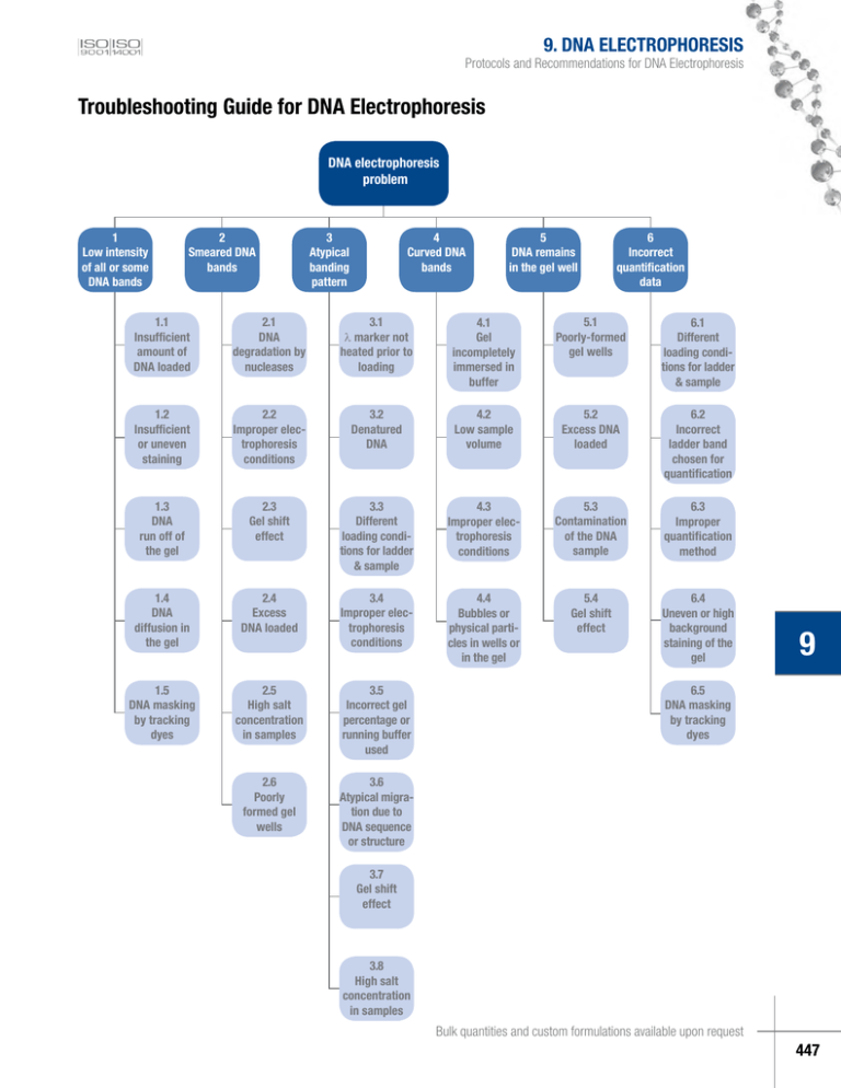
9. DNA ELECTROPHORESIS Protocols and Recommendations for DNA Electrophoresis Troubleshooting Guide for DNA Electrophoresis DNA electrophoresis problem 1 Low intensity of all or some DNA bands 2 Smeared DNA bands 3 Atypical banding pattern 4 Curved DNA bands 5 DNA remains in the gel well 6 Incorrect quantification data 1.1 Insufficient amount of DNA loaded 2.1 DNA degradation by nucleases 3.1 l marker not heated prior to loading 4.1 Gel incompletely immersed in buffer 5.1 Poorly-formed gel wells 6.1 Different loading conditions for ladder & sample 1.2 Insufficient or uneven staining 2.2 Improper electrophoresis conditions 3.2 Denatured DNA 4.2 Low sample volume 5.2 Excess DNA loaded 6.2 Incorrect ladder band chosen for quantification 1.3 DNA run off of the gel 2.3 Gel shift effect 3.3 Different loading conditions for ladder & sample 4.3 Improper electrophoresis conditions 5.3 Contamination of the DNA sample 6.3 Improper quantification method 1.4 DNA diffusion in the gel 2.4 Excess DNA loaded 3.4 Improper electrophoresis conditions 4.4 Bubbles or physical particles in wells or in the gel 5.4 Gel shift effect 6.4 Uneven or high background staining of the gel 1.5 DNA masking by tracking dyes 2.5 High salt concentration in samples 3.5 Incorrect gel percentage or running buffer used 2.6 Poorly formed gel wells 3.6 Atypical migration due to DNA sequence or structure 9 6.5 DNA masking by tracking dyes 3.7 Gel shift effect 3.8 High salt concentration in samples Bulk quantities and custom formulations available upon request 447 Table 9.8. Troubleshooting Guide for DNA Electrophoresis. Problem Possible cause and recommended solution 1. Low intensity of all or some of 1.1. Insufficient amount of ladder was loaded. the DNA bands Follow the recommendations for loading described in the certificate of analysis of the DNA ladders/markers (~0.1-0.2 µg per 1 mm gel lane width) or in the Table 9.6 on p.445. 1.2. Insufficient or uneven staining. Following electrophoresis, visualize DNA by staining in ethidium bromide solution (final concentration 0.5 μg/ml) or SYBR® Green I. Alternatively, if the DNA will not be used for cloning, add ethidium bromide to both the gel and electrophoresis buffer at a final 0.5 μg/ml concentration. After alkaline agarose gel electrophoresis the gel should be immersed for 30 min in 300 ml 0.5 M Tris-HCl buffer, pH 7.5 and only later stained in a 0.5 µg/ml ethidium bromide solution for 30 min. After denaturing polyacrylamide gel electrophoresis with urea, soak the gel for about 15 minutes in 1X TBE to remove the urea prior to staining. Stain the gel in 0.5 μg/ml ethidium bromide in 1X TBE solution for 15 min. Make sure that the gel is immersed completely in the staining solution. 9. DNA ELECTROPHORESIS 1.3. DNA run off the gel. Perform electrophoresis until the bromophenol blue dye passes 2/3 (orange G, 4/5) of the gel. Refer to the table on p.441 for migration of tracking dyes in different gels. Make sure that the entire gel is immersed completely in the electrophoresis buffer during the run. Make sure that gel and apparatus are positioned horizontally during the run. 1.4. DNA diffusion in the gel. Avoid prolonged electrophoresis or excessive staining and destaining procedures as this may cause diffusion of smaller DNA fragments in the gel. Avoid long term storage of the gel before taking a picture, as this may cause diffusion of DNA fragments and low band intensity. 1.5. DNA masking by electrophoresis tracking dyes. Do not exceed the amount of electrophoresis tracking dyes used for sample/ladder preparation. Use the loading dye solutions supplied with every Fermentas DNA ladder/marker, as these solutions contain equilibrated amount of tracking dyes which will not mask DNA under UV light. Prepare DNA ladders and probes according to recommendations on p.445. 2. Smeared DNA bands 9 2.1. DNA degradation by nucleases. Use fresh electrophoresis buffers, freshly poured gels, nuclease free vials and tips to minimize nuclease contamination of DNA solutions. 2.2. Improper electrophoresis conditions. Prepare gels according to recommendations on p.442, always use the same electrophoresis buffer for both preparation of the gel and running buffer. Make sure that the whole gel is immersed completely in the electrophoresis buffer during the run. Do not use an excessively high voltage for electrophoresis. Run the gels at 5-8 V/cm. To increase the band sharpness, use a lower voltage for several minutes at the beginning of electrophoresis. For fast electrophoresis under high voltage (up to 23 V/cm) use GeneRuler™ or O’GeneRuler™ Express DNA ladders (#SM1551/2/3 or #SM1563, p.423). An excessively low voltage during the entire run may result in diffusion of bands during electrophoresis. Excessively high voltage may result in gel heating and DNA denaturation. To calculate the optimal electrophoresis conditions (voltage) and to use the recommended V/cm value (usually 5-8 V/cm, depending on the ladder) one has to: – measure the distance between electrodes (cathode and anode) – X, cm. – and multiply that X, cm value by the recommended voltage (Y, V/cm) – the result (X, cm x recommended Y, V/cm) is Z – recommended voltage to be applied. (continued on next page) www.fermentas.com 448 9. DNA ELECTROPHORESIS Protocols and Recommendations for DNA Electrophoresis Table 9.8. Troubleshooting Guide for DNA Electrophoresis. Problem Possible cause and recommended solution 2.3. Gel shift effect. DNA binding proteins, such as ligases, phosphatases or restriction enzymes may alter DNA migration on gels and cause the DNA to remain in the gel well or gel shifting. Lambda DNA or other DNA with long complementary overhangs may anneal and migrate atypically (see p.432). To correct for the above mentioned effects, use 6X DNA Loading Dye & SDS Solution (#R1151, p.441) which is supplemented with 1% SDS to eliminate DNA-protein interactions and to prevent annealing of DNA molecules via long cohesive ends. Always heat these samples with SDS at 65°C for 10 min, chill on ice, spin down and load. 2.4. Excess DNA loaded. Follow the recommendations for loading described in the certificate of analysis of the DNA ladders/markers (~0.1-0.2 µg per 1 mm gel lane width) or in the Table 9.6 on p.445. If possible apply same requirements for DNA quantities for the samples as well. 2.5. High salt concentration in the sample. Samples containing high concentrations of salts may result in smeared or shifted band patterns. Ethanol precipitation and washing the pellet with ice cold 75% ethanol or spin column purification prior to resuspending the sample in water or TE buffer helps eliminate saltspresent in the sample. 2.6. Poorly formed (slanted) gel wells. When inserting the comb into the gel, make sure that it is vertical to the gel surface and stable during gel casting and its solidification. 3. Atypical banding pattern 3.1. Lambda DNA marker was not heated prior to loading. All DNA markers generated from Lambda DNA, as well as lambda DNA digestion products should be heated at 65°C for 5 min and chilled on ice before loading on the gel in order to completely denature the cohesive ends (the 12 nt cos site of lambda DNA) that may anneal and form additional bands. See p.445 for preparation of lambda markers for electrophoresis. 3.2. Denatured DNA. Excessively high voltage may result in gel heating and DNA denaturation. To calculate the optimal electrophoresis conditions (voltage) and to use the recommended V/cm value (often 5-8 V/cm, depending on the ladder) one has to: – measure the distance between electrodes (cathode and anode) – X, cm. – and multiply that X, cm value by the recommended voltage (Y, V/cm) – the result (X, cm x recommended Y, V/cm) is Z – recommended voltage to be applied. For non-denaturing electrophoresis use the loading dye solutions supplied with every Fermentas DNA ladder/marker, as these solutions do not contain denaturing agents. Prepare DNA ladders and probes according to recommendations on p.445. Do not heat them before loading. Heating is required only for lambda DNA markers. 9 3.3. Different loading conditions for the sample and the ladder DNA. Always use the same loading dye solution (supplied with the DNA ladder/marker) for both the sample DNA and the ladder/marker DNA. If possible always load equal or very similar volumes of the sample DNA and the ladder/marker DNA. The sample can be diluted with 1X loading dye. 3.4. Improper electrophoresis conditions. Excessive electrophoresis run times or voltage may result in migration of small DNA fragments off of the gel. Very short or slow electrophoresis may result in incompletely resolved bands. Run gels at 5-8 V/cm until the bromophenol blue passes 2/3 (orange G, 4/5) of the gel. Refer to the Table 9.4 on p.441 for migration of tracking dyes in different gels. For fast electrophoresis under high voltage (up to 23 V/cm) use GeneRuler™ or O’GeneRuler™ Express DNA ladders (#SM1551/2/3 or #SM1563, p.423). (continued on next page) Bulk quantities and custom formulations available upon request 449 Table 9.8. Troubleshooting Guide for DNA Electrophoresis. Problem Possible cause and recommended solution 3. Atypical banding pattern 3.5. Incorrect gel percentage or running buffer used. TAE buffer is recommended for analysis of DNA fragments larger than 1500 bp and for supercoiled DNA. TBE buffer is used for DNA fragments smaller tha 1500 bp and for denaturing polyacrylamide gel electrophoresis. Large DNA fragments will not separate well in TBE buffer. The correct gel percentage is important for optimal separation of the ladder DNA; prepare gels according to recommendations on p.442. When preparing agarose gels always adjust the volume of water to accommodate for evaporation during boiling. Otherwise, the gel percentage will be too high and result in bad separation of larger DNA bands. Refer to the Table 9.4 on p.441 for the range of effective separation of DNA in different gels. Ethidium bromide interferes with separation of large DNA fragments. Do not include ethidium bromide in the gel and run buffer when large DNA (more than 20 kb) or supercoiled DNA is analyzed. Stain the gel following electrophoresis in a 0.5 µg/ml ethidium bromide solution for 30 min. 9. DNA ELECTROPHORESIS 3.6. Atypical migration due to different DNA sequence or structure. During high resolution electrophoresis DNA fragments of equal size can migrate differently due to differences in DNA sequences. AT rich DNA may migrate slower than an equivalent size GC rich DNA fragment. The sequences of Fermentas DNA ladders are chosen to allow for highly accurate DNA migration according to size, however, due to differencies in nucleotide sequence or the overall DNA structure, sample migration can sometimes slightly differ from ladder band migration. DNA structures such as nicked, supercoiled or dimeric molecules will always show different mobility on gels compared to an equivalent DNA size standard. See the picture below for migration of plasmid DNA forms: bp 10000 8000 6000 5000 4000 3500 3000 2500 2000 1500 1000 750 500 250 1 GeneRuler™ 1 kb DNA Ladder (#SM0311) 2 Undigested plasmid pUC19 2,7 kb DNA, forms: – upper band (~4 kb) – dimeric plasmid – below, less visible (~3.5 kb) – nicked plasmid – lowest band (~1.9 kb) – supercoiled plasmid. 3 Linearized plasmid pUC19 (2,7 kb) – migrates according to its size 1 9 2 3 High level DNA modifications such as methylation, labeling with biotin or large fluorescent molecules also result in slower migration compared to un-modified DNA of the same size. 3.7. Gel shift effect. The presence of DNA binding proteins in the sample, such as ligases, phosphatases or restriction enzymes may alter DNA migration in the gel or cause the DNA to remain in the gel wells. Lambda DNA or other DNA with long complementary overhangs may anneal resulting in an atypical migration pattern. To eliminate these effects, use 6X DNA Loading Dye & SDS Solution (#R1151) which is supplemented with 1% SDS to eliminate DNA-protein interactions and to prevent annealing of DNA molecules via long cohesive ends. Always heat these samples with SDS at 65°C for 10 min, chill on ice, spin down and load. High salt concentration in the sample may also cause gel shift effects, see 3.8. 3.8. High salt concentration in the sample. Samples with a high salt concentration may give smeared or shifted band patterns. Ethanol precipitation and washing the pellet with ice cold 75% ethanol or spin column purification prior resuspending DNA in water or TE buffer, helps eliminate salt from the sample. (continued on next page) www.fermentas.com 450 9. DNA ELECTROPHORESIS Protocols and Recommendations for DNA Electrophoresis Table 9.8. Troubleshooting Guide for DNA Electrophoresis. Problem Possible cause and recommended solution 4. Curved DNA bands 4.1. Gel incompletely immersed in electrophoresis buffer. Electrophoresis buffer should completely cover the entire gel during sample loading and run. 4.2. Low sample volume. The sample or the ladder volume should be large enough to fill 1/3 of the total capacity of the well. Large wells should not be used with small sample volumes. If needed the sample volume can be adjusted with 1X loading dye. 4.3. Improper electrophoresis conditions. Do not use an excessively high voltage for electrophoresis. Run the gels at 5-8 V/cm. To minimize band curving, use a lower voltage for several minutes at the beginning of electrophoresis. For fast electrophoresis under high voltage (up to 23 V/cm) use GeneRuler™ or O’GeneRuler™ Express DNA ladders (#SM1551/2/3 or #SM1563, p.423). To calculate the optimal electrophoresis conditions (voltage) and to use the recommended V/cm value (which is in many cases 5-8 V/cm, depending on the ladder) one has to: – measure the distance between electrodes (cathode and anode) – X, cm. – and multiply the X value by the recommended voltage (Y, V/cm) – the result (X x Y) is the recommended voltage to be applied. 4.4. Bubbles or physical particles in the gel wells or in the gel. Use pure water, clean flasks and clean equipment for preparation of gels. Pour the gel slowly avoiding formation of bubbles. Bubbles can be removed with a pipette tip. 5. DNA remains in the gel 5.1. Poorly formed gel wells. Remove the gel comb only after complete polymerization of the gel. Pour the buffer onto the gel immediately. Rinse the wells with electrophoresis buffer to remove urea from denaturing polyacrylamide gels prior to loading the sample. 5.2. Excess DNA loaded. Follow the recommendations for loading described in the certificate of analysis of the DNA ladders/markers (~0.1-0.2 µg per 1mm gel lane width) or in the Table 9.6 on p.445. If possible load the same quantity of the sample. 5.3. Contamination of the DNA sample. Make sure that your sample DNA solution does not contain any precipitate. 5.4. Gel shift effect. The presence of DNA binding proteins in the sample, such as ligases, phosphatases or restriction enzymes may alter DNA migration in the gel and cause the DNA to remain in the gel wells. Lambda DNA or other DNA with long complementary overhangs may anneal resulting in an atypical band migration pattern. To eliminate these effects, use 6X DNA Loading Dye & SDS Solution which is supplemented with 1% SDS to eliminate DNA-protein interactions and to prevent annealing of DNA molecules via long cohesive ends. Always heat these samples with SDS at 65°C for 10 min, chill on ice, spin down and load. 6. Incorrect quantification data 9 6.1. Different loading conditions for the sample and the ladder DNA. Always use the same loading dye solution (supplied with the DNA ladder/marker) for both the sample DNA and the ladder/marker DNA. If necessary, adjust the concentration of the sample to approximately equalize it with the amount of DNA in the nearest band. If possible always load equal or very similar volumes of the sample DNA and the ladder/marker DNA. The sample can be diluted with 1X loading dye solution. 6.2. Incorrect ladder band chosen for quantification of the sample. Always compare the sample band with a similar sized ladder band. 6.3. Improper quantification method used. If possible, quantify by video-densitometry while subtracting the gel background as this method is more precise than a visual comparison of the bands. (continued on next page) Bulk quantities and custom formulations available upon request 451 Table 9.8. Troubleshooting Guide for DNA Electrophoresis. Problem Possible cause and recommended solution 6. Incorrect quantification data 6.4. Uneven staining of the gel and high background staining can also interfere with gel quantification results. Make sure that the gel is immersed completely in the staining solution. Following electrophoresis, visualize DNA by staining in ethidium bromide solution (final concentration 0.5 μg/ml) or SYBR® Green I. Do not exceed the recommended concentration of the dye for staining. Avoid prolonged staining for more than 30 min as this may result in high background. If the gel is to be stained during the run, ensure that the ethidium bromide is included in both the gel and running buffer, otherwise the staining will be uneven. After alkaline agarose gel electrophoresis the gel should be immersed for 30 min in 300 ml of 0.5 M Tris-HCl buffer, pH 7.5 and only later stained in a 0.5 µg/ml ethidium bromide solution for 30 min. After denaturing polyacrylamide gel electrophoresis with urea, soak the gel for about 15min in 1X TBE to remove the urea prior to staining. Stain the gel in 0.5μg/ml ethidium bromide in 1X TBE solution for 15 min. 9. DNA ELECTROPHORESIS 6.5. DNA masking by electrophoresis tracking dyes. Do not exceed the recommended amount of electrophoresis tracking dyes used for sample/ladder preparation. Use the loading dye solutions supplied with every Fermentas DNA ladder/marker, as these solutions contain equilibrated amount of tracking dyes which will not mask DNA under UV light. Prepare DNA ladders and probes according to the recommendations on p.445. 9 www.fermentas.com 452

