From: AAAI Technical Report SS-94-05. Compilation copyright © 1994, AAAI (www.aaai.org). All rights reserved.
Analysis of the blood flow in the aortic arc
Gian Andrea Rollandi
Istituto di Radiologia, Cattedra"R"
Universit£ di Genova, Genova(I)
Introduction
neloop techniques for MRimage acquisition make it
ssihle a non invasive investigation of the blood flow
the aorta. In this note preliminary results on the
alysis of the blood motion in the aortic arc by means
an optical flow technique are presented. Let us first
;ablish the basic anatomy of the problem, describe
~efly the cineloop MRtechnique, and then discuss
Lat can be inferred from a sequence of MRimages
visual inspection.
Alessandro Verri
Dipartimentodi Fisica
Universit£ di Genova,
Genova(I)
ment, the nth and n + lth images are compared and a
measure of the matching between points is computed,
and summedover a small region of m x m pixel s. This
can be interpreted as matching small patches from the
first image with small patches in the second. The displacement is chosen to maximize the matching measure
over all displacements (in fact, to minimize the sum of
the absolute value of the pointwise differences between
image intensities).
The accuracy was improved by interpolating the matching measure.
Experimental
/IR image sequences of the blood flow
Bowing the arc shaped aortic segment, the blood
tnges the direction of motion gradually flowing ford the head in the initial part and toward the feet
the final part of the segment. Through MRtechlues it is possible to obtain image sequences of the
od motion in the aorta. Over a single cardiac cycle,
lumber of images (typically 15 or 16) of the aortic
:, relative to the same plane of interest, are grabbed
J, by means of suitable gradient echo parameters, a
’y high contrast between the flowing blood and the
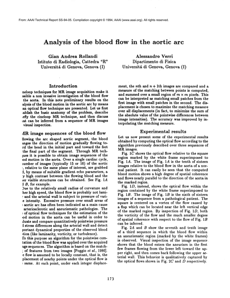
mr visible structures can be obtained. See Fig. 1A
t B, for example.
)ue to the relatively small radius of curvature and
her high speed, the blood flow is probably not iami¯ and the arterial wall is subject to pressure of varie intensity. Excessive pressure over small areas of
: aortic arc has often been indicated as a main cause
arteriosclerotic
and aneurismatic pathologies. The
: of optical flow techniques for the estimation of the
od motion in the aorta can be useful in order to
duate and compare quantitatively pointwise pressure
stress differences along the arterial wall and detect
portant dynamical properties of the observed blood
,tion (like laminarity, vorticity, or turbulence).
ro this purpose an algorithm for the pointwise cornration of the blood flow was applied over the acquired
~ge sequences. The algorithm is based on the matchof features from two images [Poggio et al., 1986].
e flow is assumedto be locally constant, that is, the
placement of nearby points under the optical flow is
: same. At each point, under each integer displace-
173
results
Let us now present some of the experimental results
obtained by computingthe optical flow according to the
algorithm previously described over three sequences of
MRimages.
Fig. 1C shows the optical flow relative to the square
region marked by the white frame superimposed to
Fig. IA. The image of Fig. 1A is the tenth of sixteen
images relative to the blood flow in the aorta of a normal patient. It can easily be seen that the computed
bloodmotionshowsa highdegreeof spatial
coherence
andflows
nearly
parallel
to thedirection
of theaortain
themarkedregion.
Fig.ID,instead,
showstheoptical
flowwithinthe
regioncontained
by thewhiteframesuperimposed
to
Fig. lB. The image of Fig. 1B is the sixth of sixteen
images of a sequence from a pathological patient. The
square is centered on a vortex of the flow caused by
a flap which can be located near the left vertical edge
of the marked region. By inspection of Fig. 1D, both
the vorticity of the flow and the much smaller degree
of spatial coherence with respect to the flow of Fig. IB
can be inferred.
Fig. 2A and B show the seventh and tenth image
of a third sequence in which the blood flow within
an aneurismatic region (marked by the white frames)
is observed. Visual inspection of the image sequence
shows that the blood enters the aneurism in the first
few frames flowing from the lower left toward the upper right, and then comes back following the upper arterial wall. This behavior is qualitatively captured by
the optical flows shown in Fig. 2C and D respectively.
A
B
C
D
>
\
l~
Figure 1: Computingthe blood flow in the aortic arc.
(A). Tenth image of an MRsequence obtained by a suitable choice of the gradient echo parameters (TE 15, FA
350, ecg gated sagittal-oblique single plane). The image
consisted of 256 x 256 pixels of 16 bits. (B). Sixth image
174
of a second MRsequence. Same parameters as in (A).
(C) and (D). Optical flow relative to the region enclosed
by the square frame of (A) and (B) respectively
tained by means of the algorithm described in the text
(with rn = lOpixels).
A
B
0
D
~v
ure 2: Computing the blood flow within an
urism. (A) and (B). Seventh and tenth images
iird MRsequence. Same parameters as in the legof Fig. I(A). (C) and (D). Optical flow relative
the region enclosed by the square frame of (A) and (B)
respectively obtained by means of the algorithm described in the text. Sameparameters as in the legend
of Fig.I(B).
175
Discussion
Although the experimental results described in the previous section appear to be very promising, much more
work needs to be done from both the theoretical and
experimental point of view. Let us briefly commenton
the main directions of future research in the analysis of
the blood flow by means of MRimage sequences.
First, a quantitative comparison of the existing algorithms for the computation of optical flow on MR.
images is needed. Preliminary results seem to indicate
that matching algorithms are more suitable than differential techniques, but a tuning or rewriting of the
equations underlying the observed flow may overturn
this qualitative impression.
Second, the obtained quantitative motion estimates
need to be correlated with dynamical properties of the
blood flow, like pressure concentration and changing
stress along the arterial wail. Third, it must be kept
in mind that the observed flow originates from a truly
three-dimensional flow. The presence of a vortex, for
example, merely indicates that the blood flow has a
component of motion in the direction orthogonal to
the acquisition plane. In order to capture all the relevant information, a reconstruction of the full threedimensional flow from a number of "different views"
may be necessary.
Finally, it would be desirable to extend the presented
single frame analysis to an intermediate or long term
analysis in which the blood flow in the aorta is observed
along the entire cardiac cycle. In a long term analysis
the features of the flow which can be tracked along the
sequence have to be determined. First order properties
of the optical flow [Campani and Verri, 1992] seem to
be good candidates to this purpose.
Conclusion
Concluding, it appears that optical flow information
can be very useful for the analysis of the blood flow
in the aorta. Preliminary results obtained on three sequences of MRimages indicate that, by means of the
computedoptical flow, it is possible to study interesting
properties of the blood flow, like laminarity, divergence,
and vorticity. Future work will correlate quantitatively
the obtained optical flow with dynamical properties of
the observed blood motion, like pressure concentration
over small areas in the proximity of aneurismatic dilatations, parietal thrombosis, and flow vortices.
References
[Campani and Verri, 1992] M. Campani and A. Verri.
Motion analysis from first order properties of optical flow. CVGIP:Image Understanding, 56:90-107,
1992.
[Poggio et al., 1986] T. Poggio, J.J. Little, and E.J.
Gamble. Parallel optical flow. Nature, 301:375-378,
1986.
176


