organic papers 2,2’-Diaminodibenzyl: a rare case of crystallo- graphically non-compliant molecular symmetry
advertisement
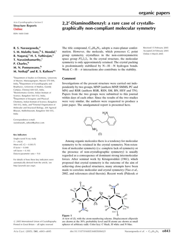
organic papers Acta Crystallographica Section E Structure Reports Online 2,2’-Diaminodibenzyl: a rare case of crystallographically non-compliant molecular symmetry ISSN 1600-5368 R. S. Narasegowda,a S. M. Malathy Sony,b S. Mondal,c B. Nagaraj,a H. S. Yathirajan,a T. Narasimhamurthy,c P. Charles,b M. N. Ponnuswamy,b M. Nethajid and R. S. Rathoree* The title compound, C14H16N2, adopts a trans-planar conformation. However, the molecule, which possesses Ci point group symmetry, crystallizes in the non-centrosymmetric space group P212121. In the crystal structure, the molecular symmetry is only approximately retained. The crystal packing is predominantly stabilized by N—H N hydrogen bonds. Weak C—H interactions also contribute to the stability. a Comment Department of Studies in Chemistry, University of Mysore, Manasagangotri, Mysore 570 006, India, bDepartment of Crystallography and Biophysics, University of Madras, Guindy Campus, Chennai 600 025, India, c Bioinformatics Centre, Indian Institute of Science, Bangalore 560 012, India, d Department of Inorganic and Physical Chemistry, Indian Institute of Science, Bangalore 560 012, India, and eOriental Organisation of Molecular and Structural Biology, 204 Agarwal Bhavan, Malleshwaram, Bangalore 560 055, India Received 15 February 2005 Accepted 24 February 2005 Online 4 March 2005 Investigations of the present structure were carried out independently by two groups, MNP (authors MNP, SMMS, PC and MN) and RSR (authors RSR, RSN, SM, BN, HSY and TN). Papers from the two groups were submitted to this journal within days of each other. Since the results of the two studies were very similar, the authors were requested to produce a joint paper. The amalgamated report is presented here. Correspondence e-mail: ravindranath_rathore@yahoo.com Key indicators Single-crystal X-ray study T = 292 K Mean (C–C) = 0.003 Å R factor = 0.040 wR factor = 0.103 Data-to-parameter ratio = 9.0 For details of how these key indicators were automatically derived from the article, see http://journals.iucr.org/e. Among organic molecules there is a tendency for molecular symmetry to be retained in the crystal symmetry. Non-retention of molecular symmetry (i.e. complete lack of symmetry or the presence of non-crystallographic symmetry) is usually regarded as a consequence of dominant strong intermolecular forces. After seminal work by Kitaigorodskii (1961), which proposed that crystal symmetry is the outcome of the aim of achieving close-packed structures, many attempts have been made to correlate molecular and crystal symmetry (Yao et al., 2002, and references cited therein). Recent work (Pidcock et Figure 1 # 2005 International Union of Crystallography Printed in Great Britain – all rights reserved Acta Cryst. (2005). E61, o843–o845 A view of (I), with the atom-numbering scheme. Displacement ellipsoids are drawn at the 30% probability level and H atoms are shown as small spheres of arbitrary radii. Color key: C black, H white and N blue. doi:10.1107/S1600536805006021 Narasegowda et al. C14H16N2 o843 organic papers Figure 2 The crystal packing in (I), showing molecules linked along the a axis via N—H N hydrogen bonds (dashed lines). Atoms labelled with an asterisk (*) or hash symbol (#) are at the symmetry positions (x, y + 12, 3 1 3 2 z) and (1 x, y 2, 2 z), respectively. Color key: C black, H white and N blue. al., 2003) has led to the conclusion that the inversion centre (Ci point group symmetry) is conserved in over 99% of cases. Higher molecular symmetries are retained in the crystal structure to a lesser extent. The present example of 2,20 diaminodibenzyl, (I), which possesses an inversion centre at the mid-point of the ethylene bridge, is a case where the molecular symmetry is only approximately conserved as pseudosymmetry in the noncentrosymmetric space group, P212121. The title compound is an intermediate in the syntheses of the anticonvulsant and antidepressant drugs carbamazepine, imipramine and desipramine (Yathirajan et al., 2004). The molecular structure of (I), which is planar, is shown in Fig. 1. Bond lengths and angles are unexceptional. The maximum deviation from the least-squares plane through all C, N and aromatic H atoms is 0.05 (1) Å for atom N1. The N1 amino group is inclined at an angle of 49 (1) to the C1–C6 ring, and the N2 amino group is inclined at 43 (1) to the C9– C14 ring. A similar observation was also found in the related structure, 2,20 -dinitrodibenzyl (Yathirajan et al., 2004). The torsion angles describing the molecular conformation are as follows: C2—C1—C7—C8 = 179.1 (2) , C7—C8—C9—C10 = 179.6 (1) , C1—C7—C8—C9 = 179.6 (2) , C9—C10— N2—H2A = 39 (2) , C1—C2—N1—H1A = 43 (2) , C9— C10—N2—H2B = 168 (2) and C1—C2—N1—H1B = 168 (2) . The r.m.s. deviation between all atoms related by the pseudo-inversion centre is 0.23 Å; if the H atoms attached to N1 and N2 are excluded this value is reduced to 0.02 Å. The maximum difference between corresponding torsion angles is 4 for C9—C10—N2—H2A and C1—C2—N1—H1A. The geometric parameters for the intermolecular interactions in (I) are listed in Table 1. The crystal structure is o844 Narasegowda et al. C14H16N2 Figure 3 A view of the crystal packing in (I), showing molecules in the (040) plane, interconnected by C—H interactions (dashed lines). Atoms labelled with an asterisk (*) or hash symbol (#) are at the symmetry positions (x 12, 32 y, 1 z) and (x + 12, 32 y, 2 z), respectively. Cg1 and Cg2 (shown in green) are the centroids of rings C1–C6 and C9–C14, respectively. stabilized primarily by N—H N hydrogen bonds and weak C—H interactions. The hydrogen bonds N1—H1B N2 and N2—H2B N1 interlink molecules along the a axis, as shown in Fig. 2. Atoms C7 and C8 of the ethylene bridge form C—H interactions with rings C9–C14 and C1–C6, on either side of the molecular plane. Significant C—H interactions are also observed for C4—H4 Cg1 (Cg1 is the centroid of the C1–C6 ring) and C11—H11 Cg2 (Cg2 is the centroid of the C9–C14 ring) (Table 1). The aromatic interactions in the crystal structure are illustrated in Fig. 3. We have examined the structures of similar compounds reported in the Cambridge Structural Database (CSD, Version 5.26; Allen, 2002). All 15 compounds containing the dibenzyl moiety and possessing Ci point group symmetry have been reported in centrosymmetric space groups. For molecules possessing Ci symmetry, crystallization in non-centrosymmetric space groups is very uncommon. In an examination of the CSDSymmetry database, a derived database of the CSD for molecular and crystallographic symmetry (Pidcock et al., 2003), out of 17 893 available compounds possessing Ci point group symmetry, 22 were found in the noncentrosymmetric space group P212121, which is generally favoured for molecules possessing C2 point group symmetry (Yao et al., 2002). Inspection of these structures reveals that an overwhelming majority of them are metal-coordination compounds and involve a large number of strong intermolecular hydrogen bonds. They include, however, the case of N,N,N0 ,N0 -tetramethyl-p-phenylenediamine (Ikemoto et al., 1979), where the crystal packing is governed purely by van der Waals forces. Acta Cryst. (2005). E61, o843–o845 organic papers Experimental The title compound was obtained from Max India Ltd. Crystals were grown by the slow-evaporation method, using both toluene and ethanol solvents. The data presented here correspond to a crystal obtained from toluene. Crystal data C14H16N2 Mr = 212.29 Orthorhombic, P21 21 21 a = 5.4716 (4) Å b = 12.480 (1) Å c = 16.695 (1) Å V = 1140.0 (2) Å3 Z=4 Dx = 1.237 Mg m3 Mo K radiation Cell parameters from 5764 reflections = 5–54 = 0.07 mm1 T = 292 (2) K Block, colourless 0.51 0.24 0.15 mm The aromatic and methylene H atoms were positioned geometrically and refined as riding on their carrier atoms, with Car—H = 0.93 Å, methylene C—H = 0.97 Å and Uiso(H) = 1.2Ueq(C). The H atoms attached to N were located in a difference electron-density map, and were refined isotropically; N—H distances are in the range 0.88 (2)–0.91 (2) Å. In the absence of significant anomalous dispersion effects, Friedel pairs were averaged. Data collection: SMART (Bruker, 1998); cell refinement: SAINTPlus (Bruker, 2001); data reduction: SAINT-Plus; program(s) used to solve structure: SHELXS97 (Sheldrick, 1997); program(s) used to refine structure: SHELXL97 (Sheldrick, 1997); molecular graphics: ORTEP3 (Farrugia, 1997) and PLATON (Spek, 2003); software used to prepare material for publication: WinGX (Farrugia, 1999) and PARST (Nardelli, 1995). Data collection 1456 independent reflections 1344 reflections with I > 2(I) Rint = 0.021 max = 27.4 h = 7 ! 6 k = 16 ! 14 l = 21 ! 21 Bruker SMART CCD area-detector diffractometer ! scans Absorption correction: multi-scan (SADABS; Sheldrick, 1996) Tmin = 0.97, Tmax = 0.98 9109 measured reflections The authors thank Max India Ltd. for providing the sample and Professor T. N. Guru Row, SSCU, Indian Institute of Science, Bangalore, for providing the CCD facility. SMMS acknowledges the CSIR, India, for financial support. Refinement Refinement on F 2 R[F 2 > 2(F 2)] = 0.040 wR(F 2) = 0.103 S = 1.11 1456 reflections 161 parameters H atoms treated by a mixture of independent and constrained refinement w = 1/[ 2(Fo2) + (0.0596P)2 + 0.1255P] where P = (Fo2 + 2Fc2)/3 (/)max < 0.001 max = 0.16 e Å3 min = 0.28 e Å3 Extinction correction: none Table 1 Hydrogen-bond geometry (Å, ). D—H A D—H H A D A D—H A N1—H1B N2i N2—H2B N1ii C7—H7A Cg2iii C8—H8B Cg1iv C4—H4 Cg1v C11—H11 Cg2vi 0.90 (2) 0.88 (2) 0.97 0.97 0.93 0.93 2.46 (2) 2.45 (2) 2.81 2.78 2.85 2.92 3.334 3.255 3.654 3.624 3.657 3.678 162 (2) 153 (2) 145 147 139 139 (2) (2) (2) (2) (2) (2) Symmetry codes: (i) x; y þ 12; 32 z; (ii) 1 x; y 12; 32 z; (iii) x 1; y; z; (iv) x þ 1; y; z; (v) x 12; 32 y; 1 z; (vi) x þ 12; 32 y; 2 z. Cg1 is the centroid of the C1– C6 ring and Cg2 is the centroid of the C9–C14 ring. Acta Cryst. (2005). E61, o843–o845 References Allen, F. H. (2002). Acta Cryst. B58, 380–388. Bruker (1998). SMART. Version 5.054. Bruker, AXS Inc., Madison, Wisconsin, USA. Bruker (2001). SAINT-Plus. Version 6.22. Bruker, AXS Inc., Madison, Wisconsin, USA. Kitaigorodskii, A. I. (1961). Organic Chemical Crystallography. New York: Consultants Bureau. Farrugia, L. J. (1997). J. Appl. Cryst. 30, 565. Farrugia, L. J. (1999). J. Appl. Cryst. 32, 837–838. Ikemoto, I., Katagiri, G., Nishimura, S., Yakushi, K. & Kuroda, H. (1979). Acta Cryst. B35, 2264–2265. Nardelli, M. J. (1995). J. Appl. Cryst. 28, 659. Pidcock, E., Motherwell, W. D. S. & Cole, J. C. (2003). Acta Cryst. B59, 634– 640. Sheldrick, G. M. (1996). SADABS. University of Göttingen, Germany. Sheldrick, G. M. (1997). SHELXS97 and SHELXL97. University of Göttingen, Germany. Spek, A. L. (2003). J. Appl. Cryst. 36, 7–13. Yao, J. W., Cole, J. C., Pidcock, E., Allen, F. H., Howard, J. A. K. & Motherwell, W. D. S. (2002). Acta Cryst. B58, 640–646. Yathirajan, H. S., Nagaraj, B., Nagaraja, P. & Lynch, D. E. (2004). Acta Cryst. E60, o2455–o2456. Narasegowda et al. C14H16N2 o845
