From: ISMB-95 Proceedings. Copyright © 1995, AAAI (www.aaai.org). All rights reserved.
Symbolic
Generation
and Clustering
of RNA 3-D Motifs
1 1’2
Marielle
Foucrault
and Francois
Major
1Ddpartement d’Informatique et de Recherche Op~rationnelle
2Montreal Joint Center for Structural Biology
Universit4 de Montr4ai
Montrdal, Qudbec, Canada H3C 3J7
major@iro.umontreal.ca
Abstract
Non canonical G.A base pairs play important
structural and functional roles e tin ribonucleic
5 -- G-- A -- 3
acids (RNA).In particular, the 3’ - A - G - ~’ motif and three of its sequencevariants have a relatively high occurrence in 16S and 23S ribosomal RNA.Extensive 3-D modelingof these variants has allowed to support a previously proposed 3-D model and to identify another series of conformations consistent with phylogenetic data. The library of 3-D conformationsgenerated by the MC-SYM
program was then used to
produce 3-D conformations of the small ribonucleotide r(GGCGAGCC)2.
This new library includes the conformation determined by nuclear
magnetic resonance spectroscopy.
Introduction
Case-based reasoning methods have been among the
most successful approaches to three-dimensional (3D) modeling of proteins mainly because of the great
similarity between the 3-D structures of two homologous peptide sequences (Hilbert, Bohm, & Jaenicke
1993). In the case of ribonucleic acids (RNA), however, the small number of experimentally determined
3-D structures limits the application of this approach
and alternative methods are sought (Malhotra, Tan,
& Harvey 1990; Major et al. 1991; Altman 1993;
Altman, Weiser, & Noller 1994).
RNAmolecules are composed of double-stranded
motifs, called double-helices,
which are built of
Watson-Crick base pairs and adopt a canonical A-RNA
conformation. The double-helices are inter-connected
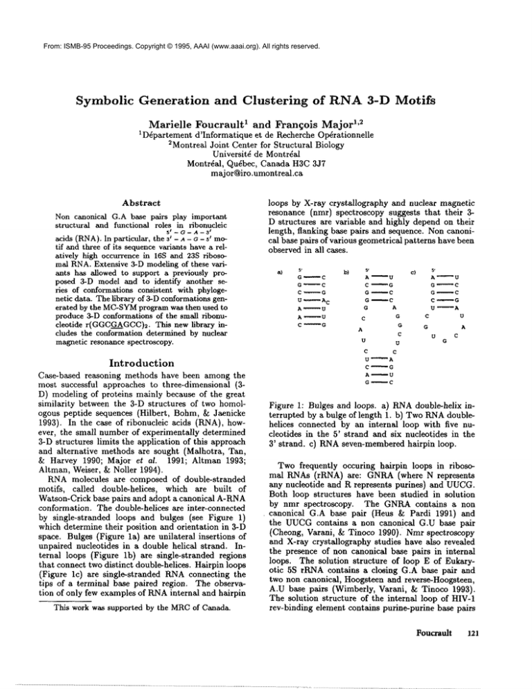
by single-stranded loops and bulges (see Figure 1)
which determine their position and orientation in 3-D
space. Bulges (Figure la) are unilateral insertions
unpaired nucleotides in a double helical strand. Internal loops (Figure lb) are single-stranded regions
that connect two distinct double-helices. Hairpin loops
(Figure lc) are single-stranded KNAconnecting the
tips of a terminal base paired region. The observation of only few examples of KNAinternal and hairpin
This work was supported by the MRCof Canada.
loops by X-ray crystallography and nuclear magnetic
resonance (nmr) spectroscopy suggests that their
D structures are variable and highly depend on their
length, flanking base pairs and sequence. Noncanonical base pairs of various geometrical patterns have been
observed in all cases.
a)
s.
O
C
G
~C
C ........
G
U ¯
-A
C
A .......... U
A .... U
C.........
G
s.
A
C
G
G
G
b)
C
’ U
G
’" C
, C
A
G
G
C
U
U
C
C
U ........
A
C.........
G
A
’" U
G
C
A
c}
s’
A........ U
G ........ C
G .........
C
C .........
G
U
~A
C
U
G
A
U
C
G
Figure 1: Bulges and loops, a) RNAdouble-helix interrupted by a bulge of length 1. b) TwoRNAdoublehelices connected by an internal loop with five nucleotides in the 5’ strand and six nucleotides in the
3’ strand, c) RNAseven-membered hairpin loop.
Two frequently occuring hairpin loops in ribosomal RNAs (rRNA) are: GNRA(where N represents
any nucleotide and R represents purines) and UUCG.
Both loop structures have been studied in solution
by nmr spectroscopy.
The GNRAcontains
a non
canonical G.A base pair (Hells & Pardi 1991) and
the UUCGcontains a non canonical G.U base pair
(Cheong, Varani, & Tinoco 1990). Nmr spectroscopy
and X-ray crystallography studies have also revealed
the presence of non canonical base pairs in internal
loops. The solution structure of loop E of Eukaryotic 5S rRNAcontains a closing G.A base pair and
two non canonical, Hoogsteen and reverse-Hoogsteen,
A.U base pairs (Wimberly, Varani, & Tinoco 1993).
The solution structure of the internal loop of HIV-1
rev-binding element contains purine-purine base pairs
Fouffault
121
(Battiste et al. 1994). Comparative sequence analysis
of rRNAhave also revealed frequently occuring tandem G.A mismatches (Gautheret, Koonings, & Gutell
1994). The tandem G.A is also present in the hammerhead (Pley, Flaherty, & McKay1994) and leadactivated (Pan & Uhlenbeck 1992) ribozymes.
group I introns and the rev-binding element of HIV1, non canonical base pairs have been deduced from
co-variation analysis and have been successfully introduced in RNA3-D modeling (Michel & Westhof 1990;
Leclerc, Cedergren, & Ellington 1994).
Here, we report on the application of the computer
modeling system MC-SYM
(Major et al. 1991) to generate a library of tandem G.A mismatches. Such libraries are necessary prior to further structural studies such as those based on energy stability, isosteric
conformations and chemical data. As a first step, the
constraint satisfaction algorithm was applied to generate all possible tandem G.A mismatches conformations and their interchangeable motifs in 16S and 23S
rRNAs (Gautheret, Koonings, & Gutell 1994). Large
numbers of conformations were generated mainly due
to the structural flexibility of non canonical base pairing and stacking of the purines. The symbolic information generated by the programwas then used to classify
the conformations into structural classes based on base
pair geometries, sugar pucker modes and torsion angle around the glycosyl bond. The library was used to
identify two series of isosteric motifs that are consistent
with phylogenetic data. The first one falls in the same
class as the solution structure of r(GGCGAGCC)2
determined by SantaLucia et al. (SantaLucia & "Ihrner
1993). The other, which we propose as a second possibility, falls in the same class as the solution structure
determined by Priv~ et al. (Priv~ et al. 1987) for
d(CCAAGATTGG)2.
Complexity
analysis
of MC-SYM
The backtracking
algorithm used in MC-SYM,although efficient, is based on a combinatorial search.
This implies that the conformational search must be
meticulously applied to avoid the combinatoric explosion. In the current implementation, the free variables
are the nucleotide units. The domainof values for each
nucleotide is defined by the Cartesian product of various pre-determined nucleotide conformations, that is,
rigid sets of atomic coordinates x spatial transformations which are used for the assembly of nucleotides.
The use of large domains allows to cover a larger
fraction of the conformational space and improves the
precision of generated models. However, the size of
the explored conformational space grows exponentially
with the size of the domains. Assuming that the
complexity of checking any given set of input constraints requires a constant amount of computer resources, then the complexity of the backtracking algorithm can be approximated with the use of three
parameters: the number of variables, N; the size of
122
ISMB-95
the domains, M; and a measure of constraint efficiency, p, that could be thought of as a probability that
any extension by one value of a partial solution will
be consistent with the input constraints, and this at
any time during the simulation. A probabilistic model
based on these three parameters has been proposed by
Haralick (Haralick 1980). In Haralick’s model, M,
and p are constant values so that the expected number of partial structures during the simulation is given
by ~"~N=I Mip(i-1)(i-2)/2; or Mg complete structures
when p = 1.
In practice however, only N, the number of nucleotides is constant and knowna priori of a simulation; Mand p vary according to available constraints.
In particular, it is extremely difficult to approximatep
prior to run time. In practice, whenp is small, combinatorial explosions are prevented and RNAstructures
are produced using reasonable amounts of computer
resources. The theoretical size of the search tree for
reproducing the yeast tRNAmotif according to input
constraints was 102s (Major, Gautheret, & Cedergren
1993). Nevertheless, available constraints were used
to sufficiently prune the search tree so that only half
a million partial constructions were evaluated. This
computation on currently available workstations takes
only about five to 20 minutes. MC-SYM
has been applied successfully to reproduce at the atomic level ~rethe ,
cision consensus structures such as the yeast tRNA
the UUCGtetranucleotide
loop (Gautheret, Major,
& Cedergren 1993) and the TYMVpseudoknot motif
(Major et al. 1991), to provide support for the Sundaralingam model of the relative position and orientation
of transfer RNAsin the ribosomal A and P sites (Easterwood et al. 1994), and using covariation data, to
model the bound conformation of the Rev-binding element of HIV-1(Leclerc, Cedergren, & Ellington 1994).
Tandem G.A mismatches
Noncanonical base pairs, and in particular those involving a guanine and an adenine (G.A), play important structural and functional roles in both deoxyribonucleic acid (DNA)and RNA.There are four different G.A base pair patterns involving two hydrogen
bonds (H-bonds). Following Saenger’s notation, the
four patterns are: VIII (A:N6-H...O6:G;A:N1...H 1 :G),
IX (A:N6-H...O6:G; A:NT...HI:G), X (A:N6-H...N3:G;
A:N1...H-N2:G) and XI (A:N6-H...N3:G; A:N7...HN2:G) (Saenger 1984).
Several structures
containing tandem G.A mismatches have been studied by nmr spectroscopy, X-ray
crystallography and molecular modeling (see Table 1).
A comparative sequence analysis reveals that tandem
G.A mismatches occur frequently in the internal loops
of 16S and 23S rRNA(Gautheret, Koonings, & Gutell
1994).
5s-A-A-31
The most frequent
5t-A-
/A--3
motifs
5e /-- G-- A -- 3
are 3’-A-~-5’,
3’ - A - G - ~’ and ~’ - G - ~ - 5’ . Onthe basis of covari-
Duplex
Structural
features
Reference
r(GGCGAGCC)2
cross-strand stacking
AG(XI)
SantaLucia ~ Turner (1993) Biochemistry 3_22:12612.
d(ATGAGCGAATA)2
cross-strand stacking
AG(XI)
Li, Zon & Wilson (1991) Proc. Natl, Acad. Sci. (USA) 888:26.
d(GCGAATAAGCG)2
cross-strand stacking
AG(XI); AA(41)
Maskos et al. (1993) Biochemistry 32:3583.
Hammerhead ribozyme
d(-AG-)2
r(-AG-)2?
cross-strand stacking
AG(XI)
AG(XI)
d(CCAAGATTGG)2?
AG(VIII)
d([Gi],)2$
AG(VIII); AG(IX)
Katahira et al. (1993) Nucleic Acids Res. 2_!1:5418.
Pley, Flaherty & McKay(1994) Nature 372:68.
Priv6 et al. (1987) Science 238:498.
Huertas et al. (1993) EMBOJ. 1-2:4029.
Table 1: A list of RNAand DNAduplexes for which nmr, X-ray crystallography?
have been done to determine 3-D structure.
5t -- G -- A I-- 3
ation analysis, the 3’ - A - G -- 5’ motif is interchange5t -- A -- A -- 3t 5t t-- G -- A -- 3
able with three motifs: 3’ - A - c - s’ , 3’ - A - A - 5’ and
and molecular modeling:~ studies
volving one H-bond were implemented and tested as
well; using the following Arabic numbering notation:
AA[35,36,37,38,39,40,41] (see Figure 2).
t51 -- A -- A -- 3
3’ - A - A - 5’ , suggesting isosteric conformations.
Implementation
All simulations were performed using "ADJACENCY
1.0 3.5", that is, the distance between the terminal nucleotide O3’ and the P atom of the next nucleotide was
required to be no shorter than 1.0/~ and no longer than
3.5/~. The following "GLOBAL"
constraints, which describe the minimumdistance in A between pairs of
atoms were used: P P 3.0; C1’ C1’ 3.0; PSE PSE 3.0;
P CI’ 2.5; P PSE 2.5; CI’ PSE 2.5; C4 C4 2.0; N1 N1
2.0; N1 C1’ 2.0; N1 C4 2.0; 02’ O4’ 2.0; 03’ C5’ 2.0;
P C3’ 2.0; 02’ C3’ 2.0; O3’ C3’ 2.0; C2 N7 1.5; and,
C2 C2’ 1.5. These constraints are used as a heuristic
control for steric avoidance and, in absence of more detailed energy function, to allow for the computation of
larger numbers of conformations in given amounts of
time.
5e -- G -- A °-- 3
For each interchangeable
motif, 3’-A-O-5’ ,
51 -- A -- A -- 31
5t -- G -- A -- 31
t
3t--A--G--51
~ 3t--A--A--5
5t t-- A -- A -- 3
and 3t--A--A--5
t
j
an
A:N6-H...N7:A
35
A:N6-H...N1:A
36
A:N6-H...N3:A
39
40
41
MC-
SYMscript was defined and used to generate libraries
of conformations. The non canonical purine-purine
base pairs from (Saenger 1984) were implemented and
systematically tested for G.A and A.A. To find isosteric substitution
for G.A, the A.A base pairs in-
Figure 2: A.A base pairing patterns composed of one
H-bond as implemented in MC-SYM.
The general script format is as follow:
Foucrault
123
; 5’-X-A-3’
; 3’-A-Y-5’
SEQUENCE
i
reference
rX
rA 2
<stack>
1
rY 3
<pair>
2
rA 4
<pair>
1
I~31ecule
<type>
<type>
<type>
<type>
The rX identifies the type of nucleotides. The integers on the right side of the nucleotide types are references in the sequence. The next column indicates the
possible spatial transformations. The integers on the
right side of the spatial transformations are references
to the nucleotides used in the application of the specified transformations. Finally, the last columnindicates
the type of nucleotide conformations that will be assigned during the simulations. The script can be read
as follow: Use nucleotide 1 as the reference. Stack the
base of A2 on the base of nucleotide 1. Append nucleotide 3 by trying all possible base pairings to form
the pair A2.Y3. Append A4 by trying all possible base
pairings to form the pair A4.X1.
In this script, rX and rY were substituted according
to the value of X and Y in the sequence, that is, rA
or rG. The reference spatial transformation is a special
one indicating a referential for the construction. The
stack transformations ensured for the stacking of bases
1 and 2 and included cross:strand stacking. The pair
transformations apply the non canonical base pairing
geometries described above. The geometries vary with
the type of nueleotides X and Y: AG_pair or AA_pair.
The conformation of all nueleotides were assigned from
a sampling of sugar puckers (C2’- and C3’-endo).
addition, all guanines were assigned a sample of conformations with anti torsion angle around the glycosyl
bond. No such constraint were applied to the adenines
so that syn torsion angles around the glycosyl bond
were also assigned.
Results
For the tandem A.G, MC-SYM
generates 893 structures that were classified into seven classes combining
the following A.G base pairing patterns: IX-IX, VIIIIX, IX-VIII, VIII-VIII, IX-XI, X-XI and XI-XI (see
Table 2). Sugar pucker modes and torsions around the
glycosyl bond were then used to regroup the structures
into 78 subclasses. The class VIII-VIII represents near
50 percent, and the class XI-XI near 30 percent of all
generated structures. The adenines are in syn and the
guanines in anti conformationsin all structures of class
IX-IX. In the symetrical VIII-IX and IX-VIII tandems,
the adenines involved in type IX base pairs are in syn
conformations. All other bases are in anti conformations. In the tandems of class VIII-VIII, IX-XI and
XI-XI, all bases are in anti conformations. Finally, in
124 ISMB-95
Class
5’~ -~ -3’
¯ ¯
3’-A-G -5’
4 3
(893)
1 2
5 ’ -G-G-C-G -A-G-C-C-3’
I I I ¯ ¯ I I I
3 ’ -C-C-G-A-G - C-G- G- 5"
4 3
(9,072)
Subclass
Occurence
ie4-2.3
(#)
(%}
IX-IX
VIII-IX
IX-VIII
V/XI-V/II
IX-XI
X-XI
XI-XI
1
4
7
34
8
4
20
0.3
3.4
ii.2
49.8
5.0
1.7
28,6
VIII-IX
~-vJ:~
VIII-VlXI
IX-X~
XI-XI
6
18
60
2
i
8.3
21.4
69,7
0.4
0.i
Table 2: Summaryof MC-SYM
simulations.
The numbers in parentheses indicate the number of models generated. Occurencepercentages are relative to the number of models generated in each case.
the X-XI tandem the adenine in the type X base pair
is in syn conformation. The other bases are in anti
conformations. The interstrand stacking observed in
the nmr structures of the class XI-XI tandem has been
reproduced properly {see Figure 3a).
The library of tandems has been used to model the
r(GGCGAGCC)2duplex of SantaLucia et al. (SantaLucia & Turner 1993). MC-SYMgenerates 9,072
such models. The models were grouped into five structural classes: VIII-IX, IX-VIII, VIII-VIII, IX-XI and
XI-XI, and further divided in 87 subclasses. Near seventy percent of the modelsfall into the class VIII-VIII.
The class XI-XI, observed by nmr spectroscopy, represents only about 0.1 percent of the models. This
class contains only one subclass; with a maximum
rms deviation between all-atoms (except hydrogens)
0.01A. The rms deviation between the MC-SYM
models and one of the nmr structures is 3.9.~. The rms deviation between a refined MC-SYM
model {by applying molecular mechanics energy minimization) and the
nmr model is 1.5/~. The most deviation comes from the
backbone atoms in the tandem which are not symmetric in MC-SYM
models. Another important characteristic of the solution model, which is no well reproduced
in the MC-SYM
models or their refinements, is the formation of H-bonds between the adenine-amino protons
and the O2’ of the guanine, and between the guanineamino protons and the adenine-phosphate oxygens.
In the context of comparative sequence analysis, two
series of isosteric conformations for
the variations of
~
5’ -- G -- A -- 3’
a’-a-G-5’
, that
5’ -- A -- A -- 3
is,
a’-a-G-5’
5’ -- G -- A -- 3"
, a’-a-A-5’
and
5’ -- A -- A -- 3’
a’-A-A- s’, havebeenidentified.
The A.G-XI;A.A41 (Figure
3b)andA.A-41;A.A-41
(Figure3c)tandems
areisosteric
to theA.G-XI;A.G-XI
(Figure
3a)tandem
observed by nmr and suggested in (Gautheret, Noonings, &Gutell 1994). Interestingly, other isosteric con-
formations were identified by our method. The A.GVIII;A.G-VIII (Figure 3d), A.G-VIII;A.A-37 (Figure
3e) and A.A-37;A.A-37(Figure 3f) are also isosteric.
d]
b)
e)
Figure 3: lsosteric motifs. Three-dimensional structures generated by MC-SYM
for the tandem of class
a) XI-XI, b) XI-41, c) 41-41, d) VIII-VIII, e) VIII-37,
and f) 37-37. Adenines substituting guanines are in
gray.
Conclusion
The goals of this research were to demonstrate the accuracy of MC-SYM
in reproducing experimental results for small RNAmotifs and to show how the modeling tool can be used to generate libraries of conformations for small RNAmotifs. Although it would be very
difficult to search the conformational space of RNA
exhaustively, the results presented here indicate that
MC-SYM
is accurate in generating libraries of structural motifs from sequence information. The finding of
isosteric conformations to the A.G-VIII;A.G-VIII tandem suggests that this conformation is also possible in
the context of 16S and 23S rRNAs. Energy evaluation
and fitness of precise chemical data would be necessary
to distinguish amongthe two possibilities.
Acknowledgments
We thank Dr. D. Gautheret
for providing
A.A
base pairing geometries involving a single H-bond.
We thank Dr. J. Santa Lucia Jr. for providing
atomic 3-D coordinates of the nmr structure of the
r(GGCGAGCC)~
duplex. FMis a fellow of the Canadian Genome Analysis and Technology program and
the Medical Research Council (MRC)of Canada.
References
Altman, R. B.; Weiser, B.; and Noller, H. F. 1994.
Constraint satisfaction techniques for modeling large
complexes: application to the central domain of 16S
ribosomal RNA. In Altman, R.; Brutlag, D.; Karp,
P.; Lathrop, Ft.; and Searls, D., eds., Proceedings,
second international conference on intelligent systems
for molecular biology. Menlo Park, CA94025: AAAI
Press. 10-18.
Altman, R. B. 1993. Probabilistic structure calculations: A three-dimensional tRNAstructure from sequence correlation data. In Hunter, L.; Searls, D.; and
Shavlik, J., eds., Proceedings, first international conference on intelligent systems for molecular biology.
Menlo Park, CA 94025: AAAIPress. 12-20.
Battiste,
J. L.; Tan, Ft.; Frankel, A. D.; and
Williamson, J. Ft. 1994. Binding of an HIV Rev
peptide to Rev responsive element RNAinduces formation of purine-purine base pairs. Biochemistry
33:2741-2747.
Cheong, C.; Varani, G.; and Tinoco, I. J. 1990. Solution structure of an unusually stable rna hairpin,
5’ggac(uucg)gucc. Nature 346:680-682.
Easterwood, T.; Major, F.; Malhotra, A.; and Harvey,
S. 1994. Orientation of transfer RNAin the ribosomal
A and P sites. Nucleic Acids Res. 22:3779-3786.
Gautheret, D.; Koonings, D.; and Gutell, R. 1994. A
major family of motifs involving G.A mismatches in
ribosomal RNA. J. Mol. Biol. 242:1-8.
Gautheret, D.; Major, F.; and Cedergren, R. 1993.
Modeling the three-dimensional structure of RNAusing discrete nucleotide conformational sets. J. Mol.
Biol. 229:1049-1064.
Haralick, It. M. 1980. Increasing Tree Search Efficiency for Constraint Satisfaction Problems. Artificial
Intelligence 14:263-313.
Heus, H., and Pardi, A. 1991. Structural features
that give rise to the unusual stability of R~IAhairpins
containing GNRAloops. Science 253:191-194.
Hilbert, M.; Bohm,G.; and Jaenicke, R.. 1993. Structural relationships of homologousproteins as a fundamental principle in homology modeling. Proteins
17:138-151.
Leclerc, F.; Cedergren, R.; and Ellington, A. D. 1994.
A three-dimensional model of the Rev-binding element of HIV-1 derived from analyses of aptamers.
Nature: Structural Biology 1:293-300.
Major, F.; Turcotte, M.; Gautheret, D.; Lapalme, G.;
Fillion, E.; and Cedergren, It. 1991. The combination of symbolic and numerical computation for threedimensional modeling of RNA. Science 253:12551260.
Major, F.; Gautheret, D.; and Cedergren, Ft. 1993.
Reproducing the three-dimensional structure of a
transfer RNAmolecule from structural constraints.
Proc. Natl. Acad. Sci. (USA)90:9408-9412.
Malhotra, A.; Tan, Ft. K.; and Harvey, S. C. 1990.
Prediction of the three-dimensional structure of esFoucrault
125
cherichia coli 30s ribosomal subunit: A molecular
mechanics approach. Proc. Natl. Acad. Sci. (USA)
87:1950-1954.
Michel, F., and Westhof, E. 1990. Modelling of the
three-dimensional architecture of group I catalytic introns based on comparative sequence analysis. J. Mol.
Biol. 216:585-610.
Pan, T., and Uhlenbeck, O. C. 1992. A small metalloribozyme with a two-step mechanism. Nature
358:560-563.
Pley, H. W.; Flaherty, K. M.; and McKay, D. B.
1994. Three-dimensional structure of a hammerhead
ribozyme. Nature 372:68-74.
PrivY, G. G.; Heinemann, U.; Chandrasegaran, S.;
Kan, L.-S.; Kopka, M. L.; and Dickerson, R. E. 1987.
Helix geometry, hydratation and G.A mismatch in a
b-dna decamer. Science 238:498-504.
Saenger, W. 1984. Principles of Nucleic Acid Structure. New-York: Springer-Verlag.
SantaLucia, J. J., and Turner, D. 1993. Structure
of (rGGCGAGCC)2in solution
from NMRand restrained molecular dynamics. Biochemistry 32:1261212623.
Wimberly, B.; Varani, G.; and Tinoco, I.J. 1993.
The conformation of loop E of eukaryotic ribosomal
RNA. Biochemistry 32:1078-1087.
126
ISMB-95

