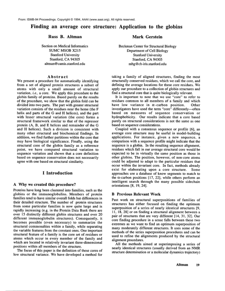
From: ISMB-94 Proceedings. Copyright © 1994, AAAI (www.aaai.org). All rights reserved.
Finding an average core structure:
Russ
B. Altman
Section on MedicalInformatics
SUMC MSOB X215
Stanford University
Stanford, CA94305
altman @camis.stanford.edu
Abstract
Wepresent a procedure for automatically identifying
from a set of aligned protein structures a subset of
atoms with only a small amount of structural
variation, i.e.. a core. Weapply this procedureto the
globin family of proteins. Based purely on the results
of the procedure, we showthat the globin fold can be
divided into two parts. The part with greater structural
variation consists of the residues near the heme(the
helix and parts of the G and H helices), and the part
with lesser structural variation (the core) forms
structural frameworksimilar to that of the repressor
protein (A, B, and E helices and remainder of the
and H helices). Such a division is consistent with
manyother structural and biochemical findings. In
addition, wefind further partitions within the core that
mayhave biological significance. Finally, using the
structural core of the globin family as a reference
point, we have compared structural variation to
sequence variation and shownthat a core definition
based on sequence conservation does not necessarily
agree with one basedon structural similarity.
I Introduction
A Why we created this procedure?
Proteins have long been clustered into families, such as the
globins or the immunoglobulins. Membersof protein
families tend to havesimilar overall folds but differences in
their detailed structure. The numberof protein structures
from some particular families is now quite large and is
rapidly increasing (e.g. in the Protein Data Bankthere are
over 15 distinctly different globin structures and over 20
different immunoglobulinstructures). Consequently, it
becomes possible (even necessary) to summarize the
structural commonaltieswithin a family, while separating
the variable features from the constant ones. Oneimportant
structural feature of a family is the core set of residues or
atoms which occur in every memberof the family and
which are located in relatively invariant three-dimensional
positions within all membersof the structure.
The focus of this paper is the definition of these cores of
low structural variance. Wehave developed a method for
Application to the globins
Mark Gerstein
BeckmanCenter for Structural Biology
Departmentof Cell Biology
Stanford University
Stanford, CA94305
mbg@cb-iris.stanford.edu
taking a family of aligned structures, finding the most
structurally conservedresidues, whichwe call the core, and
defining the average locations for these core residues. We
apply our procedureto a collection of globin structures and
find a structural core that is quite biologicallyrelevant.
It is important to note that we use "core" to refer to
residues commonto all membersof a family and which
have low variance in a-carbon position.
Other
investigators have used the term "’core" differently--often
based on measures of sequence conservation
or
hydrophobicity. Our results indicate that a core based
purely on structural considerations is not the sameas one
based on sequenceconsideratiOns.
Coupled with a consensus sequence or profile [6], an
average core structure maybe useful in model-building
applications.
For instance, given a new sequence, a
comparisonwith a sequence profile might indicate that the
sequence is a globin. In the resulting sequence alignment,
residues which fall in our average structural core wouldbe
expected to be in virtually the same position as those in
other globins. The position, however, of non-core atoms
could be adjusted to adapt to the particular residues that
occur within the invariant core. In fact, methodsalready
exist for elaborating upon a core structure.
Some
approaches use a database of knowsegments to match to
the t~-carbon positions [17, 22], while others perform an
intelligent search through the manypossible sidechain
orientations [8, 19, 24].
B Previous Relevant Work
Past work on structural superpositions of families of
structures has either focused on finding the optimum
superposition of a series of nearly identical structures [9,
11, 18, 28] or on finding a structural alignment betweena
pair of structures that are very different [14, 31, 32]. Our
core finding procedure in a sense falls betweenthese two
extremes as we want to find an optimumsuperposition of
manymoderately different structures. It uses someof the
methodsof the series superposition procedures and can be
used to refine the alignments produced by the structural
alignment procedures.
All the methods aimed at superimposing a series of
nearly identical structures (usually derived from an NMR
structure determination or a molecular dynamicstrajectory)
Airman
19
start by assumingthat there is an alignment pairing each
atom in one structure with an equivalent atom in the
others. After movingthe centroids of all the structures to
the origin, they try to find a rotation for each structure that
minimizesthe sumof squares of the coordinate differences
betweenall pairs of aligned atoms. That is, they seek to
minimize:
2
E(n)=~,jN<~_,i’i.(R,xii-R,xki)
(I)
4.
Apply formula (I) to the ensemble of average
structures to computeE(~*), the sumof squares of the
coordinate differences between all pairs of aligned
positions. (N2 distance calculations.)
5.
If E(D*) is below somesmall threshold, then all the
structures in D*are the sameto within this threshold.
Consequently, one of the structures can be picked and
returned as the unbiased average. Otherwisego back to
step 1 using f~* as the newstarting ensemble.
where
thefirst
sumisoverallpairs
j,k oftheN structures
in theensemble
~, thesecondsumis overtheM aligned
All pairwise fits are done using the method of Arun &
positions
in eachstructure,
andRjxjiis therotated
Huang[2]. The unbiased nature of the methodis apparent
coordinates
ofstructure
j.
on inspection, since the order of the N structures can be
randomlyshuffled at the outset, with no difference in the
result. Using the unbiased average, we have a way to put
II Methods
all the proteins into the same coordinate system without
favoring any of them.
A Overview of the Core Finding Algorithm
C Calculating the Structural Variation of a given
Ona high level, our core finding algorithm has 4 steps,
position (step 3)
whichwe will subsequentlydiscuss in detail:
After the original N structures are fit to the average
1. Start with an aligned ensembleof structures. Initially
structure determined in the above step, it is possible to
consider each aligned residue position to be a possible
look at the structural variation of each aligned position. We
core position.
havedescribedelsewhere[ 1 ], a representation of a structure
2. Calculate an unbiased average of all core positions and
that summarizesvariability about a meanposition (for an
fit each memberof the original ensemble to this
atom) in terms of a variance/covariance matrix C. The
averagestructure.
"~ 2 9
eigenvalues of this matrix (cx, Oy, Oz) give the variance of
3. Calculate the structural variation of each aligned atom
position (measured in terms of the ellipsoid volume
the distribution in its three principal directions. Thcy
relating to the spread of coordinate positions) and
roughly correspond to the axes of an "’ellipsoid of errors"
removethe position with the largest variation from the
centered at the mean. Wecan average these eigenvalues in
list of candidatecore atoms.
either a geometricalor arithmetic fashion and calculate the
volumeof the ellipsoid or its averageaxis length:
4. Go back to step 2 until all atoms have been removed
from the core list. Simple analysis of the statistics
V=417~G2.~
(41)
’nr~ Cry~0-~
~ 0"
resulting from this procedure allows the core to be
2 ,
identified.
R = -~-~/0-~ + 0-~. + :
(5)
For the procedure as applied here, we have reported the
B Calculation of an Unbiased Average (step 2)
volumeof the ellipsoid as a measureof structural variation.
We have developed a simple method for averaging
(We
get virtually the same results using the mean axis
structures in an unbiasedfashion. It proceedsas follows:
length). Atomswhichappear in the samerelative locations
1. Start with an ensembleof N structures :
over all the structures in the ensemble will have small
~2 ={abcd...}
(2)
volumes,whereasatomswhichare variable in their relative
location will have large volumes.
2. Pickthe first structure (in this case a) as a referenceand
fit the remaining structures to it to create a newly
oriented ensemble.Thenaverage the coordinates of this
D Defining a Core Cutoff (step 4)
ensembleto create an average structure, denoteda*.
The atom with the largest ellipsoid volume is, in some
3. Repeat step 2 using each structure in turn as a
sense, the least likely atomto be a memberof a structural
reference. This will create a new ensembleof average
core. In our procedure, it is therefore discarded as "nonstructures. (Steps 2 and 3 require a total of N(N-1) fits
core." The entire averaging, fitting and volume-calculation
and N averaging operations).
procedure is repeated until all atoms are movedfrom the
candidate"core" list to the "non-core"list.
~* = {a* b* c* d*...}
(3)
2O ISMB-94
1MBD
V L S E G E W Q L V L H V W A K V E A
2° Structure
A161
I A1
Aligned
I
I
ThrowOut
23 15 3~ 5310410(3102105101 9~ 92 91 90 67 49 43
Core
’’ I
I
Repressor
~~\\\\\\\\\\\\\\\\\\~
llvBD
D V A G H G Q D I L I R L F K S H P E T L E K F D R F K H L
2° Structure
I B1
B161
-C:tC71 CO1CO2CO4
Aligned
I
I
ThrowOut
106 114 113 112 110 108 107 115 111 109 84 82 B3 80 69 79 77 68 86 .54 7
25
Repressor
~\\\\\\\\\\\\\\\\\\"~
1MBD
K T E A E M K A S E
2° Structure
l-D1
071
51
Aligned
I
Throw
Out
1 2
Core
Repressor
llV~D
K G H H E A E L K P
2° Structure
I F1
Aligned
I
Throw
Out
20 17 8 27 13 30 29 28
Core
Repressor
llV~D
I P I K Y L E F I S
2° Structure
FG4~
G1
AJkjned
I
D L K K H G V T V L T A L G A
I
L K K
~ EEl
I
10 55 24 14 58 57 55 60 81 59 51 68 50.48 52 47 11 12 26
L A Q S H A T
K H
~1~1FC~
K
I
41 38 36 35 42 22 6 5 3
E A I
I
H V L H S R H P G D F
G19
I
I
ThrowOut 76 75 44 72 89 .87 71 68 85 70 63 65 64 46 45 61 62 16 9 4
Core
"
q’ ’r
Repressor
~~\~\_\\\\\\\\\\\\\\\\\\\\\\\\\~,3
1MBD
G A D A Q G A M N K A L E L F R K D I A A K Y K E
2° Structure
[ H1
H24]
Al~jned
I
Throw
Out
96 99 100 94 95 97 93 74 76 73 34 37 40 33 21 32 31 18 19
....
H
!
Bore
..
!
Repressor
k.\\~X\\\\\\~\X\\\\\\\\\\\\",~\\\\N
.......................
Figure 1. (LEFT) Cylinders representation
of a globin
(1MBD). (ABOVE)Listing of the various subsets of globin
residues.
From an entire globin (1MBD, which has 153
residues, is shown as an example), the set of 115 residue
positions that were structurally
aligned with the other
globins were extracted
(ALIGNEDrow). These aligned
residues roughly correspond to the helices in the globins
with the exception of the D helix (2 ° STRUCTURE
row).
The core finding procedure was applied to these 115 aligned
residues to produced a core of 73 residues (CORErow). The
iteration
at which each of the 115 aligned residues was
deleted from the putative
core is shown (THROWOUT
row). Finally, we compare our 73 core positions to the 52
positions in the repressor protein which are aligned to
myoglobin (REPRESSOR row).
Altman
21
There are many properties of the core and non-core
regions definedat each step in the iteration that can be used
to define a cutoff. Conceptually, we are seeking a measure
that has the property of being optimizedat the point in the
process at which there is an optimal separation between
core atoms and non-core atoms. Our procedure starts with
an empty list of non-core atoms, and initially adds atoms
with largest structural variability to this list (and removes
them from the core list). At somepoint in this process,
atomsthat are appropriately considered core are transferred
to the non-core list. This point can be readily identified by
looking at the variance of the volumes of the non-core
atomsafter they have been fit to one another. The success
of this measure depends on the fact that core atoms (by
definition) will tend to have small volumesthat cluster
tightly around some mean value. Non-core atoms, on the
other hand, will have a broader range of volumes, and so
the distribution of their volumeshas high variance. Now,
consider the situation wben core atoms (small average
volume, low variance) are added to the list of non-core
atoms (high average volume, high variance). The variance
of volumes will nowtend to decrease, since a cohort of
relatively uniform volumesare nowbeing added. Thus, at
the point whencore atomsstart to be transferred to the noncore list, there should be a drop in the variance of the
distribution of volumesin non-core regions. This is exactly
whatis observed(Figure 3).
E Measuring Sequence Variation
To measure the sequence diversity of the globins we used
the structurally based alignment of 577 globin sequences
reported in Gerstein et al. [12]. At a given position in this
alignment, we can calculate the frequencies of occurrenceof
the different amino acids and then measure sequence
variability through the computation of an informationtheoretical entropy 126, 27, 29]. However,because of the
biases in the sampling of sequences within the databanks,
there is an over-representation of certain globins, such as
Figure2. The ellipsoids around the 73 atoms classified as belonging to the core (LEFF)and 42 atoms classified as not
belonging to the core (RIGHT).The view is roughly the same as in Figure I. To form each ellipsoid, we created a 3x3
variance/covariance matrix from the spread of Ct~ coordinates at each aligned position in the 8 globin structures. Then we
perform a Jacobi decomposition[25] whichprovides the lengths of the three ellipsoid semi-axes (from the eigenvalues) and
the orientation of ellipsoid (from the eigenvectors).
22
ISMB-94
Identification of Core Subunits
VolumeDistributions for Core/Non-Core
24
25
----O
23-
Variance of volumes
of non-core atoms.
20o
2215-
2120
---
¯---
Core Volumes
Non-core Volumes
10-
19
5 -
18
I
I
-( ........ I ........ I
17 L I
30 40 50 60 70 80 90 100
I
l!\..
0
0
4
2
3
5
6
Volumeof Residue Variation (1SDellipsoid)
Core Finding Cycle
Figure3. (LEFT)The variance in the ellipsoid volumedistribution for non-core atoms (after being optimally fit to
another) is used to identify the core. This measurepeaks at a "core" threshold. For the globins theprimarythreshold between
core and non-core is at cycle 42, with secondarycutoffs at cycle 64 and 84. (RIGHT)
Distribution of ellipsoid volumes(at
standard deviation) at cycle 42, the primary threshold. Eachof the 73 core positions has an error ellipsoid of volume-5/~3
while the 42 non-core positions have larger ellipsoids (of volume-16/~3 ) that are morewidely varied in volume.
Figure 4. Graph of sequence diversity
7
6
-----13¯
CoreN°n’c°re
"" " "i "
~~~
"-~L~
54
"53
¢=
"i
~,, =
¯
¯
"%=
.~ 2
i
N
i..i
nun
rk~
¯
==
¯IB
n
¯
¯
¯
_ rl O=
versus structural diversity for each globin
position. At a particular position, sequence
variation is measuredby information content
relative to that of unaligned sequences
Rsequence(i) (in bits per residue)
structural diversity by the volumeV of the
covariancematrixellipsoid (in ,~3). There are
115 positions represented in total here and
the overall Pearsoncorrelation coefficient is
0.12. The 73 core positions are highlighted
by white boxes. The correlation between
information content and ellipsoid volumefor
just the core positions is 0.25.
¯
o__1
0
1
2
3
4
InformationContentCompared
to a Random
Sequence
Altman
23
mammalianhemoglobins, and under-representation of other
globins, such as the plant globins, and this bias will affect
the entropy calculation significantly. In constructing the
alignment, a tree-based weighting schemewas developed to
correct for this over- and under-representation [12]. (For
general discussion of weighting schemessee [33]). In this
work in order to more accurately compare sequence
variability with structure variability, we have incorporated
weights into the calculation of entropy. To measure the
entropy of a particular position i in a sequencealignment,
we use the standard formula for Shannonentropy:
~-,21~ .
H(i) = -2_.,i=~f(t, t) log2 f(i, t)
(6)
However, instead of the frequency of amino acid t at
position i, we takef(i,t) to be the normalized sum of the
weights for sequences with a residue of type t in
position i :
f(i,t)
~,,j~°,w(s)
= ~.~ w(s)
(7)
where the denominator sum is over all sequences s in the
alignment and the numerator sum is over just those
sequences that have a residue of type t at position i. We
report sequencevariability as information content relative
to that if the sequenceswere aligned randomly:
where ~(t) is the average frequency of type t in the
alignment(i.e.f(i,t) averagedover i ).
III Application to the Globins
A Choice and Alignment of Structures
Fromthe Protein databank [5], we chose 8 structures from
the globin family which have been the subject of previous
investigations [4, 12, 20]: 1ECD, 1MBA,IMBD,2HBG,
2LH4, 2LHB and the A and B chains of 3HHB. (All
structures are of the deoxy form except for I MBAand
2LHB.) Using a canonical numbering scheme first
developed by a Kendrew, Lesk & Chothia had previously
aligned these structures by eye [20]. The 115 common
positions in their structural alignment are shown in
Figure 1.
B Progress of the Iteration
Figure 3 shows the variance of non-core volumes as a
function of cycle. Wedefined peaks by performing a 5-atom
moving average in order to smooth the curve, and then
selected local maxima.The primary peak of this curve is at
cycle 42 (the first 42 atoms have been removedfrom the
original 115, and 73 putative core atoms remain). Wetook
this peak to indicate the that the primary core segmentof
the globin family contains 73 atoms, as shownin Figure
I. Wealso noted secondary peaks at cutoffs of 64 and 84
which are discussed below.
24
ISMB--94
In order to further validate the choice of our cutoff at
iteration 42, we also plotted the distribution of ellipsoid
volumesfor the core residues and tbr the non-core residues
in Figure 3. The twodistributions are fairly well separated.
The core residues clearly have a muchsmaller meanvolume
than the non-core residues. Moreimportantly, they have a
muchsmaller variance in volume.
C A Core Containing 73 Positions
The ellipsoids of variation lor the 73 core and 42 non-core
atomsare showngraphically in Figure 2. lfa core is to be
used as a starting point for modeling,then the quality of its
structure becomesa matter of importance. Weassessed the
"’quality" of our ~-carbonstructure by looking at the stereochemistry of the connected core residues. Three parameters
characterize the geometryof an o~-carbonstructure [21]; for
each of these parameterswe comparedthe range of values in
our structure with established norms. (1) The most
important parameter is the distance betweentwo connected
C~ atoms, which should be 3.8 ,~. We find the mean
distance between the 67 connected core C~ atoms to be 3.8
.~ with a standard deviation of 0.(13 A. (2) The next most
important parameter is the angle "t betweenthree connected
Co~ atoms. This angle can range between approximately
80° and 135°. For our core atoms we find 62 "~ angles
defined, which range between 58° and 122° with a meanof
92°. (3) The third parameter, the pseudo-torsion angle o’.
between four connected C~ atoms, can acceptably range
from -180° to +!80° and so does not form a meaningful
constraint on o~-carbonstructure.
D No correlation between sequence and
structural diversity
Figure 4 shows the relationship
between sequence
variation, measuredby our weightedentropy, and structural
variation, measuredby ellipsoid volume. As discussed in
the figure caption, there is no significant correlation
betweenthemfor either all 115 aligned positions or for just
the core positions.
IV Discussion
A Comparison with other methods to find an
unbiased average
Central to our procedure is the finding of an unbiased
average of a ensembleof structures. This is essentially the
same as what is accomplished by the four previously
proposed methodsto perform an unbiased superposition of
multiple structures (since after superposition all one need
do is average the coordinates) [9, 11, 18, 281. Our method
to find an unbiased average relies solely on pairwise
superpostions. Consequently,we believe it is simpler than
the four methods previously proposed and easier to
implement. For the globins we have extensively compared
the average structure producedby our procedurea~ainst that
produced by Diamond’s procedure. Wefind that the RMS
deviation between them is less than 0.001 A/atom.
Our overall procedure would be unaffected if the step
involving the calculation of an unbiased average were done
with any of the above four methods. For a large numberof
structures it may be advisable to use Diamond’smethod
since its calculations increase linearly with the numberof
structures while ours increase quadratically. However,the
calculations in our procedureare easily parallelizable in a
coarse-grainedfashion.
B Relationship to methods that perform a
structural alignment
Our procedurerequires that there exist an initial structural
alignment between the structures in the ensemble. Our
procedure then refines this initial alignment by throwing
away atoms that superpose badly. Wehave designed our
methodto workon families of relatively similar structures,
such as the globins or immunoglobulins.In these cases, it
maybe possible to get an initial structural alignment by
eye or by sequence alignment (e.g. the Kendrewsequence
numbering for the globins or the Kabat sequence
numbering for the immunoglobulins implies a structural
alignment). Obviously, the scope of our method would
increase if it could use alignments between dissimilar
structures generated by automatic structure alignment
procedures, such as have been previously reported [14, 31,
32]. At present there is no known method for doing
multiple structural alignment. Considering the analogy
with some of the known methods for multiple sequence
alignment [30], we believe our method for finding and
refining an unbiased average of manystructures could be a
useful part of an iterative multiple structural alignment
algorithm.
C Practical Use of Our Procedure in Model
Building
The average structures generated by our procedure exhibit
acceptable stereo-chemistry. This is because we restrict
ourselves to only averaging the most similar parts of the
structures, and because we only average or-carbon positions
and thus need never worry about averaging over a peptide
flip. Because of their acceptable stereo-chemistry the
structures producedby our procedureprovide ideal, unbiased
points to build models from. Furthermore, the structural
variances that our procedure assigns to each position
indicate the degree that the backbone structure in a
particular region must be maintained in model-building.
D Sequence-Structure Implications
Wefind that the structure variation is not correlated with
sequencevariation. This lack of correlation is true whether
we consider all 115 aligned positions, the 72 core
positions, or just the 31 core positions that are buried in all
the globin structures. This result has strong implications
for the modeling of proteins. Manymodeling studies
assume that sequence conservation implies structural
conservation. While this is probably true on the level of
the overall fold, it is not correct in the globins to assume
that the positions that are most conserved in sequence are
those mostinvariant in structure.
The lack of correlation between sequence conservation
and structure conservation mayresult from the wayprotein
structure accommodatessequence changes. One point of
views says the structural accommodation is local: a
mutation in one residue is accommodatedby sidechain
torsion angle changes in this residue and complementary
mutations in neighboring residues. The backbone stays
fixed throughout. A contrasting perspective says that the
structural accommodationis more global: a mutation in
one residue is accommodated
by backboneshifts throughout
the protein (see for example the T4 lysozyme work [3,
10]). Our result provides clear evidence for this second
perspective.
E Significance of the identified globin core
The most striking aspect of the core we identified in the
globins is that it does not include any atoms from the F
helix (or any atoms from the end of H helix that contacts
the F helix). As shown in Figure 1, the heme group is
bound between the E and F helices. Consequently, the
pocket between these two helices is in a sense the active
site of the globins. The movement
of the F helix relative
to the rest of globin core maybe part of the mechanismby
which the different globins modulate the environment of
the hemeand achieve different oxygenbinding affinities.
Our finding that the core does not include the F helix is
consistent with a number of recent NMRspectroscopy
experiments [7, 16]. These experiments have shown that
the F and D helices are the most mobile parts of
myoglobinin solution and the last to fold. Furthermore,
the folding experiments[16] indicate that the A,B,G,and H
helices forma stable association before the remainderof the
protein folds. This is consistent with our finding that our
secondarycore found at iteration 84 does not include the E
helix and only includes one residue from the C helix. Thus,
our core finding procedure is able to identify the most
stable parts of the globin fold.
Our finding that the core does not include the F helix is
also consistent with the way globin helices have been
mappedonto ideal polyhedra. Muzin & Finkelstein [23]
found that the helix packing geometry of most all-or
proteins roughly follows the edges of ideal polyhedra. The
globins, however, were an exception. They wouldfit ideal
polyhedraonly if the F helix is ignored.
F Relationship between the Globin Core and the
Repressor Protein
Recently, it has been pointed out that someparts of the
globin structure are similar to that of helix-turn-helix
(HTH)proteins [31]. The HTHmotif is one of the most
commonfolds for DNAbinding [13]. For the bacteriophage
434 repressor protein Subbiahet al. [3 I] pointed out that
the two helices in the HTHmotif align well with globin
Altman
25
helices B and E and that the other three helices in the
protein align well with globin helix A and parts of helices
G and H (Figure l ).
The 73 positions in our globin core coincide closely
with the 52 positions wherethe repressor aligns well to the
globins. As shownin Figure l, there is only one position
(G17) where an aligned position in the repressor does not
correspondto a position in the globin core. The structural
similarity of the globins to other proteins beside the
repressor, such as the phycocyanins and colicin A, has
been pointed out [ 15]. Thesesimilarities, however,involve
almost all of the globin fold and not distinct subsets.
Consequently,they are not suitable to compareto the core.
4.
Bashford, D.; Chothia, C. & Lesk, A. M. 1987.
Determinantsof a Protein Fold: UniqueFeatures of the
Globin AminoAcid Sequences. J. Mol. Biol. 196:
199-216.
5.
Bernstein, F. C.; Koetzle, T. F.; Williams, G. J. B.;
Meyer, E. F., Jr; Brice, M. D.; Rodgers, J. R.;
Kennard, O.; Shimanouchi, T. & Tasumi, M. 1977.
The protein data bank: a computer-basedarchival file
for macromolecular structures. J. Mol. Biol. II 2:
535-542.
6.
Bowie, J. U.; Liithy, R. & Eisenberg, D. 199l. A
Methodto Identify Protein SequencesThat Fold into a
KnownThree-Dimensional Structure. Science 253:
164-170.
7.
Cocco, M. J. & Lecomte, J. T. J. 1990.
Characteriztion
of Hydrophobic
Cores in
Apomyoglobin: A proton NMRSpectroscopy Study.
Biochemistry 29:11067-11072.
8.
Desmet, J.; Maeyer, M. D.; Hazes, B. & Lasters, I.
1992. The dead-end elimination theoremand its use in
protein side-chain positioning. Nature 356: 539-542.
9.
Diamond,R. D. 1992. On the multiple simultaneous
superposition of molecular structures by rigid-body
transformations. Protein Science 1 : 1279-1287.
V Conclusion
We have presented a method for finding an average
structural core. Wehave applied this methodto the globin
family and found that the division between core and noncore correlates very well with other information about the
globin structure, such as NMR
data and the alignment with
the 434 repressor. Wehave also shownthat for the globins
a core based purely on structural conservation is not the
same as one based on sequence conservation.
Acknowledgments
RBA is a Culpeper Medical Scholar.
Computing
environment provided by the CAMISresource under NIH
grant LM-05305. MGis supported by a Damon-Runyon
Walter-Winchell fellowship (DRG-1272). We thank
Levitt and S Subbiahfor discussions.
Availability of Results
Coordinatesof the 73 residue globin core as well as further
documentation in hypertext form are accessible at the
following location (URL)
tip ://cb-iri s. stanford, edu/pub/mbg/ISMB-94-60/.
References
1.
2.
3.
26
Altman, R. & Jardetzky, O. 1989. The Heuristic
Refinement Method for the Determination of the
Solution Structure of Proteins from NMR
Data. Meth,
Enzym. 177: 177-218.
Arun, K. S.; Huang, T. S. & Blostein, S. D. 1987.
Least-squares fitting of two 3-D point sets. IEEE
Transactions on Pattern Analysis and Machine
hztelligence 9: 698-700.
Baldwin, E. P.; Hajiseyedjavadi, O.; Baase, W. A. &
Matthews, B. W. 1993. The Role of Backbone
Flexibility in Accomodationof Variants that Repack
the Core of T4 Lysozyme. Science 262:1715-1718.
ISMB-94
10. Eriksson, A. E.; Baase, W.A.; Zhang, X. J.; Heinz,
D. W.; Blaber, M.; Baldwin, E. P. & Matthews, B.
W. 1992. Response of a protein structure to cavity
creating mutations and its relation to the hydrophobic
effect. Science 255: 178-183.
I1. Gerber, P. R. & Miiller, K. 1987. Superimposing
Several Sets of Atomic Coordinates. Acta Crvst.
A43: 426-428.
12. Gerstein, M.; Sonnhammer,E. & Chothia, C. 1994.
VolumeChanges on Protein Evolution. J. Mol. Biol.
236: 1067-1078.
13. Harrison, S. C. 1991. A structural taxonomyof DNAbinding domains. Nature 353: 715-720.
14. Holm, L. & Sander, C. 1993. Protein Structure
Comparison by Alignment of Distance Matrices. J.
Mol. Biol. 233: 123-128.
15. Holm, L. &Sander, C. 1993. Structural alignment of
globins, phycocyanins and colicin A. FEBS Lett.
315: 301-306.
16. Jennings, P. A. & Wright, P. E. 1993. Formation of
a Molten Globule Intermediate Early in the Kinetic
Folding Pathway of Apomyoglobin. Science 262:
892-896.
17. Jones, T. A. & Thirup, S. 1986. Using known
substructures
in protein model building and
crystallography. EMBO
J. 5: 819-822.
26. Schneider, T. D. & Stephens, R. M. 1991. Sequence
logos: a new way to display consensus sequences.
Nucl. Acids Res. 18: 6097-6100.
18. Kearsley, S. K. 1990. An Algorithm for the
SimultaneousSuperposition of a Structural Series. J.
Comp. Chem. 11: 1187-1192.
27. Schneider, T. D.; Stormo, G. D.; Gold, L. &
Ehrenfeucht, A. 1986. Information Content of Binding
Sites on Nucleotide Sequences. J. Mol. Biol. 188:
415-431.
! 9. Lee, C. &Levitt, M. 1991. Accurate prediction of the
stability and activity effects of site-directed
mutagenesis on a protein core. Nature 352: 449-451.
20. Lesk, A. M. & Chothia, C. H. 1980. HowDifferent
Amino Acid Sequences Determine Similar Protein
Structures: The Structure and Evolutionary Dynamics
of the Globins. J. Mol. Biol. 136: 225-270.
21. Levitt, M. 1976. A simplified representation of
protein conformations for rapid simulation of protein
folding. J. Mol. Biol. 104: 59-107.
22. Levitt, M. 1992. Accurate Modeling of Protein
Conformation by Automatic Segment Matching. J.
MoLBiol. 226: 507-533.
23. Murzin, A. G. & Finkelstein, A. V. 1988. General
Architecture of the or-Helical Globule. J. Mol. Biol.
204: 749-769.
24. Ponder, J. W. & Richards, F. M. 1987. Tertiary
templates for proteins: use of packing criteria in the
enumeration of allowed sequences for different
structural classes. J. Mol. Biol. 193: 775-791.
25. Press, W. H.; Flannery, B. P.; Teukolsky, S. A. &
Vetterling, W. T. 1992. Numerical Recipes in C.
Cambridge: CambridgeUniversity Press.
28. Shapiro, A. & Botha, J. D. 1992. A Method for
Multiple Superposition of Structures. Acta Cryst.
A48: 11-14.
29. Shenkin, P. S.; Erman, B. & Mastrandrea, L. D.
1991. Information-Theoretical Entropy as a Measureof
Sequence Variability. Proteins: Struc. Func. Genet.
11: 297-313.
30. Subbiah, S. & Harrison, S. C. 1989. A Method for
Multiple Sequence Alignment With Gaps. J. Mol.
Biol. 209: 539-548.
31. Subbiah, S.; Laurents, D. V. & Levitt, M. 1993.
Structural similarity of DNA-binding domains of
bacteriophage repressors and the globin core. Curr.
Biol. 3: 141-148.
32. Taylor, W. R. & Orengo, C. A. 1989. Protein
Structure Alignment. J. Mol. Biol. 208: 1-22.
33. Vingron, M. & Sibbald, P. R. 1993. Weighting in
sequence space: A comparison of methods in terms of
generalized sequences. Proc. Natl. Acad. Sci. USA90:
8777-8781.
Airman
27


