1 of 4 Massachusetts Institute of Technology Department of Biology
advertisement

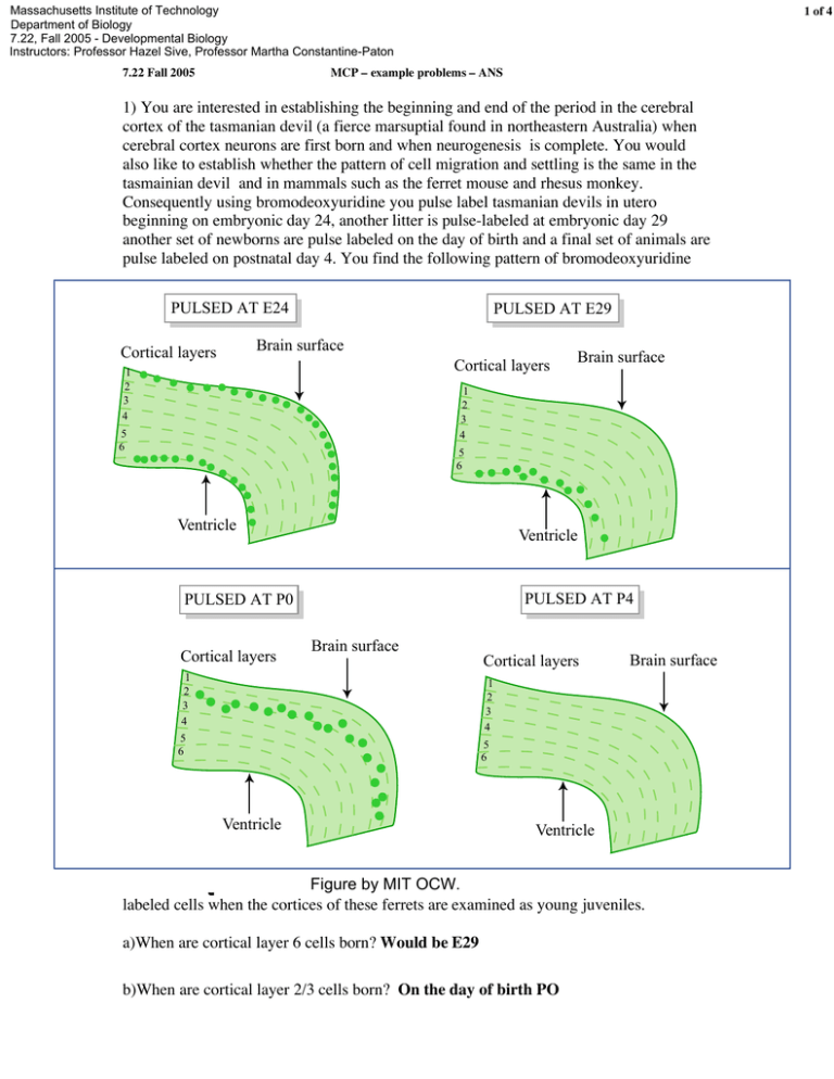
Massachusetts Institute of Technology Department of Biology 7.22, Fall 2005 - Developmental Biology Instructors: Professor Hazel Sive, Professor Martha Constantine-Paton 7.22 Fall 2005 1 of 4 MCP – example problems – ANS 1) You are interested in establishing the beginning and end of the period in the cerebral cortex of the tasmanian devil (a fierce marsuptial found in northeastern Australia) when cerebral cortex neurons are first born and when neurogenesis is complete. You would also like to establish whether the pattern of cell migration and settling is the same in the tasmainian devil and in mammals such as the ferret mouse and rhesus monkey. Consequently using bromodeoxyuridine you pulse label tasmanian devils in utero beginning on embryonic day 24, another litter is pulse-labeled at embryonic day 29 another set of newborns are pulse labeled on the day of birth and a final set of animals are pulse labeled on postnatal day 4. You find the following pattern of bromodeoxyuridine PULSED AT E24 PULSED AT E29 Brain surface Cortical layers 1 2 3 4 Cortical layers Brain surface 1 2 3 4 5 6 5 6 Ventricle Ventricle PULSED AT P4 PULSED AT P0 Cortical layers Brain surface 1 2 3 4 Cortical layers Brain surface 1 2 3 4 5 6 5 6 Ventricle Ventricle Figure by MIT OCW. labeled cells when the cortices of these ferrets are examined as young juveniles. a)When are cortical layer 6 cells born? Would be E29 b)When are cortical layer 2/3 cells born? On the day of birth PO 7.22 Fall 2005 MCP – example problems – ANS 2 of 4 c)Which cells have the longest migration path? The cells born on P0 You compare your pulse labeling pattern with that of ferrets pulsed at comparable stages of development. d)You find that young juvenile ferrets have no labeled cells above layer 2/3 or below layer 6 when pulsed at a stage comparable to E24 in the tasmanian devils. However you are surprised to find when you examine younger (P6) ferrets pulsed at the “E24” stage that they show the same bilayer pattern of expression as the tasmanian devil. Can you suggest an explanation for what is going on between the P6 and young juvenile stage in the ferret cerebral cortex that is not going on in the young tasmanian devils cortex? One of 2 things either the top and bottom cells are dying between P6 and the young juvenile or they start to divide in early postnatal life and dilute out their label. e)Suggest an experiment that would test your hypothesis. If they started to divide between P6 and the young juvenile a pulse given at say P12 should produce some heavily labeled cell when the cortex is examined a day later. If cells were undergoing programed cell death they might express vertebrate orthologs of c elegans cell death genes. f)Would you have detected the bilayer pattern of cell genesis after and E24 pulse of BrdU if you had used a retrovirus with a reporter gene to label a few progenitors at E24? No because the retrovirus will label the progeny of dividing cells but because it will not become diluted out with subsequent cell division all progeny of the stem cell will be labeled not just those cell that leave the mitotic cycle soon after labeling. 2) Exposure of Xenopus embryos to retinoic acid (RA) results in expansion of the Xhox 6 gene into presumptive midbrain regions of the animal and a “posteriorization” of the embryos. You obtain the upstream flanking region of the Xhox 6 gene and use restriction enzymes R, N K and B to produce the constructs illustrated below that you introduce into Xenopus neural tube stage embryos bilaterally using electroporation. You next take the groups of these embryos transfected with these constructs and divide each construct group into 2 experimental groups: those treated with exogenous RA MB = midbrain r = rhombdomere (w/RA) and those not treated with exogenous RA (w/o RA). You obtain the following 35 KB R N N K B MB-r2 w/RA K R N R B Figure by MIT OCW. w/o RA w/RA r3 r4-r8 w/o RA w/RA w/o RA 7.22 Fall 2005 MCP – example problems – ANS 3 of 4 data on LacZ expression in the midbrain and forebrain. a) What region of this 5’ sequence appears to confer restriction of Xhox6 to r4-r8 in the ‘normal’ embryos (the embryos untreated with RA)? Explain your reasoning. K-B because expression in animals without RA treatment is restricted to r4-r6 only when the fragment R-K is present not when either R-N or N-K is present. b) Where in this regulatory region is the retinoic acid responsive element (RARE)? Explain your reasoning. RN because given that there is negative regulatory element in KB R-K should not show expression in either MB-r2 or r3 with RA unless their was a positive RARE in one of the other regulatory fragment. The only regulatory fragment that shows Xhox 6 expression in anterior segments in the absence of the inhibitory fragment KB is RN 3). A variety of reagents and approaches are frequently used by developmental biologists to understand the tissue interactions and molecular signaling pathways involved in particular aspects of development. Several of these approaches or reagents are listed below. To determine how well you understand “how we know what we know” in systems where the forward genetics used in fruit flys and worms is difficult (at best) find one example from your reading and lecture notes where this type of approach was used to demonstrate and important developmental principle. Give the example and the developmental principle illustrated. a) Over-expression of a mRNA in the Xenopus zygote. Over expression of BMP4 ventralizes the embryo; overexpression of goosecoid dorsalizes the embryo; overexpression of a mutant FGF receptor subunit anteriorizes the embryo etc. b) Trans-species injection of an orthologous gene from Drosophila into a developing vertebrate embryo. Injection of Sog into the belly region of of Xenopus early gastrula stage embryos induces a second embryonic axis. Sog and Chordin are orthologous protein that have also retained a similar function. Could also use the Dickkopf example that is stated in guestion 4 below. c) Transgenic mouse carrying the entire regulatory region of Hoxb1 in front of the reporter gene LacZ. LacZ product is obvious in an anterior rhobdomere and then in the last 2 rhombomeres with a slowly decreasing expression down the spinal cord. 7.22 Fall 2005 MCP – example problems – ANS 4 of 4 d) Knockout mouse for a known patterning gene. Lim1 knockout mouse blocks head formation. Pups are born headless and then die. Hoxc8 knockout shows a homeotic transformation of the first lumbar vertebrae into a rib-bearing chest vertebrae. (etc) e) An incapacitated retrovirus with a strong promotor driving LacZ or green flourescent protein genes. A non-diluting lineage tracer. Could label cells in the posterior marginal zone of early chick embryos and show that the label winds up in the notochord and paraxial mesoderm. Could label a cell in the expanding progenitor population of the cerebral lobes in a mouse embryo and find that a large “wedge” of cortex is labeled when the animals are examined after birth, f) Exposure of a specified but undetermined tissue to different “inducing” tissues hypothesized to change the prospective fate of cells. Xenopus “animal cap” exposed to ventral/ventral endoderm blastomeres. Get ventral to ventral lateral mesoderm differentiating eg blood, kidney.


