Document 13386089
advertisement
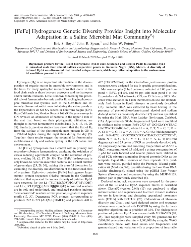
APPLIED AND ENVIRONMENTAL MICROBIOLOGY, July 2009, p. 4620–4623 0099-2240/09/$08.00⫹0 doi:10.1128/AEM.00582-09 Copyright © 2009, American Society for Microbiology. All Rights Reserved. Vol. 75, No. 13 [FeFe] Hydrogenase Genetic Diversity Provides Insight into Molecular Adaptation in a Saline Microbial Mat Community䌤† Eric S. Boyd,1 John R. Spear,2 and John W. Peters1* Department of Chemistry and Biochemistry and Astrobiology Biogeocatalysis Research Center, Montana State University, Bozeman, Montana 59717,1 and Division of Environmental Science and Engineering, Colorado School of Mines, Golden, Colorado 804012 Received 10 March 2009/Accepted 29 April 2009 Degenerate primers for the [FeFe] hydrogenase (hydA) were developed and used in PCRs to examine hydA in microbial mats that inhabit saltern evaporative ponds in Guerrero Negro (GN), Mexico. A diversity of deduced HydA was discovered that revealed unique variants, which may reflect adaptation to the environmental conditions present in GN. 427 (TGGVMEAA) in the Clostridium pasteurianum protein sequence, were designed for use in specific gene amplifications. Mat core samples (1 by 6 cm) were collected at 2:00 pm from pond 4 (35°C, pH 8.0, and 80 ppt salt) near pond 5 at the Exportadora de Sal saltworks, GN, on 13 February 2005. Mat cores were sectioned in 1-mm increments on site and immediately flash frozen in liquid nitrogen as previously described (16). Genomic DNA was extracted by bead beating in the presence of phenol-chloroform-isoamyl alcohol and sodium dodecyl sulfate as previously described (7) and was quantified by using the High DNA Mass Ladder (Invitrogen, Carlsbad, CA). Approximately 500-bp fragments of hydA were amplified in triplicate using primers FeFe-272F (5⬘-GCHGAYMTBAC HATWATGGARGA-3⬘, where H ⫽ A, C, T; Y ⫽ C, T; M ⫽ A, C; B ⫽ C, G, T; W ⫽ A, T; R ⫽ A, G; 432-fold degeneracy) and FeFe-427R (5⬘-GCNGCYTCCATDACDCCDCCNGT-3⬘, where N ⫽ A, C, T, G; Y ⫽ C, T; D ⫽ A, G, T; 864-fold degeneracy) and 35 cycles of PCR as previously described (4). An empirically determined annealing temperature of 56.5°C, a MgCl2 concentration of 1.5 mM, and a primer concentration of 1 M for each forward and reverse primer were utilized in 50-l PCR mixtures containing 10 ng of genomic DNA as the template. Equal 40-l volumes of three replicate PCR products were pooled, purified using the Promega Wizard purification kit (Madison, WI), quantified using the Low Mass DNA Ladder (Invitrogen), cloned using the pGEM Easy Vector System (Promega), and sequenced by using the M13F-M13R primer pair as previously described (5). Deduced amino acid sequences were screened for the presence of the L1 and L2 HydA sequence motifs as described above. ClustalX (version 2.0.8) (15) was employed to align inferred amino acid sequences and to create distance matrices for use in identifying and clustering operational taxonomic units (OTUs) with DOTUR (26). Calculations of Shannon diversity and Chao1 and Ace1 deduced amino acid sequence richness were completed with DOTUR by using the furthestneighbor algorithm and a precision of 0.01. The phylogenetic position of putative HydA was assessed with MRBAYES (10, 25). Tree topologies were sampled every 500 generations for 2,000,000 generations (burnin ⫽ 1,000,000) by using the WAG evolutionary model with fixed amino acid frequencies and gamma-shaped rate variation with a proportion of invariable Hydrogen (H2) is an important intermediate in the decomposition of organic matter in anaerobic environments and is the basis for many syntrophic interactions that occur in the food chain such as those between acetogens and methanogens and/or sulfate reducers. Little is known concerning the potential role of fermentative bacteria and/or H2 cycling in phototrophic microbial mat systems, such as the 6-cm-thick and extremely diverse microbial mats inhabiting the saline ponds at the Exportadora de Sal SA saltern in Guerrero Negro (GN), Baja California Sur, Mexico. Recent 16S rRNA gene surveys in GN revealed an abundance of bacteria in the upper 2 mm of the mat that, based on their phylogenetic affiliation, are thought to harbor fermentative metabolisms (6, 8, 13, 16, 20, 28). Similarly, previous studies have shown that the flux of H2 from the surface of the phototrophic mats present in GN is ⬃150-fold higher during the night than during the day (9). Together, these results suggest the potential for fermentative metabolisms in H2 and carbon cycling in the GN saline mat environment. The [FeFe] hydrogenase has a central role in primary and secondary substrate fermentations, catalyzing the oxidation of excess reducing equivalents coupled to the reduction of protons, yielding H2 (2, 17, 29, 30). The [FeFe] hydrogenase is only known to occur in anaerobic bacteria and a small number of green algae (23, 29, 30), making this a useful biomarker for examining the diversity and distribution of this functional class of organism. Eighty-two putative [FeFe] hydrogenase largesubunit protein sequences (HydA) present in the GenBank database that represent the known diversity of putative HydA were screened for the L1 ([FLI]TSC[C/S]P[GAS]W[VIQH]) and L2 ([IVLF]MPCx[ASRD]K[KQ]xE) (conserved residues are in bold and underlined, and bracketed positions indicate “semiconserved” residues at that position) signature sequence motifs (17, 30). Degenerate PCR primers, corresponding to positions 272 to 279 (AD[M/L]TIMEE) and positions 420 to * Corresponding author. Mailing address: Department of Chemistry and Biochemistry, 103 Chemistry Research Building, Montana State University, Bozeman, MT 59717. Phone: (406) 994-7211. Fax: (406) 994-5407. E-mail: John.peters@chemistry.montana.edu. † Supplemental material for this article may be found at http://aem .asm.org/. 䌤 Published ahead of print on 8 May 2009. 4620 VOL. 75, 2009 [FeFe] HYDROGENASE DIVERSITY IN A SALINE MAT 4621 FIG. 1. (A) Phylogenetic tree based on deduced putative HydA amino acid sequences from GN and reference HydA sequences. Nodes with posterior probabilities of ⬍50% were collapsed in this analysis (posterior probabilities of 100 are denoted by asterisks). Bar, one substitution per 10 sites. (B) Partial-length deduced amino acid sequences of environmental and reference deduced HydA sequences illustrating the phylogenetic coherence of novel substitutions and insertions in and upstream of L1 sequence motif. Cluster designations (C1 to C7) correspond to those presented in Table 1. sites as recommended by ProtTest (1). The Saccharomyces cerevisiae Narf protein, a homolog of HydA, was used as the outgroup. The phylogenetic tree was projected using TreeView (version 1.6.6) (21). An unexpectedly diverse assemblage of putative HydA variants was present in the top 1 mm of the GN microbial mat, corresponding to the photic zone. At a sequence identity threshold (SIT) of 100%, the predicted Shannon diversity index was 3.84 and the mean Chao1 HydA richness was 187 unique OTUs (see Fig. S1a in the supplemental material). When a 99% SIT was applied, the predicted Chao1 richness estimate decreased 56% to 82 unique OTUs and the Shannon 4622 BOYD ET AL. APPL. ENVIRON. MICROBIOL. TABLE 1. Phylogeny of partial-length putative HydA sequence clusters Sequence clustera No. of clones in cluster Maximal intracluster sequence divergence (%)b C1 C2 C3 C4 C5 C6 C7 22 16 6 9 3 7 2 49.4 41.6 28.8 38.6 38.6 51.8 32.9 Most closely related sequencec Phylumd Mean % sequence identity (range)e Moorella thermoacetica ATCC 39073 Opitutus terrae PB90-1 Bacteroides thetaiotaomicron VPI-5482 Alkaliphilus oremlandii OhILAs Moorella thermoacetica ATCC 39073 Heliobacillus mobilis Thermoanaerobacterium saccharolyticum JW/SL-YS485 Firmicutes Verrucomicrobia Bacteroidetes Firmicutes Firmicutes Firmicutes Firmicutes 61.8 (56.4–67.3) 64.3 (56.3–71.1) 72.1 (69.8–73.2) 65.1 (61.4–66.4) 61.4 (NA) 50.3 (47.5–54.6) 61.2 (58.5–64.2) a Sequence clusters correspond to those presented in Fig. 1. Maximum intracluster sequence divergence among sequences within a cluster as calculated by the ClustalX sequence identity matrix following alignment using the Gonnet protein weight matrix (gap opening penalty of 13, gap extension penalty of 0.05). c The most closely related sequence is defined as the taxon with the HydA sequence most closely related to that of members within the cluster as presented in Fig. 1. d Phylum designation of most closely related HydA sequence as defined in Fig. 1. e Sequence identity means and ranges were calculated by using the ClustalX sequence identity matrix as described in footnote b. NA, not available. b diversity index decreased slightly to 3.55 (data not shown). A further decrease in the SIT from 99 to 85% resulted in a slight decrease (23%) in the Chao1 predicted richness and a modest decrease in the Shannon diversity index from 3.55 to 3.28, suggesting that the majority of the putative HydA OTUs present in the top 1 mm of the mat is a result of recent evolution or speciation. The putative HydA variants from GN resolved into 42 OTUs which clustered into one of seven distinct phylogenetic clusters (Fig. 1A), although clusters 5 and 7 could not be adequately resolved. Intriguingly, the mean identity scores determined from sequence alignment matrices for members of each of the seven sequence clusters recovered from GN, when compared to sequences in the GenBank database, were very low (47.5 to 73.2%) (Table 1), suggesting that the putative HydA sequences recovered from GN represent novel sequence space. The highest proportion of putative HydA sequences recovered from the top 1-mm transect of the GN microbial mat were related to acetogens most closely affiliated with the Firmicutes (66.2% of the clones) and the Verrucomicrobia (24.6%), with a lower proportion related to the Bacteroidetes (9.2%). These observations support those of previous studies that identified the highest abundance of 16S rRNA genes related to fermentative/acetogenic organisms within the phyla Firmicutes, Verrucomicrobia, and Bacteroidetes in the upper few millimeters of microbial mat (16), coinciding with the mat transect where nighttime H2 concentrations were also elevated (9). The recovery of putative HydA sequences in the photic zone of the GN microbial mat is consistent with the results of previous studies conducted in the phototrophic mats in Yellowstone National Park, WY, which documented the potential for the secondary fermentation of cyanobacterial primary fermentation products in the top photic layers of the mat at night (3). Importantly, cyanobacterial fermentation may also contribute to the production of H2 in the mats during periods of anoxia (9). Deduced amino acid sequences from GN had significantly lower (P ⫽ ⬍0.01 for both GRAVY and aliphatic indices) hydropathy indices than nonhalotolerant HydA sequences from GenBank (see Table S1 in the supplemental material), a feature which may reflect molecular adaptation to salinity. These observations are consistent with the results of previous investigations of deduced amino acid sequences from a variety of halophilic organisms that indicate hydrophobicity indices lower than those of their nonhalophilic counterparts, which is hypothesized to reduce the effects of salt-driven protein misfolding and/or aggregation (11, 12, 14, 24, 27, 31). In addition, many of the putative HydA sequences recovered from GN exhibited previously unobserved substitutions in the L1 motif and novel insertion domains upstream from the L1 motif (Fig. 1B). Residues within L1 are involved in the coordination of the oxygen-labile [4Fe-4S] subcluster of the H cluster of HydA (18, 19, 22). Substitutions in this region may have implications for the redox properties of this [4Fe-4S] cluster and thus may be important in conferring stability in the presence of oxygen (23). The discovery of a rich assemblage of putative HydA variants in the top 1 mm of the GN microbial mat underscores the utility of these primers in examining the natural diversity of putative HydA and fermentative organisms in complex microbial ecosystems. The composition of putative HydA sequences from the top 1 mm of the microbial mat suggests adaption to the conditions present in the evaporative salterns in GN. Nucleotide sequence accession numbers. A total of 65 putative hydA gene sequences determined in the present study have been deposited in the GenBank, DDBJ, and EMBL databases under accession numbers FJ623894 to FJ623958. We gratefully thank the Exportadora de Sal, GN, Baja California Sur, Mexico, for access to their saltworks, as well as the NASA Ames GN researchers who facilitated the permitting process and gave advice. We are also grateful to Laura Beer for helpful comments during the preparation of the manuscript. This work was supported by Air Force Office of Scientific Research Multidisciplinary University Research Initiative Award FA9550-05-010365 and the NASA Astrobiology Institute Funded Astrobiology Biogeocatalysis Research Center (NNA08C-N85A to J.W.P). E.S.B. was supported by a NASA Postdoctoral Fellowship awarded by the NASA Astrobiology Institute. J.R.S. was supported by a National Science Foundation Microbial Biology Postdoctoral start-up award (0624789) and a U.S. Air Force Office of Scientific Research award (R-8196-G1). REFERENCES 1. Abascal, F., R. Zardoya, and D. Posada. 2005. ProtTest: selection of best-fit models of protein evolution. Bioinformatics 21:2104–2105. VOL. 75, 2009 2. Adams, M. W. W. 1990. The structure and mechanism of iron hydrogenases. Biochim. Biophys. Acta 1020:115–145. 3. Anderson, K. L., T. A. Tayne, and D. M. Ward. 1987. Formation and fate of fermentation products in hot spring cyanobacterial mats. Appl. Environ. Microbiol. 53:2343–2352. 4. Boyd, E. S., D. E. Cummings, and G. G. Geesey. 2007. Mineralogy influences structure and diversity of bacterial communities associated with geological substrata in a pristine aquifer. Microb. Ecol. 54:170–182. 5. Boyd, E. S., S. King, J. K. Tomberlin, D. K. Nordstrom, D. P. Krabbenhof, T. Barkay, and G. G. Geesey. 2009. Methylmercury enters an aquatic food web through acidophilic microbial mats in Yellowstone National Park, Wyoming. Environ. Microbiol. 11:950–959. 6. Canfield, D. E., and D. J. Des Marais. 1993. Biogeochemical cycles of carbon, sulfur, and free oxygen in a microbial mat. Geochim. Cosmochim. Acta 57:3971–3984. 7. Dojka, M. A., P. Hugenholtz, S. K. Haack, and N. R. Pace. 1998. Microbial diversity in a hydrocarbon- and chlorinated-solvent-contaminated aquifer undergoing intrinsic bioremediation. Appl. Environ. Microbiol. 64:3869– 3877. 8. Feazel, L. M., J. R. Spear, A. B. Berger, J. K. Harris, D. N. Frank, R. E. Ley, and N. R. Pace. 2008. Eucaryotic diversity in a hypersaline microbial mat. Appl. Environ. Microbiol. 74:329–332. 9. Hoehler, T. M., B. M. Bebout, and D. J. Des Marais. 2001. The role of microbial mats in the production of reduced gases on the early Earth. Nature 412:324–327. 10. Huelsenbeck, J. P., and R. Ronquist. 2001. MRBAYES: Bayesian inference of phylogeny. Bioinformatics 17:754–755. 11. Hutcheon, G. W., N. Vasisht, and A. Bolhuis. 2005. Characterization of a highly stable ␣-amylase from the halophilic archaeon Haloarcula hispanica. Extremophiles 9:487–495. 12. Joo, W. A., and C. W. Kim. 2005. Proteomics of halophilic archaea. J. Chromatogr. B Analyt. Technol. Biomed. Life Sci. 815:237–250. 13. Kunin, V., J. Raes, J. K. Harris, J. R. Spear, J. J. Walker, N. Ivanova, C. von Mering, B. M. Bebout, N. R. Pace, P. Bork, and P. Hugenholtz. 2008. Millimeter-scale genetic gradients and community-level molecular convergence in a hypersaline microbial mat. Mol. Syst. Biol. 4:198. 14. Lanyi, J. K. 1974. Salt-dependent properties of proteins from extremely halophilic bacteria. Bacteriol. Rev. 38:272–290. 15. Larkin, M. A., G. Blackshields, N. P. Brown, R. Chenna, P. A. McGettigan, H. McWilliam, F. Valentin, I. M. Wallace, A. Wilm, R. Lopez, J. D. Thompson, T. J. Gibson, and D. G. Higgins. 2007. Clustal W and Clustal X version 2.0. Bioinformatics 23:2947–2948. [FeFe] HYDROGENASE DIVERSITY IN A SALINE MAT 4623 16. Ley, R. E., J. K. Harris, J. Wilcox, J. R. Spear, S. R. Miller, B. M. Bebout, J. A. Maresca, D. A. Bryant, M. L. Sogin, and N. R. Pace. 2006. Unexpected diversity and complexity of the Guerrero Negro hypersaline microbial mat. Appl. Environ. Microbiol. 72:3685–3695. 17. Meyer, J. 2007. [FeFe] hydrogenases and their evolution: a genomic perspective. Cell. Mol. Life Sci. 64:1063–1084. 18. Nicolet, Y., B. J. Lemon, J. C. Fontecilla-Camps, and J. W. Peters. 2000. A novel FeS cluster in Fe-only hydrogenases. Trends Biochem. Sci. 25:138–143. 19. Nicolet, Y., C. Piras, P. Legrand, C. E. Hatchikian, and J. C. FontecillaCamps. 1999. Desulfovibrio desulfuricans iron hydrogenase: the structure shows unusual coordination to an active site Fe binuclear center. Structure 7:13–23. 20. Nübel, U., F. Garcia-Pichel, M. Kuhl, and G. Muyzer. 1999. Quantifying microbial diversity: morphotypes, 16S rRNA genes, and carotenoids of oxygenic phototrophs in microbial mats. Appl. Environ. Microbiol. 65:422–430. 21. Page, R. D. M. 1996. TREEVIEW: an application to display phylogenetic trees on personal computers. Comput. Appl. Biosci. 12:357–358. 22. Peters, J. W., W. N. Lanzilotta, B. J. Lemon, and L. C. Seefeldt. 1998. X-ray crystal structure of the Fe-only hydrogenase (CpI) from Clostridium pasteurianum to 1.8 angstrom resolution. Science 282:1853–1858. 23. Posewitz, M. C., D. W. Mulder, and J. W. Peters. 2008. New frontiers in hydrogenase structure and biosynthesis. Curr. Chem. Biol. 2:178–199. 24. Reistad, R. 1970. On the composition and nature of the bulk protein of the extremely halophilic bacteria. Arch. Mikrobiol. 71:353–360. 25. Ronquist, F., and J. P. Huelsenbeck. 2003. MRBAYES 3: Bayesian phylogenetic inference under mixed models. Bioinformatics 19:1572–1574. 26. Schloss, P. D., and J. Handelsman. 2005. Introducing DOTUR, a computer program for defining operational taxonomic units and estimating species richness. Appl. Environ. Microbiol. 71:1501–1506. 27. Spahr, P. F. 1962. Amino acid composition of ribosomes from Escherichia coli. J. Mol. Biol. 4:395–406. 28. Spear, J. R., R. E. Ley, A. Berger, and N. R. Pace. 2003. Complexity in natural microbial ecosystems: the Guerrero Negro experience. Biol. Bull. 204:168–173. 29. Vignais, P. M., and B. Billoud. 2007. Occurrence, classification, and biological function of hydrogenases: an overview. Chem. Rev. 107:4206–4272. 30. Vignais, P. M., B. Billoud, and J. Meyer. 2001. Classification and phylogeny of hydrogenases. FEMS Microbiol. Rev. 25:455–501. 31. Visentin, L. P., C. Chow, A. T. Matheson, M. Yaguchi, and F. Rollin. 1972. Halobacterium cutirubrum ribosomes. Properties of the ribosomal proteins and ribonucleic acid. Biochem. J. 130:103–110.
