7.88 Lecture Notes - 4 7.24/7.88J/5.48J The Protein Folding and Human Disease
advertisement
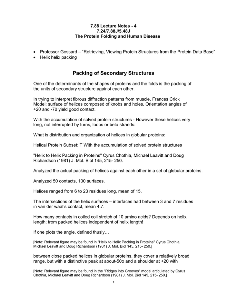
7.88 Lecture Notes - 4 7.24/7.88J/5.48J The Protein Folding and Human Disease • • Professor Gossard – “Retrieving, Viewing Protein Structures from the Protein Data Base” Helix helix packing Packing of Secondary Structures One of the determinants of the shapes of proteins and the folds is the packing of the units of secondary structure against each other. In trying to interpret fibrous diffraction patterns from muscle, Frances Crick Model: surface of helices composed of knobs and holes. Orientation angles of +20 and -70 yield good contact. With the accumulation of solved protein structures - However these helices very long, not interrupted by turns, loops or beta strands: What is distribution and organization of helices in globular proteins: Helical Protein Subset; T With the accumulation of solved protein structures "Helix to Helix Packing in Proteins" Cyrus Chothia, Michael Leavitt and Doug Richardson (1981) J. Mol. Biol 145, 215- 250. Analyzed the actual packing of helices against each other in a set of globular proteins. Analyzed 50 contacts, 100 surfaces. Helices ranged from 6 to 23 residues long, mean of 15. The intersections of the helix surfaces – interfaces had between 3 and 7 residues in van der waal’s contact, mean 4.7. How many contacts in coiled coil stretch of 10 amino acids? Depends on helix length; from packed helices independent of helix length! If one plots the angle, defined thusly… [Note: Relevant figure may be found in "Helix to Helix Packing in Proteins" Cyrus Chothia, Michael Leavitt and Doug Richardson (1981) J. Mol. Biol 145, 215- 250.] between close packed helices in globular proteins, they cover a relatively broad range, but with a distinctive peak at about-50o and a shoulder at +20 with [Note: Relevant figure may be found in the "Ridges into Grooves" model articulated by Cyrus Chothia, Michael Leavitt and Doug Richardson (1981) J. Mol. Biol 145, 215- 250.] 1 This kind of packing was explained by: "Ridges into Grooves" model articulated by Cyrus Chothia, Michael Levitt, and David Richardson (1981) J. Mol. Biol., 145, 215-250. If you examine the surface of an alpha helix you see that residues on the surface from ridges made of residues 4 apart in sequence, 3 apart, and one apart. The +/- 4n rows form most prominent feature. • Adjacent - Cβ atoms 6.4A apart and as proceed along row • rotated 40oC. If side chains bigger than alanine, likely be in contact. In the +/-3n row atoms are a little closer, 5.6 A, but rotated 60oC, so ridge less prominent. Even side chains still separated because of rotation angle. Bigger angle separated by grooves. In the ridges into grooves model, helices pack together by the ridges of one helix packing into the grooves of the other and vice versa. The ridges on the helix surface are usually formed by the side chains of residues four separate in the sequence, i i+4, I + 8, .....i+1, i+5, i+9, etc. (the +-4n ridges). Occasionally can be formed by residues separated by three residues If both helices intercalate through their 4n ridges and grooves, omega is close to 50o. If one helix uses the 4n groove and the other the 3n groove, the resulting angle is close to 20o. The use of particular ridges depends upon the size and conformation of the side chains, and upon the surface of the other helix. This can be violated if have a very small residue, for example glycine or serine forming a gap in the ridge, in which case a ridge can cross a ridge!?. This packing angle encoded in amino acid sequence in comprehensible manner! In helix packings, the axis to axis distance varies between 6.8 Angstrom and 12.0 Angstra. This variation is principally a function of the size of the side chains at the center of the interface. Mean interaxial distances for packed helices is 9.4 A. The mean interpenetration of atoms at the interface is 2.3A?! Thus the contacts between packed helices mainly involved the ends of the side chains. In particular backbone atoms not involved in helix packing Now we have enough information to begin formulating formal models; 2 Chain appears sequentially, we will assume initiation of a helix, propagation, termination Soluble, unstructured initiate helix propagate, terminate Diffuse and collide and Dock. This is reasonable Newtonian model; Alternate formulations; helix might nucleate in model and propagate bidrectionality. One problem; if these are going to form buried hydrophobic patches expect to be hydrophobic patches. This should be very unstable conformation as free helix. So perhaps they are not; perhaps at these spots ion pairs concentrated or hbonded across gap Table 4: "Amino Acid Composition of the Ten Proteins and of the Residues at the Helix to Helix Interfaces" Total Residues: Contact Residues: Name Gly Ala Val Leu Ile Pro Phe Tyr Trp His ½Cys Met Ser Thr Asp Asn Glu Gln Lys Arg Total 182 191 151 148 114 67 68 87 35 45 21 29 165 132 112 113 94 70 125 76 % Total 9 9 7 7 6 3 3 6 2 2 1 1 8 7 6 6 5 3 6 4 Of the interface residues, • 50% are Ala, Val, Leu, Ile and Phe • 25% = Asp, Asn, Glu, Gln, Lys and Arg. 3 At contacts 15 49 46 48 36 41 25 14 7 18 3 10 19 21 14 13 13 12 19 13 % at contacts 4 12 12 12 9 1 6 4 2 5 1 3 5 5 4 3 3 3 5 3 Latter group tends to be at edge with non polar group involved in packing and polar group accessible to the solvent. Below: Major contact subset pulled out from Table 4 above: Name Ala Val Leu Ile Phe 32% of total Total 191 151 148 114 68 % Total 9 7 7 6 3 At contacts 49 46 48 36 25 % at contacts 12 12 12 9 6 51% of contact Other interesting numbers: • of 50 interfaces, 17 had either H-bonds or salt bridges across the interface • Twelve had one H-bond or salt bridge; • four have two, • one has three. • Nineteen of these between two side chains; • four are between a side chain and a main chain atom. Factors Contributing to Stability of Correctly Folded Native State 1) Major source of stability = removal of hydrophobic side chains atoms from the solvent and burying in environment which excludes the solvent (Entropic contribution from water structure). 2) Formation of hydrogen bonds between buried amide and carbonyl groups is maximized 3) Retention of backbone conformations close to the minimal energies. 4) Close packing means optimal Van der Waals interactions. 4 MIT OpenCourseWare http://ocw.mit.edu 7.88J / 5.48J / 10.543J Protein Folding and Human Disease Spring 2015 For information about citing these materials or our Terms of Use, visit: http://ocw.mit.edu/terms.
