Readout of chromatin marks by histone-binding modules
advertisement
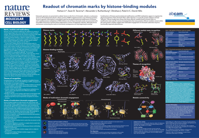
Readout of chromatin marks by histone-binding modules Haitao Li*, Sean D. Taverna*, Alexander J. Ruthenburg*, Dinshaw J. Patel & C. David Allis Combinations of histone posttranslational modifications and DNA methylation appear to regulate the physical properties of the chromatin fibre, either directly or via specific protein adaptors termed ‘effectors’. Recent studies have shown that these effector modules bind to histone tails in a modification state-specific manner. Emerging hints that many putative effector modules coexist within the same protein complex suggest that multivalent engagement of chromatin substrates may be a functionally important phenomenon. Eukaryotic genomes are presented to cellular factors in the form of chromatin, wherein a nucleosome serves as the fundamental subunit, which consists of DNA wrapped around a core of histone proteins. Access to genetic information is controlled in part by the posttranslational modification of histones and 5-methylcytosine methylation of DNA. Distinct sets of these chromatin marks are associated with most DNA transactions and have been implicated as carriers of epigenetic identity, although precise mechanisms connecting the marks to functional consequences are only beginning to emerge. Marks, modules and multivalency Histones can be covalently modified by the addition of a variety of chemical appendages that create binding sites for specific protein modules. The top panel illustrates the chemical structures of the small modifications that have been most intensively studied. Many examples of histone-binding, or effector, modules are known; shown in the middle panel are representative examples of each different class of protein fold (grey) in complex with its cognate modified histone-binding partner (yellow); see also Table 1. In most cases, residues that line the binding pocket (red) of a module dictate the modification state of a mark that is preferentially bound, while residues outside the binding pocket contribute to much of the histone sequence specificity. In several instances, similar folds bind different marks: for example, tudor domains can bind Kme3 (JMJD2A) or Kme2 (53BP1), and PHD fingers can bind exclusively Kme2/3 (BPTF) or unmodified K (BHC80). The structural underpinnings of this methyl-state specific readout (K, Kme1, Kme2 or Kme3) are demonstrated by comparing the four different structures displayed in the ‘Different methyl state recognition’ panel. In many cases, there is no longer a clear correlation between a single histone mark and its function. For example, the Kme3 mark, when in the context of the N terminus of H3 (K4me3), can be bound by PHD fingers of proteins in complexes that either activate (for example, the BPTF subunit of the NURF complex in homeotic gene remodelling enhances transcription) or repress gene expression (for example, the ING2 subunit of the Sin3a–HDAC complex is involved in repression following DNA damage). Analyses of native histone modification states has increased our appreciation that histone PTMs occur in various combinations rather than in isolation (see Table 2), which may help account for this paradox. Perhaps multivalent interactions with discrete patterns dictate composite specificity and enhance the affinity of chromatin-associated complexes. Histone marks + + Different methyl state recognition + + + + + PHD finger (BHC80) MBT (L3MBTL1) – K Kme1 Kme2 Kme3 Kac Cit R Rme1 Rme2s Rme2a Yph K Tph Kme1 Histone-binding modules Kac Kme2 Kme3 K Kme2 PHD finger (BHC80) Kme3 Themes of recognition R Kme3 Tudor (53BP1) Free N-terminus interactions Bromo (Gcn5) Kme3 WD40r (WDR5) N+2 or N–2 residue recognition Aromatic cage PHD finger (ING2) Sph Kme2 Kme3 MBT (L3MBTL1) Tudor (JMJD2A) Induced β-strand for histone peptide tandem BRCT (MDC1) 14-3-3 (14-3-3ζ) Tudor H3K4me2/3, H3K9me2/3, H3K27me2/3 H3K4me3, H4K20me2 MBT WD40 repeats Bromodomain PHD 14-3-3 BRCT H3K4me1, H4K20me1/2, H1K26me1 R2/H3K4me2? Kac H3K4, H3K4me3, H3K9me3, K36me3 H3S10ph H2A.XS139ph Intranucleosomal Oligovalent complex BAH 2 BPTF H4 4 H3 H4 H3 6 PWWP H4 7 SANT H3 H3 Trans-histone HP1 Adjacent bridging Histones and Transcription at Abcam Abcam is able to deliver a comprehensive portfolio of all the very best and most up to date histone and transcription factor antibodies. We are constantly increasing the number of ChIP grade and batch tested antibodies enabling the research community to deconstruct nuclear signaling pathways. Three of our very best ChIP grade transcription antibodies are for RNA polymerase II: • RNA pol II CTD repeat (ab26721) • RNA pol II CTD repeat phospho S2 (ab5095) • RNA pol II CTD repeat phospho S5 (ab5131) HP1 2 22 3 H3 HP1 2 MBD Chromo 8 2 1 Tudor Discontinuous bridging Further examples of our highly regarded antibodies for transcription factors: • Ctip2 monoclonal Zinc finger protein (ab18465) • HEXIM1 ChIP Grade (ab25388) All our transcription factor antibodies are highly characterized and our datasheets provide a library of relevant information. Investigate these and other transcription and Histone antibodies further at: www.abcam.com/transcription and www.abcam.com/Histones The Abpromise If our antibodies do not perform as described on the datasheet, notify us within 4 months of delivery so we can help you or offer a replacement or a refund. Transcriptionally active chromatin Mitogen-stimulated transcription H3R17me1/me2a + H3K18ac Oestrogen-stimulated transcription Chaperone association H4K5ac + H4K12ac Pre-deposition Stem cell transcriptional plasticity H3K4me3 + H3K27me3 ‘Bivalent domains’ at key developmental genes Transcriptional repression H3K9me3 + H3K27me3 Silent loci + CpG 5-MeC H3K27me3 + H2AK119ub1 Silent homeotic genes Heterochromatin H3K9me3 + H4K20me3 + CpG 5-MeC H3K9me2/3 + H4K20me1 Inactive X-chromosome + H4K27me3 + CpG 5-MeC Contact information: Bromo PHD HP1 H3K4me2/3 + H3K9/14/18/23ac H3S10ph + H3K14ac Transcriptionally active homeotic genes 3 4 H3 Locus/chromatin state WD40r Static effectormodule pockets Internucleosomal Oligovalent protein Histone marks Transcriptional activation H3K4me2/3 + H4K16ac 53BP1, p53 binding protein-1; 5-MeC, 5-methylcytosine; BAH, bromo-adjacent homology domain; BHC80, BRAF-HDACcontaining protein; BPTF, bromodomain PHD finger transcription factor; BRCT, breast cancer carboxy-terminal domain; Bromo, bromodomain; Chromo, chromodomain; Cit, citrulline; CpG, the DNA sequence that is often targeted for epigenetic 5-cytosine methylation; Double chromo, double chromodomain; HP1, heterochromatin protein 1; ING2, inhibitor of growth protein-2; JMJD2A, jumonji domain-containing protein 2A; K, lysine; Kac, acetylated lysine; Kme1, mono-methylated lysine; Kme2, di-methylated lysine; Kme3 tri-methylated lysine; MBT, malignant brain tumour; PHD finger, plant homeodomain finger; PTM, posttranslational modification; R, arginine; Rme1, mono-methylated arginine; Rme2a, asymmetric dimethylated arginine; Rme2s, symmetric di-methylated arginine; Sph, phosphorylated serine; Tph, phosphorylated threonine; Yph, phosphorylated tyrosine; WD40r, WD40 repeat. Module connectivity Modes of multivalent chromatin engagement Cis-histone Known histone marks Abbreviations Sph Double chromo (CHD1) hTaf1 Histone-binding or effector module Chromodomain Table 2 | Functional associations of coexisting marks Royal family Chromo (HP1) Table 1 | Recognition of histone marks by modules PHD finger (BPTF) Tudor (53BP1) Modes of multivalent chromatin engagement Histone-binding modules are often found in the same protein with other binding modules, suggesting a means to simultaneously interact with particular combinations of modifications and a mechanism for increasing the affinity of complexes for chromatin (lower panel). The number of proteins with multiple potential histone-binding modules in the human proteome (http://smart.embl-heidelberg.de) are shown schematically in the ‘Module connectivity’ panel. We would like to articulate nomenclature to describe the possible modes of multivalent recognition, as distinguishing among these permutations is important for future studies. Intranucleosomal: mechanisms for simultaneous histone-tail binding in the same nucleosome. • Cis-histone: Tandem modules bind to marks that are present on the same histone tail. • Trans-histone: Modules in a protein or complex bind to marks that are present on different histone tails. Internucleosomal: mechanisms for simultaneous histone-tail binding in different nucleosomes. • Adjacent bridging: Multiple modules engage marks on adjacent nucleosomes. • Discontinuous bridging: Multiple modules engage marks on nonadjacent nucleosomes. – + Sph Themes of recognition Emerging themes in the molecular recognition of modified histones are illustrated in the context of the PHD finger of BPTF, and appear to be the product of convergent evolution. • The histone peptide often engages the binding module in an induced β-strand backbone conformation, whereas the protein-binding modules remain relatively static. • Aromatic residues in the module envelop histone methylation in a cation-π cage as also depicted in the ‘Different methyl state recognition’ panel. • Residues in the N+2 or N-2 position with respect to the histone mark are often crucial determinants of binding sequence specificity. • Additional specificity may be imparted by recognition of a free N terminus. – Linked review article: Ruthenburg, A.J., Li, H., Taverna, S.D., Patel, D.J. & Allis, C.D. Multivalent engagement of chromatin modifications by linked binding modules. Nature Rev. Mol. Cell Biol. 8, xx–xx (2007). Further reading: Bernstein, B.E. et al. A bivalent chromatin structure marks key developmental genes in embryonic stem cells. Cell 125, 315–326 (2006). Cheng, X., Collins, R.E. & Zhang, X. Structural and sequence motifs of protein (histone) methylation enzymes. Annu. Rev. Biophys. Biomol. Struct. 34, 267–294 (2005). S. D. Taverna, A. J. Ruthenburg, and C. D. Allis are at the Laboratory of Chromatin Biology and Epigenetics, The Rockefeller University, 1230 York Ave, Room 1501, New York, NY, 10065, USA. | C. D. Allis’ homepage: www.rockefeller.edu/labheads/allis/index.php | H. Li and D. J. Patel are at the Structural Biology Program, Memorial Sloan-Kettering Cancer Center, New York, NY, 10021, USA. | D. J. Patel’s homepage: www.mskcc.org/mskcc/html/10829.cfm *Authors contributed equally to this work. Poster design by Vicky Askew. Edited by Arianne Heinrichs. © 2007 Nature Publishing Group. www.nature.com/nrm/poster/xxx Hodawadekar, S.C. & Marmorstein, R. Chemistry of acetyl transfer by histone modifying enzymes: structure, mechanism and implications for effector design. Oncogene 26, 5528–5540 (2007). Krishnamurthy, V.M., Estroff, L.M. & Whitesides, G.M. in Fragmentbased Approaches in Drug Discovery (eds Jahnke, W. & Erlanson, D.A.) 11–53 (Wiley-VCH, Weinheim, 2006). Shi, Y. & Whetstine, J.R. Dynamic regulation of histone lysine methylation by demethylases. Mol. Cell 25, 1–14 (2007). Strahl, B.D. & Allis, C.D. The language of covalent histone modifications. Nature 403, 41–45 (2000).
