Document 13310408
advertisement
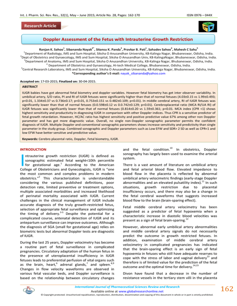
Int. J. Pharm. Sci. Rev. Res., 32(1), May – June 2015; Article No. 28, Pages: 162-170 ISSN 0976 – 044X Research Article Doppler Assessment of the Fetus with Intrauterine Growth Restriction 1 2* 3 1 4 5 Ranjan K. Sahoo , Sibananda Nayak , Sitansu K. Panda , Pravkar B. Pati , Sahadev Sahoo , Mahesh C Sahu Department of Radiology, IMS and Sum Hospital, Siksha O Anusandhan University, K8-Kalinga Nagar, Bhubaneswar, Odisha, India. 2 Dept of Obstetrics and Gynaecology, IMS and Sum Hospital, Siksha O Anusandhan Univ, K8-KalingaNagar, Bhubaneswar, Odisha, India. 3 Department of Anatomy, IMS and Sum Hospital, Siksha O Anusandhan University, K8-Kalinga Nagar, Bhubaneswar, Odisha, India. 4 Department of Obstetrics and Gynaecology, Hi-tech Medical College, Bhubaneswar, Odisha, India. 5 Central Research Laboratory, IMS and Sum Hospital, Siksha O Anusandhan University, K8-Kalinga Nagar, Bhubaneswar, Odisha, India. *Corresponding author’s E-mail: nayak_sibananda@yahoo.com 1 Accepted on: 17-03-2015; Finalized on: 30-04-2015. ABSTRACT IUGR babies have got abnormal fetal biometry and doppler variables. However fetal biometry has get inter observer variability. In umbilical artery, S/D ratio, PI and RI of IUGR fetuses were significantly higher than that of normal fetuses (4.03±0.15 vs 1.99±0.493; p<0.01, 1.334±0.37 vs 0.736±0.17; p<0.01, 0.753±0.151 vs 0.482±0.109; p<0.01). In middle cerebral artery, RI of IUGR fetuses was significantly lower than that of normal fetuses (0.0.598±0.12 vs 0.0.742±0.129; p<0.01). Cerebroplacental ratio (MCA RI/UA RI) of IUGR fetuses was significantly lower than that of normal fetuses (0.814±0.20 vs 1.59±0.361; p<0.01. MCA index (CPR <1) shows highest sensitivity and productivity in diagnosing IUGR in comparison with other Doppler indices. Thus CPR is a sensitive predictor of fetal growth retardation. However, HC/AC ratio has highest sensitivity and positive predictive value 67% among other non Doppler parameter and has got more diagnostic value. Overall, no single non-Doppler sonographic parameter permits the confident diagnosis of IUGR. Multiple Doppler and conventional sonographic parameters shows increase sensitivity and predictivity than single parameter in the study group. Combined sonographic and Doppler parameters such as Low EFW and SDR> 2 SD as well as CPR<1 and low EFW have better sensitive and predictive value. Keywords: Cerebro placental ratio, Doppler, Fetal biometry, IUGR. INTRODUCTION I ntrauterine growth restriction (IUGR) is defined as sonographic estimated fetal weight<10th percentile for gestational age.1 According to the American College of Obstetricians and Gynecologists, IUGR is “one the most common and complex problems in modern obstetrics.”2 This characterization is understandable considering the various published definitions, poor detection rate, limited preventive or treatment options, multiple associated morbidities and increased likelihood of perinatal mortality associated with IUGR. Current challenges in the clinical management of IUGR include accurate diagnosis of the truly growth-restricted fetus, selection of appropriate fetal surveillance and optimizing the timing of delivery.3-5 Despite the potential for a complicated course, antenatal detection of IUGR and its antepartum surveillance can improve outcomes. Although the diagnosis of SGA (small for gestational age) relies on biometric tests but abnormal Doppler tests are diagnostic of IUGR. During the last 25 years, Doppler velocimetry has become a routine part of fetal surveillance in complicated pregnancies. Circulatory adaptation of the human fetus in the presence of uteroplacental insufficiency in IUGR fetuses leads to preferential perfusion of vital organs such 6 7 8,9 as the brain, heart, adrenal glands, and spleen. Changes in flow velocity waveforms are observed in various fetal vascular beds, and Doppler surveillance is based on the relationship between circulatory changes and the fetal condition.10 In obstetrics, Doppler sonography has largely been used to examine the arterial system. There is a vast amount of literature on umbilical artery and fetal arterial blood flow. Elevated impedance to blood flow in the placenta is reflected by abnormal umbilical artery velocimetric findings (early-stage Doppler abnormalities and an elevated pulsatility index).11 In such situations, growth restriction due to placental insufficiency occurs, and there may also be a change in the fetal cerebral waveforms that suggests increased blood flow to the brain (brain-sparing effect). Fetal middle cerebral artery velocimetry has been suggested as a predictor of fetal hypoxemia when a characteristic increase in diastolic blood velocities was present as a sign of fetal brain-sparing.12 However, abnormal early umbilical artery abnormalities and middle cerebral artery signals do not necessarily predict the outcome in growth restricted fetuses. In addition, examination of middle cerebral artery velocimetry in complicated pregnancies has indicated that the brain-sparing effect is an early sign of fetal hypoxemia in fetuses who still have adequate reserves to 13 cope with the stress of labor and vaginal delivery and therefore is of limited value for the prediction of the fetal 14,15 outcome and the optimal time for delivery. Divon have found that a decrease in the number of resistance vessels in the tertiary stem villi in the placenta International Journal of Pharmaceutical Sciences Review and Research Available online at www.globalresearchonline.net © Copyright protected. Unauthorised republication, reproduction, distribution, dissemination and copying of this document in whole or in part is strictly prohibited. 162 © Copyright pro Int. J. Pharm. Sci. Rev. Res., 32(1), May – June 2015; Article No. 28, Pages: 162-170 causes an increase in resistance, leading to decreased flow through the UA and an increase in the UA PI. This is described as umbilical placental insufficiency.18 Fleischer and Schulman have found that in IUGR complicated by pregnancy-induced hypertension, there is inadequate trophoblastic invasion of the spiral arteries, leading to increased resistance in the spiral arteries and decreased blood flow in the placental vascular bed and in the UA, thereby resulting in an increase in the UA PI. This 19 is described as uteroplacental insufficiency. In pregnancies with chronic fetal hypoxia, the blood volume in the fetal circulation is redistributed in favor of vitally important organs, i.e., the heart, kidneys and brain. Vasodilatation of the MCA, with an increase in diastolic flow through it, results in a decrease in its PI. Thus, in asymmetrical growth retardation, there is a high UA PI and low MCA PI. As a result, the C/U ratio is lower than normal in growth-retarded fetuses. A significant association between the C/U ratio and the HC/AC ratio can be seen. The C/U ratio remains constant during the last 10 weeks of gestation and provides better diagnostic accuracy than either vessels’ PI considered alone.20 The purpose of this study was to assess the accuracy of the middle cerebral to umbilical artery resistance index ratio (C/U RI) in predicting IUGR and to find the combined sensitivity of C/U ratio and foetal biometry indiagnosis of IUGR in high-risk pregnancies. MATERIALS AND METHODS The study was conducted in Department of obstetrics and gynaecology, IMS and sum hospital, during January 2013 to December 2014. A total, 50 pregnant women were randomly selected for the study. Among them, 30 cases were in study group with the risk factors of hypertension, diabetes or heart disease and the rest 20 cases were in control group. The consent from all the patients was taken. A complete history, general physical and obstetrical examination was carried out in all patient and the cases were assigned to control and study group. The control group had following criteria: ISSN 0976 – 044X iii) Poor maternal weight gain. iv) Fundal height of the uterus, on clinical evaluation, lagging behind by at least 4 weeks of the expected height according to gestational age. Ultrasound and Doppler studies were carried out at department of Radiodignosis, IMS and sum hospital, to determine composite ultrasound gestational age, HC/AC ratio, estimated fetal weight (EFW) and Doppler velocity waveforms as well as Doppler velocimetry such as S/D ratio, PI and RI of umbilical artery and middle cerebral artery of the foetus. Fetal weight was estimated according to Haddlock16. IUGR was defined as estimated fetal weight of less than the 10th percentile for gestational age, according to Sabbagha and Minogue growth curves17. Umbilical artery Doppler indices were estimated on a free loop of cord. Waveforms of good quality were collected and analyzed in the absence of fetal breathing movements; on average, 3 separate readings were performed. During the examination, the women were in a semirecumbent position with the head and chest slightly elevated. The recording was performed during periods of fetal apnea, because of a potential effect of fetal breathing movements on waveform variability. For measurements of the middle cerebral artery indices, an axial view of the fetal head was obtained at the level of the cerebral peduncles. Color Doppler was used to visualize the circle of Willis. The Doppler sample volume was placed within 1 cm of the origin of the middle cerebral artery that was easily identified as a major branch running in anterolateral direction from the circle of Willis towards the lateral edge of the orbit. While angle of correction is not necessary when measuring the middle cerebral artery pulsatility index (PI), peak systolic velocity measurement should use angle correction and the angle of incidence should be <30 degrees; optimally as close to 0 degrees as possible. For the waveform analysis, maximum and minimum values of the velocity waveforms on the frozen image were measured by use of electronic calipers of the machine. i) There was close relation (±2 weeks) between gestational age and Clinical evaluation. ii) There were no maternal complications known to affect the normal fetal growth e.g. chronic hypertension, diabetes, heart disease. The Pourcelot resistance index (systole-diastole/dystole) was calculated by a built-in microcomputer. iii) There was no patient with multiple gestations and at risk for family history dwarfism. The C/U RI was estimated with a cut-off value of 1.0. Only C/U RI<1.0 were considered abnormal. Statistical analysis was performed using SPSS software. The Study Group included basing upon following criteria: i) History of previous growth retarded fetuses. ii) History of chronic hypertension, insulin dependent diabetes mellitus and other maternal diseases known to affect fetal growth. Observation The patients of control and study group were analyzed with respect to age of the patient. It was found that there was no statistically different in between these groups (P=0.225). Most of the International Journal of Pharmaceutical Sciences Review and Research Available online at www.globalresearchonline.net © Copyright protected. Unauthorised republication, reproduction, distribution, dissemination and copying of this document in whole or in part is strictly prohibited. 163 © Copyright pro Int. J. Pharm. Sci. Rev. Res., 32(1), May – June 2015; Article No. 28, Pages: 162-170 patients in both control and study group were within the maternal age of 20-25 years. The umbilical artery indices such as S/D ratio decreased from 2.63 to 1.96. In addition, PI decreased from 1.24 to 0.60 & RI decreased from 0.62 to 0.48 from 32 weeks to term respectively. Doppler indices of MCA decrease after 32 weeks till term. The middle cerebral artery indices such as PI decreased from 2.16 to 1.36 and RI decreased from 0.89 to 0.62 from 32 weeks to term respectively. In control group CPR was more than 1 (Table 1). Out of 30 cases of the study group, 25 cases shows PI of Umbilical artery more than 2 SD, 23 cases with th SDR of umbilical artery > 3, 20 cases with SDR > 95 percentile whereas in middle cerebral artery 15 cases with SDR > 95th percentile and 27 cases with CPR < 1 (Table 2). About 96% of the babies of the patients in the study group were below 2500gram with a mean birth weight of 1806gram compared to 10% in the control group were the mean birth weight was 2749g, which is statistically significant at P=0.0001 (Table 3). Each of these sonographic parameters (HC/AC Ratio, AFI and EFW) has been found to have a statistically significantly different mean value or frequency of occurrence, in growth-retarded as compared with normal fetuses. Mean value of AFI and EFW of study ISSN 0976 – 044X group is lower than that of control group where as Mean value of HC/AC of study group is higher than control group (Table 4). In umbilical artery, S/D ratio, PI and RI of IUGR fetuses were significantly higher than that of normal fetuses (4.03±0.15 vs 1.99±0.493; p<0.01, 1.334±0.37 vs 0.736±0.17; p<0.01, 0.753±0.151 vs 0.482±0.109; p<0.01). In middle cerebral artery, RI of IUGR fetuses was significantly lower than that of normal fetuses (0.0.598±0.12 vs 0.0.742±0.129; p<0.01). Cerebroplacental ratio (MCA RI/UA RI) of IUGR fetuses was significantly lower than that of normal fetuses (0.814±0.20 vs 1.59±0.361; p<0.01 (Table 5). Umblical artery shows 73% sensitivity, 85% specificity, 35% positive predictive value with 96% negative predictive value when S/D ratio was more than 3 (Table 6). Sonography parameter such as HC/AC ratio shows 93% sensitivity, 95% specificity, 67% positive predictive value and 99% negative predictive value for IUGR. Low EFW shows 83% sensitivity and 85% specificity, 38% positive predictive value and 97% negative predictive value for estimation of IUGR. Out of sonographic parameters HC/AC ratio has better predictive value(67%) (Table 7). Combined sonographic and Doppler parameters such as Low EFW and SDR> 2 SD as well as CPR<1 and low EFW have better sensitive and predictive value (Table 8). Table 1: Doppler indices of umbilical and middle cerebral artery of the control group S. No Middle cerebral artery Umbilical artery Gestation(wk) PI RI SDR PI RI CPR (RI of MCA/RI of UA) 1 32 1.24 0.62 2.63 2.16 0.89 1.44 2 32 1.26 0.65 2.71 2.23 0.88 1.35 3 32 1.22 0.59 2.79 2.16 0.90 1.53 4 35 1.14 0.64 2.48 1.46 0.86 1.34 5 35 1.15 0.69 2.56 1.35 0.80 1.16 6 35 1.11 0.67 2.39 1.57 0.93 1.39 7 36 0.77 0.56 2.48 0.90 0.71 1.27 8 36 0.76 0.57 2.47 1.45 0.78 1.37 9 36 0.82 0.53 2.60 1.47 0.76 1.43 10 36 0.75 0.55 2.34 1.56 0.75 1.36 11 37 0.76 0.51 2.39 1.55 0.70 1.37 12 37 0.76 0.56 2.40 1.54 0.70 1.25 13 37 0.82 0.54 2.28 1.57 0.67 1.24 14 37 0.77 0.53 2.32 1.35 0.69 1.30 15 37 0.70 0.50 2.12 1.36 0.65 1.30 16 38 0.61 0.48 2.11 1.36 0.65 1.35 17 38 0.60 0.49 1.99 1.38 0.67 1.37 18 38 0.59 0.50 1.98 1.43 0.64 1.28 19 39 0.60 0.48 1.96 1.46 0.62 1.29 20 39 0.57 0.48 1.97 1.39 0.63 1.31 International Journal of Pharmaceutical Sciences Review and Research Available online at www.globalresearchonline.net © Copyright protected. Unauthorised republication, reproduction, distribution, dissemination and copying of this document in whole or in part is strictly prohibited. 164 © Copyright pro Int. J. Pharm. Sci. Rev. Res., 32(1), May – June 2015; Article No. 28, Pages: 162-170 ISSN 0976 – 044X Table 2: Doppler indices of umbilical and middle cerebral artery of the study group Umbilical Artery Gestational age Middle Cerebral Artery CPR PI RI SDR PI RI 30 1.39 0.79 4. 71 1.10 0.70 0.88 32 0.80 0.57 2.33 1.10 0.70 1.22 32 0.97 0.49 2.30 0.86 0.58 1.18 33 1..92 0.85 6.6 1.13 0.64 0.75 34 1.34 1 Inf 0.89 0.61 0.61 34 1.24 0.82 5.48 1.12 0.62 0.75 34 1.40 1.0 inf 0.83 0.40 0.40 34 2.58 1.00 inf 0.94 0.49 0.49 35 1.6 0.71 3.47 0.99 0.64 0.90 35 1.23 0.76 4.3 0.99 0.57 0.75 35 1.7 0.81 5.3 0.85 0.64 0.79 35 1.5 0.76 4. 31 0.89 0.61 0.84 35 1.26 0.51 2.06 0. 59 0.37 0.72 36 0.92 0.60 2..25 0.98 0.44 0.73 36 0.89 0.49 1.97 0.89 0.38 0.77 36 1.47 0.85 6.59 2.24 0.79 0.92 36 1.36 0.62 2.63 0.78 0.45 0.72 36 1.20 0.61 2.57 1.77 0.88 1.44 37 1..5 0.77 4.29 1. 2 0.74 0.96 37 1.12 0.72 3.56 0.81 0.57 0.79 37 1..23 0.82 5.63 0. 98 0.72 0.87 37 1.47 0.85 6.59 0.98 0.74 0.87 38 1.45 1 inf 1.14 0.67 0.67 38 1..27 0. 93 5.8 0.90 0. 58 0.62 38 1.15 0.69 3. 2 0.96 0.60 0.86 39 1..41 1 inf 1. 3 0.50 0.86 40 1.81 079 4.76 0.87 0.56 0.70 40 1.3 0.68 3.16 0.99 0.51 0.75 43 1.63 0.83 5.8 1.0 0.63 0.75 46 0.98 0.70 3.33 1.2 0.62 0.86 Table 3: Birth weight in control and study group Birth Weight Weight < 2500gm (%) Weight > 2500gm (%) Mean Birth Weight Std deviation Study group 29(96%) 01(4%) 1806±408gm Control group 02(10%) 18(90%) 2749±203gm GROUP Chi-sqare value=82.3302, P=0.0001, DF = 1 Table 4: Comparison of mean value of sonographic parameters between study and control group S. No. fetal biometry 1 Study Group 2 Control Group HC/AC ratio AFI EFW mean 1.2007 4.9967 1783.83 Standard Deviation 0.07 2.1573 479.43 mean 1.0745 11.100 2731.75 Standard Deviation 0.077 1.9973 306.7 <0.0001 <0.0001 <0.0001 Test of Significance(P value) International Journal of Pharmaceutical Sciences Review and Research Available online at www.globalresearchonline.net © Copyright protected. Unauthorised republication, reproduction, distribution, dissemination and copying of this document in whole or in part is strictly prohibited. 165 © Copyright pro Int. J. Pharm. Sci. Rev. Res., 32(1), May – June 2015; Article No. 28, Pages: 162-170 ISSN 0976 – 044X Table 5: Comparison of mean value of doppler indices between study and control group S. No Umbilical artery Type of Wave Form 1 Study Group 2 Control Group Middle cerebral artery PI RI SDR PI RI CPR mean 1.334 0.753 4.039 1.0423 0.5983 0.814 Standard Deviation 0.370 0.151 1.582 0.3057 0.1208 0.20 mean 0.7365 0.482 1.9960 1.3810 0.7425 1.5910 Standard Deviation 0.1756 0.109 0.4938 0.6142 0.129 0.361 <0.001 <0.001 <0.001 <0.013 =0.002 <0.001 Test of Significance (P- value) Table 6: Doppler criteria for intrauterine growth retardation: performance characteristics (present study) Sensitivity (%) Specificity (%) Positive Predictive Value (%) Negative Predictive Value (%) 67 90 42 96 S/D Ratio >3 73 85 35 96 Absent/reverse diastolic flow 20 95 30 91 PI > 2SD above mean 83 95 64 98 MCA CPR <1 90 95 6 98 Criterion Umbilical artery th S/D ratio >95 percentile Computed using Bayes’ theorem and assuming an IUGR prevalence rate of 10%. Benson CB, Doubilet PM. Doppler criteria for intrauterine growth retardation: predictive values. J Ultrasound Med, 7, 1988, 655-65947. Table 7: Conventional sonographic criteria for IUGR:-performance characteristics (in present study). Criterion Sensitivity (%) Specificity (%) Positive Predictive value Negative Predictive value Low EFW (<2500gm) 93 90 50 99 Decrease AFI (<8cm) 83 85 38 97 Elevated HC/AC Ratio 93 95 67 99 Compared with Benson CB, Doubilet PM, Saltzman DH: Intrauterine Growth retardation: Predictive value of US criteria for antenatal diagnosis, Radiology, 160, 1986, 415. Computed using Bayers’ theorem and assuming an IUGR prevelance rate is 10%. Table 8: Combined doppler and conventional sonographic parameter performance characteristics. Criterion Sensitivity (%) Specificity (%) Positive Predictive value Negative Predictive value Low EFW + UA-SDR>2SD 100 95 68.5 100 HC/AC ratio + SDR>2SD 97 95 68.3 99.6 CPR<1 + Low EFW 100 95 68.5 100 DISCUSSION IUGR and a compromised placenta are usually linked with abnormal Doppler variables. One of these parameters is the proportion of fetal cardiac output distributed to the placenta. Human foetuses normally direct one-third of their cardiac output to the placenta during the second half of pregnancy and one-fifth during the last couple of months.21 In contrast, IUGR foetuses with early umbilical artery abnormalities direct a reduced volume of blood toward the placenta, both in absolute and relative terms, while maintaining relatively normal cardiac output.22 Low cardiac output to the placenta may already be present at a stage of the disease that precedes the appearance of clinical evidence of a reduction in fetal growth and changes in impedance to flow in the fetal arterial and venous circulation.23 This condition may suggest that, at an early stage of placental compromise, the volume of fetal blood flow toward the placenta is reduced, and more extensive recirculation of umbilical blood in the fetal body develops in an attempt to achieve more efficient extraction of oxygen and nutrients.22–24 In control group 90% cases had BW> 2.5 kg and the mean birth weight is 2749 gm., which is normal according to Indian standards. In study group the mean birth weight is 1806 gm and 96% shows birth weight < 2500 gm. In the study by Mallikarjunappa cases of PIH with IUGR had an International Journal of Pharmaceutical Sciences Review and Research Available online at www.globalresearchonline.net © Copyright protected. Unauthorised republication, reproduction, distribution, dissemination and copying of this document in whole or in part is strictly prohibited. 166 © Copyright pro Int. J. Pharm. Sci. Rev. Res., 32(1), May – June 2015; Article No. 28, Pages: 162-170 average birth weight of 1708 g. which correlates with this study.25 Sonographic parameters (HC/AC Ratio, AFI and EFW) have been found to have a statistically significantly different mean value or frequency of occurrence, in growthretarded as compared with normal fetuses (1.2007±0.07 vs 1.0745±0.07; P<0.0001, 4.9967±2.1573 vs 11.10±1.99; P<0.0001, 1783.83±479.43 vs 2731±306.7; P<0.0001) in the present study. Crane JP, Kopta MM. Prediction of intrauterine growth retardation via ultrasonically measured head/abdominal circumference ratios. Obstet Gynecol, 54, 1979, 597-601 has similar statistical significant different mean in IUGR and normal pregnancy.26 When the fetus is hypoxic, the cerebral arteries tend to become dilated in order to preserve the blood flow to the brain. In the middle cerebral artery, the systolic to diastolic S/D ratio will decrease (due to an increase in diastolic flow) in the presence of chronic hypoxic insult to the fetus. This increase in blood flow can be evidenced by Doppler ultrasound of the middle cerebral artery. This effect has been called “brain sparing effect” and is demonstrated by a lower value of the pulsatility index and resistive index. The cerebro-placental ratio (R.I of MCA/ R.I of umbilical Artery) becomes less than one. Banu (1998) studied the PI and RI in umbilical and middle cerebral arteries and compared ratio of PI and RI in UA and MCA. They found that ever though the measurement of PI value in the umbilical artery is enough to detect IUGR per se, the ratio of these indices between the UA and MCA is more accurate than independent evaluations in identifying fetuses developing fetal.27 KW Fong 1999 evaluated the usefulness of MCA to umbilical artery RI ratio (C/U ratio) as a predictor of adverse perinatal outcome. They concluded that the C/U ratio is a good predictor of neonatal outcome and can be used to identify fetuses at high risk of morbidity and mortality.28 Present study shows that in umbilical artery, S/D ratio, PI and RI of IUGR fetuses were significantly higher than that of normal fetuses (4.03±0.15 vs 1.99±0.493; p<0.01, 1.334±0.37 vs 0.736±0.17; p<0.01, 0.753±0.151 vs 0.482±0.109; p<0.01). In middle cerebral artery, RI of IUGR fetuses was significantly lower than that of normal fetuses (0.0.598±0.12 vs 0.0.742±0.129; p<0.01). Cerebroplacental ratio (MCA RI/UA RI) of IUGR fetuses was significantly lower than that of normal fetuses (0.814±0.20 vs 1.59±0.361; p<0.01 (Table 6). Similar study in the year 2000 by Yoon Ha Kim/Seok Mo Kim/Tae Bok Song/Ji Soo Byun revealed that in umbilical artery, S/D ratio, PI and RI of IUGR fetuses were significantly higher than that of normal fetuses (3.34±0.69 vs 2.29±0.29; p<0.01, 1.27±0.27 vs 0.81±0.14; p<0.01, 0.70±0.07 vs 0.55±0.06; p<0.01). In middle cerebral artery, RI of IUGR fetuses was significantly lower than that of normal fetuses (0.72±0.09 vs 0.82±0.07; p<0.01). ISSN 0976 – 044X Cerebroplacental ratio (MCA RI/UA RI) of IUGR fetuses was significantly lower than that of normal fetuses (0.93±0.25 vs 1.35±0.16; p<0.01). They concluded that umbilical blood flow was affected but middle cerebral blood flow was maintained in IUGR fetuses. So fetal blood flow redistribution in favor of the brain at development of IUGR may be present and detectable by Doppler ultrasonography using Cerebroplacental ratio.29 In 2005, Alaa Ebrashy, Osama Azmy, Magdy Ibrahim, Mohamed Waly, Amira Edris, Obstetrics and Gynecology Department, Kasr El Aini Hospital, Cairo University; Reproductive Medicine Unit, National Research Center; and Neonatology Department, Kasr El Aini Hospital, Cairo University, Cairo, Egypt (Croat Med J, 46(5), 2005, 821825) performed prospective case-control study of 50 pregnant women with preeclampsia with or without intrauterine growth restriction (IUGR) middle cerebral/umbilical artery resistance index (C/U RI) ratio < 1 is considered abnormal. In the preeclampsia group, C/U RI was abnormal in 32 out of 38 fetuses with IUGR, and in only 5 out of 12 of fetuses without IUGR.30 In the present study, although sensitivity was lower for the systolic to diastolic ratio (73%) of the umbilical artery than for the sonographic estimation of fetal weight (93%), the umbilical artery studies had a higher specificity (90 versus 85 %) (Table- 8 & 9).These findings indicate that sonographic biometry is a more sensitive technique for identifying IUGR but that the umbilical artery waveforms are a valuable adjunct for improving the diagnostic accuracy for the prenatal detection of IUGR. Berkowitz GS, Mehalek KE also reported the same finding in Sonographic estimation of fetal weight and Doppler analysis of umbilical artery velocimetry in the prediction of intrauterine growth retardation: a prospective study.31 The most common determination of fetal growth restriction is based on the estimated fetal weight, EFW, determined from a combination of BPD and AC (Campbell, 1975). Cerebroplacental ratio (CPR) less than one is seen in 90% of IUGR cases with positive predictive value 66% in the present study (Table-8). Article by Tatjana Reihs and Matthias Hofer in chapter (6) of teaching manual of color duplex sonography by Matthias Hofer mentioned that this index (CPR) is very sensitive predictor (80%) of fetal growth retardation.32 The middle-cerebral-to-umbilical-artery ratio remains relatively constant (mean ± SD 1.33 ± 0.19) between 27 and 37 weeks. A cutoff value of 1.0 (sensitivity 57.9%, specificity 75.6%, false-positive rate 24.4%) was selected from the receiver-operator characteristic curve analysis. This cutoff value successfully identified a population at significant risk of fetal growth retardation (relative risk 3.07, 95% confidence interval 1.73 to 5.45, exact twotailed p = 0.0009). A middle-cerebral-to-umbilical-artery ratio of ≤ 1.0 identifies a subgroup of patients at high risk for fetal growth retardation and severe neonatal 33 morbidity. International Journal of Pharmaceutical Sciences Review and Research Available online at www.globalresearchonline.net © Copyright protected. Unauthorised republication, reproduction, distribution, dissemination and copying of this document in whole or in part is strictly prohibited. 167 © Copyright pro Int. J. Pharm. Sci. Rev. Res., 32(1), May – June 2015; Article No. 28, Pages: 162-170 Alaa Ebrashy, Osama Azmy1, Magdy Ibrahim, Mohamed Waly, Amira Edris et al found C/U RI was abnormal (<1) in 32 out of 38 fetuses with IUGR in 50 preeclampsia 34 pregnancy group. In the present study (Table-8) sensitivity, specificity, positive and negative predictive values are evaluated for umbilical as well as middle cerebral artery. Umbilical artery shows 73% sensitivity, 85% specificity, 35% positive predictive value and 96% negative predictive value when S/D ratio is more than 3. Overall, however, positive predictive values of Doppler criteria for IUGR were found to be poor. Benson CB, Doubilet PM have observed 35,36 similar finding in their study. Sonographic parameters such as HC/AC Ratio shows 93% sensitivity, 95%specificity, 67% positive predictive value and 99% negative predictive value. Low EFW shows 93% sensitivity, 90% specificity, 50% positive predictive value and 99% negative predictive values. Ninety percent of fetuses are not growth-retarded, so any reasonable test will have a negative predictive value of at least 90%. Out of sonographic parameter HC/AC Ratio has better positive predictive value (67%) (Table-9). Overall, no single nonDoppler sonographic parameter permits the confident diagnosis of IUGR. Benson CB, Doubilet PM, Saltzman DH observed the similar findings in their study of intrauterine growth retardation: predictive value of ultrasound criteria for antenatal diagnosis. (Radiology, 160, 1986, 415-417).37 Multiple Doppler and conventional sonographic parameters shows increase sensitivity and predictivity than single parameter in the study group. Low EFW combined with Umbilical SDR >2 SD, HC/AC ratio combined with SDR >2 SD and CPR<1 with low EFW parameters shows increased predictivity and sensitivity (Table-10). Berkowitz GS, Chitkara U, Rosenberg J, Cogswell C, Walker B, Lahman EA, Mehalek KE, Berkowitz RL concluded that sonographic biometry is a more sensitive technique for identifying IUGR but that the umbilical artery waveforms are a valuable adjunct for improving the diagnostic accuracy for the prenatal 38 detection of IUGR. Ott WJ concluded that when used in combination, abdominal circumference and Doppler, or estimated fetal weight and Doppler resulted in the best predictive values. Either estimated fetal weight or abdominal circumference (alone) is accurate predictors of IUGR. Combined with Doppler studies of the umbilical artery either method will provide accurate evaluation of suspected IUGR. Above two studies support the findings.39 Our study also showed that C/U RI had a better specificity than either middle cerebral or umbilical artery resistance indices as measured by Doppler in predicting poor neonatal outcome. pregnancy. The study was conducted in a total of 50 pregnant women of whom 30 cases had clinically suspected IUGR or high risk of developing IUGR (study group) and the rest 20 cases with normal pregnancy (control group). We arrive at following inferences. Out of 30 cases of IUGR pregnancy cases 73% showed abnormal umbilical artery indices. Umbilical artery PI more than 2 SD and S/D ratio more than 3 show higher sensitivity and positive predictive value in comparison to other umbilical Doppler indices in diagnosing IUGR. This indicates that umbilical artery indices are more sensitive indicator in IUGR. In middle cerebral artery evaluation of fetus, RI of IUGR fetuses was significantly lower than that of normal fetuses (0.0.598±0.12 vs 0.0.742±0.129; p<0.01). Cerebroplacental ratio (MCA RI/UA RI) of IUGR fetuses was significantly lower than that of normal fetuses (0.814±0.20 vs 1.59±0.361; p<0.01). MCA index (CPR <1) shows highest sensitivity and productivity in diagnosing IUGR in comparison with other Doppler indices. Thus CPR is a sensitive predictor of fetal growth retardation. Conventional fetal sonographic biometry study of control and study groups revealed that sonographic parameters (HC/AC Ratio, AFI and EFW) have been found to have a statistically significantly different mean value or frequency of occurrence, in growth-retarded as compared with normal fetuses (1.2007±0.07 vs 1.0745±0.07; P<0.0001, 4.9967±2.1573 vs 11.10±1.99; P<0.0001, 1783.83±479.43 vs 2731±306.7; P<0.0001). HC/AC ratio has highest sensitivity and positive predictive value 67% among other non Doppler parameter and has got more diagnostic value. Overall, no single non-Doppler sonographic parameter permits the confident diagnosis of IUGR. Multiple Doppler and conventional sonographic parameters shows increase sensitivity and predictivity than single parameter in the study group. Low EFW combined with Umbilical SDR >2 SD and HC/AC ratio combined with SDR >2 SD parameters shows increased predictivity and sensitivity. Considering that C/U RI reflects not only the circulatory insufficiency of the placenta by alteration in the umbilical resistance index, but also the adaptive changes resulting in modification of the middle cerebral resistance index, it seemed to be a potentially useful tool in predicting adverse perinatal outcome in high risk cases. Our results support the correlation between abnormal fetal C/U RI and adverse perinatal outcome in patients with preeclampsia with or without IUGR. REFERENCES 1. Battaglia FC, Lubchenko LO. A practical classification of newborn infants by weight and gestational age. J Pediatr, 71, 1967, 159-163. 2. Stern W, Farmakides G, Jagani N, Blattner P. American College of Obstetricians and Gynaecologists. Intrauterine growth restriction, ACOG practice bulletin no-12, Washington, DC, ACOG, 2000. 3. Baschat AA. Arterial and venous Doppler in the diagnosis and management of early onset fetal growth restriction, Early Hum Dev, 81, 2005, 877-887. SUMMARY AND CONCLUSION The foregoing study was conducted to assess the Doppler sonographic evaluation of the umbilical and middle cerebral arteries of intrauterine growth retardation ISSN 0976 – 044X International Journal of Pharmaceutical Sciences Review and Research Available online at www.globalresearchonline.net © Copyright protected. Unauthorised republication, reproduction, distribution, dissemination and copying of this document in whole or in part is strictly prohibited. 168 © Copyright pro Int. J. Pharm. Sci. Rev. Res., 32(1), May – June 2015; Article No. 28, Pages: 162-170 4. Baschat AA, Gembruch U, Reiss I, Gortner L, Weiner CP, Harman CR. Relationship between arterial and venous Doppler and perinatal outcome in fetal growth restriction. Ultrasound Obstet Gynecol, 16, 2000, 407-413. 5. Bilardo CM, Wolf H, Stigter RH. Relationship between monitoring parameters and perinatal outcome in severe, early intrauterine growth restriction. Ultrasound Obstet Gynecol, 23, 2004, 119-125. 6. Campbell S, Vyas S, Nicolaides KH. Doppler investigation of fetal circulation. J Perinat Med, 19, 1991, 21–26. 7. Mari G, Uerpairojkit B, Abuhamad AZ, Copel JA. Adrenal artery velocity waveforms in the appropriate and small-forgestational-age fetuses. Ultrasound Obstet Gynecol, 8, 1996, 82–86. 8. 9. Abuhamad AZ, Mari G, Bogdan D, Evans ET. Doppler flow velocimetry of the splenic artery in the human fetus: is it a matter of chronic hypoxia? Am J Obstet Gynecol, 172, 1995, 820–825. Hecher K, Campbell S, Doyle P, Harrington K, Nicolaides K. Assessment of fetal compromise by Doppler ultrasound investigation of the fetal circulation: arterial, intracardiac, and venous blood flow velocity studies. Circulation, 91, 1995, 129–138. 10. Baschat AA, Gembruch U, Reiss I, Gortner L, Weiner CP, Harman CR. Relationship between arterial and venous Doppler and perinatal outcome in fetal growth restriction. Ultrasound Obstet Gynecol, 16, 2000, 407–413. 11. Divon MY. Umbilical artery Doppler velocimetry: clinical utility in high risk pregnancies. Am J Obstet Gynecol, 174, 1996, 10–14. 12. Mari G, Deter RL. Middle cerebral artery flow velocity waveforms in normal and small-for-gestational-age fetuses. Am J Obstet Gynecol, 166, 1992, 1262–1270. 13. Hofstaetter C, Gudmundsson S, Dubiel M, Marsál K. Ductus venosus velocimetry in high-risk pregnancies. Eur J Obstet Gynecol Reprod Biol, 70, 1996, 135–140. 14. Gudmundsson S, Dubiel M, Gunnarson G, Stale H, Maesel A, Marsál K. Middle cerebral artery velocimetry as a predictor of hypoxemia in foetuses with increased pulsatility index in the umbilical artery. J Matern Fetal Invest, 4, 1994, 19. ISSN 0976 – 044X Obstet Gynecol, 148, 1984, 985-990. 20. Giles WB, Trudinger BJ, Baird PJ. Fetal umbilical flow velocity waveform and placental resistance pathological correlation. Br J Obstet Gynaecol, 92, 1985, 31-38. 21. Robinson JS, Kingston EJ, Jones CT, Thorburn GD. Studies on experimental growth retardation in sheep: the effect of removal of endometrial caruncles on fetal size and metabolism. J Dev Physiol, 1, 1979, 379–398. 22. Kiserud T, Ebbing C, Kessler J, Rasmussen S. Fetal cardiac output, distribution to the placenta and impact of placental compromise. Ultrasound Obstet Gynecol, 28, 2006, 126– 136. 23. Rizzo G, Capponi A, Cavicchioni O, Vendola M, Arduini D. Low cardiac output to the placenta: an early hemodynamic adaptive mechanism in intrauterine growth restriction. Ultrasound Obstet Gynecol, 32, 2008, 155–159. 24. Bellotti M, Pennati G, De Gasperi C, Bozzo M, Battaglia FC, Ferrazzi E. Simultaneous measurements of umbilical venous, fetal hepatic, and ductus venosus blood flow in growth-restricted human fetuses. Am J Obstet Gynecol, 190, 2004, 1347–1358. 25. B. Mallikarjunappa, H. Harish, S. R. Ashish, Ravindra S. Pukale. Doppler Changes in Pre-Eclampsia. JIMSA, 26, 2013, 4. 26. Crane JP, Kopta MM. Prediction of intrauterine growth retardation via ultrasonically measured head/abdominal circumference ratios. Obstet Gynecol, 54(5), 1979, 597601. 27. Banu AA. “Doppler velocimetry in the umbilical and middle cerebral arteries in fetuses with intrauterine growth retardation or fetal distress.” Fukuoka igaku zasshi= Hukuoka acta medica, 89(5) 1998, 133-144. 28. Fong K W. “Prediction of Perinatal Outcome in Fetuses Suspected to Have Intrauterine Growth Restriction: Doppler US Study of Fetal Cerebral, Renal, and Umbilical Arteries 1.” Radiology, 213(3), 1999, 681-689. 29. Kim YH. “Umbilical Venous Blood Gases, Middle Cerebral and Renal Arterial Blood Flow Velocity Waveforms in Intrauterine Growth Restriction Fetuses.” Korean Journal of Perinatology, 12(2), 2001, 145-154. 15. Dubiel M, Gudmundsson S, Gunnarson G, Marsál K. Middle cerebral artery velocimetry as a predictor of hypoxemia in fetuses with increased resistance to blood flow in the umbilical artery. Early Hum Dev, 47, 1997, 177–184. 30. Ebrashy A. “Middle Cerebral/Umbllical Artery Resistance Index Ratio as Sensitive Parameter for Fetal Well-being and Neonatal Outcome in Patients with Preeclampsla: Casecontrol Study.” Croatian medical journal, 46(5), 2005, 821. 16. Hadlock FP. “Estimating fetal age: computer-assisted analysis of multiple fetal growth parameters.” Radiology, 152.2, 1984, 497-501. 31. Berkowitz GS. “Doppler umbilical velocimetry in the prediction of adverse outcome in pregnancies at risk for intrauterine growth retardation.” Obstetrics & Gynecology, 71(5), 1988, 742-746. 17. Macgregor S N. “Underestimation of gestational age by conventional crown-rump length dating curves.” Obstetrics & Gynecology, 70(3) 1987, 344-348. 18. Divon M. “Intrauterine Growth Retardation-a Prospective Study of the Diagnostic Value of Real-Time Sonography Combined With Umbilical Artery Flow Velocimetry.” Obstetrics & Gynecology, 72(4), 1988, 611-614. 19. Schulman H, Fleischer A, Stern W, Farmakides G, Jagani N, Blattner P. umbilical wave ratios in human pregnancy. Am J 32. Hofer M. Ultrasound teaching manual: the basics of performing and interpreting ultrasound scans. Thieme, 2005. 33. Fernando A. Fetus-Placenta-Newborn: Accuracy of the middle-cerebral-to-umbilical-artery resistance index ratio in the prediction of neonatal outcome in patients at high risk for fetal and neonatal complications, American Journal of Obstetrics and Gynecology, 171, 1994, 1541–1545. International Journal of Pharmaceutical Sciences Review and Research Available online at www.globalresearchonline.net © Copyright protected. Unauthorised republication, reproduction, distribution, dissemination and copying of this document in whole or in part is strictly prohibited. 169 © Copyright pro Int. J. Pharm. Sci. Rev. Res., 32(1), May – June 2015; Article No. 28, Pages: 162-170 ISSN 0976 – 044X 34. Ebrashy A. “Middle Cerebral/Umbllical Artery Resistance Index Ratio as Sensitive Parameter for Fetal Well-being and Neonatal Outcome in Patients with Preeclampsla: Casecontrol Study.” Croatian medical journal, 146(5), 2005, 821. 37. Benson, Carol B., Peter M. Doubilet, and Daniel H. Saltzman. “Intrauterine growth retardation: predictive value of US criteria for antenatal diagnosis.” Radiology, 160(2), 1986, 415-417. 35. Benson CB. “Intrauterine growth retardation: diagnosis based on multiple parameters--a prospective study.” Radiology, 177(2), 1990, 499-502. 38. Berkowitz, Gertrud S. “Sonographic estimation of fetal weight and Doppler analysis of umbilical artery velocimetry in the prediction of intrauterine growth retardation: a prospective study.” American journal of obstetrics and gynecology, 158(5), 1988, 1149-1153. 36. Raio L. “Umbilical Cord Morphologic Characteristics and Umbilical Artery Doppler Parameters in Intrauterine Growth–Restricted Fetuses.” Journal of ultrasound in medicine, 22(12), 2003, 1341-1347. 39. Ott, William J. “Intrauterine growth restriction and Doppler ultrasonography.” Journal of ultrasound in medicine, 19(10), 2000, 661-665. Source of Support: Nil, Conflict of Interest: None. International Journal of Pharmaceutical Sciences Review and Research Available online at www.globalresearchonline.net © Copyright protected. Unauthorised republication, reproduction, distribution, dissemination and copying of this document in whole or in part is strictly prohibited. 170 © Copyright pro

