Document 13309074
advertisement
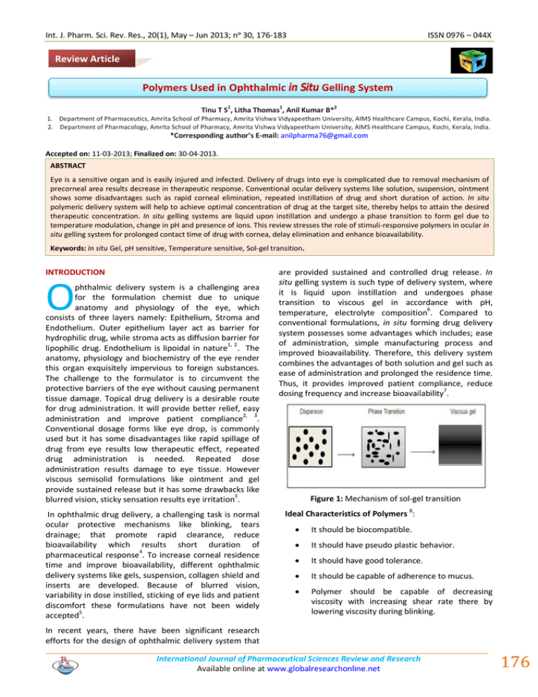
Int. J. Pharm. Sci. Rev. Res., 20(1), May – Jun 2013; nᵒ 30, 176-183 ISSN 0976 – 044X Review Article Polymers Used in Ophthalmic in Situ Gelling System 1 1 2 Tinu T S , Litha Thomas , Anil Kumar B* 1. Department of Pharmaceutics, Amrita School of Pharmacy, Amrita Vishwa Vidyapeetham University, AIMS Healthcare Campus, Kochi, Kerala, India. 2. Department of Pharmacology, Amrita School of Pharmacy, Amrita Vishwa Vidyapeetham University, AIMS Healthcare Campus, Kochi, Kerala, India. *Corresponding author’s E-mail: anilpharma76@gmail.com Accepted on: 11-03-2013; Finalized on: 30-04-2013. ABSTRACT Eye is a sensitive organ and is easily injured and infected. Delivery of drugs into eye is complicated due to removal mechanism of precorneal area results decrease in therapeutic response. Conventional ocular delivery systems like solution, suspension, ointment shows some disadvantages such as rapid corneal elimination, repeated instillation of drug and short duration of action. In situ polymeric delivery system will help to achieve optimal concentration of drug at the target site, thereby helps to attain the desired therapeutic concentration. In situ gelling systems are liquid upon instillation and undergo a phase transition to form gel due to temperature modulation, change in pH and presence of ions. This review stresses the role of stimuli-responsive polymers in ocular in situ gelling system for prolonged contact time of drug with cornea, delay elimination and enhance bioavailability. Keywords: In situ Gel, pH sensitive, Temperature sensitive, Sol-gel transition. INTRODUCTION O phthalmic delivery system is a challenging area for the formulation chemist due to unique anatomy and physiology of the eye, which consists of three layers namely: Epithelium, Stroma and Endothelium. Outer epithelium layer act as barrier for hydrophilic drug, while stroma acts as diffusion barrier for lipophilic drug. Endothelium is lipoidal in nature1, 2. The anatomy, physiology and biochemistry of the eye render this organ exquisitely impervious to foreign substances. The challenge to the formulator is to circumvent the protective barriers of the eye without causing permanent tissue damage. Topical drug delivery is a desirable route for drug administration. It will provide better relief, easy administration and improve patient compliance2, 3. Conventional dosage forms like eye drop, is commonly used but it has some disadvantages like rapid spillage of drug from eye results low therapeutic effect, repeated drug administration is needed. Repeated dose administration results damage to eye tissue. However viscous semisolid formulations like ointment and gel provide sustained release but it has some drawbacks like blurred vision, sticky sensation results eye irritation3. In ophthalmic drug delivery, a challenging task is normal ocular protective mechanisms like blinking, tears drainage; that promote rapid clearance, reduce bioavailability which results short duration of pharmaceutical response4. To increase corneal residence time and improve bioavailability, different ophthalmic delivery systems like gels, suspension, collagen shield and inserts are developed. Because of blurred vision, variability in dose instilled, sticking of eye lids and patient discomfort these formulations have not been widely 5 accepted . are provided sustained and controlled drug release. In situ gelling system is such type of delivery system, where it is liquid upon instillation and undergoes phase transition to viscous gel in accordance with pH, temperature, electrolyte composition6. Compared to conventional formulations, in situ forming drug delivery system possesses some advantages which includes; ease of administration, simple manufacturing process and improved bioavailability. Therefore, this delivery system combines the advantages of both solution and gel such as ease of administration and prolonged the residence time. Thus, it provides improved patient compliance, reduce dosing frequency and increase bioavailability7. Figure 1: Mechanism of sol-gel transition Ideal Characteristics of Polymers 8: It should be biocompatible. It should have pseudo plastic behavior. It should have good tolerance. It should be capable of adherence to mucus. Polymer should be capable of decreasing viscosity with increasing shear rate there by lowering viscosity during blinking. In recent years, there have been significant research efforts for the design of ophthalmic delivery system that International Journal of Pharmaceutical Sciences Review and Research Available online at www.globalresearchonline.net 176 Int. J. Pharm. Sci. Rev. Res., 20(1), May – Jun 2013; nᵒ 30, 176-183 Table 1: Polymers used for ocular in situ gelling system 9, 10 Polymer Origin Charge Solubility Mucoadhesive capacity Carbomer Synthetic Anionic Insoluble +++ Polyacrylic acid Natural Anionic Insoluble +++ Chitosan Natural Cationic Soluble ++ Xanthan gum Natural Anionic Insoluble + Methyl cellulose Natural Nonionic Soluble + ISSN 0976 – 044X Polymers used in pH sensitive in situ gelling system Carbomer Xyloglucan Natural Anionic Soluble + Poloxamer Synthetic Nonionic Soluble ++ Sodium alginate Natural Anionic Soluble ++ Properties HPMC Natural Nonionic Soluble + Carbomer is a high molecular weight, cross linked polyacrylic acid derivative and has strongest mucoadhesive property. It is water soluble vinyl polymer. In aqueous solution, it shows sol to gel transition, when the pH is raised above its pKa of about 5.515. As the concentration of carbomer increases, its acidic nature may cause irritation to eye. Addition of cellulose will reduce polymer concentration as well as will improve gelling property. Different grades of carbomer are available in market which includes carbopol 934(lowest cross linking density), carbopol 940 (highest cross linking density), and carbopol 981 (intermediate cross linking density). Carbopol is used as gelling, emulsifying and suspending agent16. Mucoadhesive capacity : Excellent(+++), Good(++), Poor(+) Classification of in situ gelling system11 1. pH sensitive in situ gelling system 2. Temperature sensitive in situ gelling system 3. Ion sensitive in situ gelling system 1. pH sensitive in situ gelling system In this system, gelling of the solution is triggered by change in pH, when pH is raised from 5-7.412. At higher pH, polymer forms hydrogen bond with mucin, which leads to hydrogel formation. Cellulose acetate phthalate latex, Carbopol, Polyacrylic Acid, Polyethylene Glycol are pH dependent polymers. Mechanism for pH sensitive gelling System All pH sensitive polymers contain pendant acidic or basic groups that can either accept or release protons in response to changes in environmental pH. In case of weakly acidic group, swelling of hydrogel increases as the external pH increases, while decreases in case of weakly basic groups 13. Figure 2: Mechanism of pH sensitive in situ gelling system14 Scheme 1: Structure of Carbomer Mechanism Mucoadhesive property is due to hydrogen bonding, electrostatic interaction or hydrophobic interaction17. Carbopol molecule is tightly coiled acidic molecule. Once dispersed in water, carboxylic group of the molecule partially dissociates to form flexible coil. Being a pH sensitive polymer, increase in solution pH results swelling of polymer. In acidic medium, it is in collapsed state due to hydrogen bonding, as the pH increases, electrostatic repulsion occur between the anionic groups, results gel swelling18. The gelling effect is activated in two stages: Dispersion and hydration of carbopol, neutralizing the solution by addition of sodium hydroxide, Triethanolamine, or potassium hydroxide. As the concentration of carbopol increases, due to its acidic nature it causes irritation to the eye. Addition of viscosity enhancer like HPMC, MC will reduce the concentration 17, 18 without affecting its gelling property . Srividya etal., developed a pH triggered ophthalmic delivery of ofloxacin by using carbopol and HPMC, results indicated that it produce sustained release over a period of 8 hours 19. Lin HR et al., formulated a carbopol/pluronic based ocular in situ gelling system. The mixture of 0.3% carbopol/14% pluronic solution showed significant enhancement in gel strength and bioavailability 20. Figure 3: Graphical representation of pH sensitive in situ gelling system Pandey et al., developed ocular in situ gel of levobunolol hydrochloride. The combination of mucoadhesive International Journal of Pharmaceutical Sciences Review and Research Available online at www.globalresearchonline.net 177 Int. J. Pharm. Sci. Rev. Res., 20(1), May – Jun 2013; nᵒ 30, 176-183 ISSN 0976 – 044X carbopol and viscosity enhancer HPMC provide sustained action over a period of time 21. Mohanambal et al., developed carbpol/HPMC based pH triggered ocular in situ gel of levofloxacin. The developed formulation was stable, non irritant and sustained release over a period of time 22. Polycarbophil23 Polycarbophil is lightly cross linked polyacrylic acid having excellent mucoadhesive property. Figure 5: Graphical representation of temperature sensitive in situ gelling system Mechanism Polymers used in temperature sensitive gelling system It is insoluble in water but its swelling capacity in neutral medium permits the entanglement of polymer chain with mucus layer. The carboxylic acid group of polycarbophil binds to mucin by hydrogen bonds. Noveon®AA-1 polycarbophil, is a high molecular weight polyacrylic acid polymer cross linked with divinyl glycol and exhibit sol-gel transition 23, 24. Poloxamer Cellulose acetate latex (CAP latex) another pH sensitive Polymers these are flowing liquid at pH 4.8 and gel at pH 7.4. Scheme 2: Structure of Poloxamer 2. Temperature sensitive in situ gelling system25 These are liquid solutions at room temperature (25-27oC) and undergo gelation when in contact with body fluid (3537oC) due to change in temperature. Temperature sensitive gels are three types; positive temperature sensitive gel, negative temperature sensitive gel, thermally reversible gel. Negative temperature sensitive gel has Lower Critical Solution Temperature (LCST), such gel contracts on heating above LCST25, 26. Positive temperature sensitive gel has an Upper Critical Solution Temperature (UCST) such gel contracts on cooling below UCST. Mechanism: The sol-gel phase transition occurs upon increasing temperature is due to three mechanisms: Desolvation of the polymer, increased micellar aggregation and increased entanglement of polymeric network27. When temperature increases polymeric chain degraded, leads to the formation of hydrophobic domain and phase 28 transition (liquid to hydrogel) occurred . Poloxamer are water soluble tri-block copolymer consisting of two polyethylene oxide (PEO) and polypropylene oxide (PPO) core in an ABA configuration29. Properties It is commercially available as Pluronic® and has good thermal setting property and increased drug residence time. It is used as gelling agent, emulsifying agent and solubilizing agent. Poloxamer gives colourless, transparent gel 30. Depending upon the ratio and distribution of hydrophilic and hydrophobic chain several molecular weights available, having different gelling property30. Table 2: Classification of poloxamer Poloxamer Molecular Weight 124 2200 188 8400 237 7959 338 14600 407 12600 Mechanism of gelling action Figure 4: Mechanism of Temperature sensitive in situ gelling system 14 It consists of central hydrophobic part (polypropylene oxide) surrounded by hydrophilic part (polyethylene oxide). At room temperature (25oC), it behaves as viscous liquid and is transformed to transparent gel when temperature increases (37oC) 31. At low temperature, it forms small micellar subunit in solution and increase in temperature results increase in viscosity leads to swelling to form large micellar cross linked network 14. International Journal of Pharmaceutical Sciences Review and Research Available online at www.globalresearchonline.net 178 Int. J. Pharm. Sci. Rev. Res., 20(1), May – Jun 2013; nᵒ 30, 176-183 ISSN 0976 – 044X Mechanism Gelation of cellulose solution is caused by hydrophobic interactions between molecules containing methoxy substitution. At low temperature, molecules are hydrated and little polymer-polymer interaction occurs, whereas at high temperature, polymers lose their water of hydration37. Figure 6: Gelling mechanism of Poloxamer 14 Xyloglucan Kamel et al., developed a Pluronic F 127 based in situ gelling system containing timolol maleate for sustained ocular delivery. In vivo study showed that ocular bioavilability of Pluronic F127gel based formulation increased by 2.5 fold as compared with aqueous timolol 32 solution . Qui et al., developed a pluronic and carbopol based ocular in situ gelling containing puerarin. Incorporatoion of carbopol enhance mucoadhesive force and provide sustained drug release over a period of 8hrs 33. Qian Y et al., formulated temperature sensitive poloxamer based in situ gelling system of methazolamide, for increasing corneal residence time and bioavilability. From the study, in vitro release shows that diffusion controlled release of drug from poloxamer solution over a period of 10 hours 34. Cao F et al., developed poloxamer/carbopol based ophthalmic in situ gelling system of azithromycin. Addition of carbopol 974 could increase the solubility of azithromicin by salt effect and enhance mucoadhesive property. The formulation exhibited 24 hour sustained release 35. Cellulose derivative Scheme 4: Structure of Xyloglucan Xyloglucan is water soluble hemicelluloses obtained from vascular plants and it exhibit thermally responsive behavior when more than 35% galactose residues are removed. It composed of (1,4)-β-D- glucan back bone chain(GLC) with (1,6)-α-D- xylose branches (XYL), partially substituted by (1-2)-β-D-galactoxylose (GAL) 37,38. Properties Xyloglucan is consists of three different oligomers like heptasaccharide, octasaccharide, nonsaccharide, which differ in number of galactose side chain. It is widely used in oral, rectal, ocular drug delivery due to its non- toxicity, biodegradable and biocompatible property. Like, poloxamer it exhibit gelation on heating refrigerator temperature or cooling from a higher temperature. But the difference is xyloglucan forms gel at lower concentration (1-2%wt) 39. Mechanism In native form of xyloglucan does not show gelation, its dilute solutions form so-gel transition on heating due to partial degradation of β-galactosidase. The transition temperature is inversely related to galactose removal ratio and polymer concentration 40. Scheme 3: Structure of HPMC Cellulose is composed of glucan chain with repeating β(1,4)-D-glucopyranose unit. Natural polymers like HPMC, MC, and EC exhibit temperature sensitive sol-gel phase 36 transition . Cellulose material will increases its viscosity when temperature is decreases while its derivatives like HPMC, MC, will increase its viscosity when temperature is increased 37. Miyasaki S et al., developed xyloglucan based ocular in situ gelling system of pilocarpine. That results degree of enhancement of miotic response followed by sustained 41 release of pilocarpine . Chitosan MC is composed of native cellulose with alternate methyl substitution group on its chain. At low temperature (30oC) solution is in liquid form and when temperature is o increases (40-50 C) gelation occurred. Scheme 5: Structure of Chitosan International Journal of Pharmaceutical Sciences Review and Research Available online at www.globalresearchonline.net 179 Int. J. Pharm. Sci. Rev. Res., 20(1), May – Jun 2013; nᵒ 30, 176-183 Chitosan is a cationic polysaccharide consisting copolymers of glucosamine and N-acetyl glucosamine, these are natural polymer obtained by deacetylation of chitin. Chitosan has mucoadhesive property due to electrostatic interactions between positively charged amino group and negatively charged mucin. It is non toxic, biocompatible, biodegradable polysaccharide and having bioadhesive, antibacterial activity 42. ISSN 0976 – 044X Polymers used for ion sensitive in situ gelling system Deacetylated gellan gum (Gelrite) Mechanism The mucoadhesive property is due to the formation of ionic interaction between the positively charged amino groups of chitosan and negatively charged sialic acid 43 residues of mucins, depends on environmental pH . Because of its bioadhesive, hydrophilic, good spreading properties, is used as viscosifying agent in artificial tear 44, 45 formulations . Gupta H etal., was developed ion and pH sensitive in situ gelling system for sustained delivery of timolol maleate. Chitosan and gellan gum were used as gelling agent, it enhance transcorneal drug permeation 46. Scheme 5: Structure of Gelrite Gellan gum is an anionic hetero polysaccharide, secreted by microbe Sphingomonas elodea. It consists of glucose, rhamnose, glucuronic acid and are linked together to give a tetrasaccharide unit 51. Properties Felt O et al., developed chitosan based ophthalmic gel for enhancing corneal residence time when compared to tobrex® 48. Gelrite is deacetylated gellan gum, obtained by treating gellan gum with alkali to remove the acetyl group in the molecule. Upon instillation, gelrite forms gel due to the presence of calcium ions52. The gelation involves the formation of double helical junction zones followed by aggregation of double helical segment to form three dimensional networks by complexaton with cations and hydrogen bonding with water53. Because of its thixotropy, thermo plasticity, pseudo plasticity are widely use in ophthalmology. In food industry, is used as suspending and stabilizing agent 23. 3. Ion Sensitive Gelling System Mechanism Gelation is triggered by the presence of cations (Na+, Mg++, Ca++) in the tear fluid. These can be achieved by polymers like sodium alginate, gellan gum 49. Gellan gum produce a cation induced in situ gelation (Ca2+, Mg 2+, K+, Na+) due to the cross linking between negatively charged helices and mono or divalent cations (Na+, Ca+, Mg+). Divalent ions superior to promoting gelation as compared to monovalent cations. Gelation prolongs the residence time of drug at absorption site and bioavailability of the drug is increased 54. Gratieri T et al., developed chitosan/poloxamer based in situ gelling system. The results indicated that chitosan improves the mechanical strength of poloxamer and increase mucoadhesive activity 47. Gelation is occurred by ionic interaction of polymer and divalent ions of tear fluid. When anionic polymers come in contact with cationic ions, it converts to form gel 50. Vodithala S et al., was developed gelrite based ion activated in situ gel of keterolac tromethamine and concluded that formulation produces sustained action over a period of 6 hours 55. Figure 7: Mechanism of Ion sensitive in situ gelling system14 Geethalakshmi A et al., developed gelrite based in situ gelling system of brimonidine tartarate and produce sustained release of drug 56. Balasubramaniam J et al., formulated gelrite based ion activated in situ gelling system of Indomethacine. Gelrite forms gels in the presence of mono or divalent cations present in the lacrimal fluid and it produce sustained 57 release over a period of 8 hours . Rajas N J et al., developed gelrite based ocular in situ gel of levofloxacin hemihydrate for bacterial infections. The developed formulation shows better corneal residence 58 time and sustained release of the drug . Figure 8: Graphical representation of ion activated in situ gelling system International Journal of Pharmaceutical Sciences Review and Research Available online at www.globalresearchonline.net 180 Int. J. Pharm. Sci. Rev. Res., 20(1), May – Jun 2013; nᵒ 30, 176-183 Sodium Alginate ISSN 0976 – 044X situ gel formulation makes acceptable and controlled drug delivery system. Thus sustained and prolonged release of drug, biocompatibility characteristics makes in situ gel dosage form reliable. In recent technology, polymer combinations focus the development of safe ophthalmic delivery system. REFERENCES Scheme 9: Structure of sodium alginate Sodium alginate is a gum extracted from brown algae. It is a salt of alginic acid. It is a linear block polysaccharide consisting of two type monomers β –D- Mannuronic acid and α-L-glucuronic acid residues joined by 1,4 glycosidic 59 linkages . It exhibit good mucoadhesive property due to its carboxylic group. It is biodegradable and non toxic 60. 1. Katariya Dhiraj kumar Champalal, Poddar Sushil Kumar S, Current status of ophthalmic in-situ forming hydrogel, International Journal of Pharma and Bio Sciences, 3, 2012, 372 – 388. 2. Kumar manish, Kulkarni GT, Recent Advances in ophthalmic drug delivery system, International journal of pharmacy and pharmaceutical sciences, 4, 2012, 387-394. 3. Dudinski O, Finnin BC, Reed BL, Acceptability of thickened eye drops to human subjects, Curr Ther Res, 33, 1983, 322337. 4. Rathore KS, Nema RK, Ishibashi Tejraj, Yokoi N,Born JA, Tiffany MJ, Komuro A, Review on ocular inserts, International Journal of Pharm Tech Research, 1, 2009, 164-169. 5. Jothi M, Harikumar SL and Geeta Aggarwal, In-situ ophthalmic gels for the treatment of eye diseases, International Journal of Pharmaceutical Sciences and Research, 3, 2012, 1891-1904. 6. Rathore KS, In situ Gelling ophthalmic drug delivery system: An overview, Int J Pharmacy and Pharm Sci, 2, 2010, 30-34. 7. Raval Sanjay, Vyas Jigar, Parmar Vijay, Raval Dhaval, International journal of drug formulation and research, A review on novel in situ polymeric drug delivery system, 2, 2011, 143-173. 8. Van M, In biopharmaceutics of ocular drug delivery. Ed. P. Edman, CRC press, Boca Raton, Fla, 1993, 27-42. 9. Lehr CM, Bouwstra JA, Schacht EH, In vitro evaluation of mucoadhesive properties of chitosan and some other natural polymers, Int J Pharm, 78, 1992, 43-48. Mechanism The monomers of alginate (β-D-mannuronic acid (M) and α-L- glucuronic acid (G) are arranged as M-M block or G-G block with alternating sequence (M-G) block. Upon interaction of G block of polymer with calcium moieties resulting in the formation of homogenous gel. Mechanical strength and porosity of hydrogel depends on G: M ratio, type of cross linker used and concentration of alginate solution 61, 62. Liu Z et al., developed ophthalmic gelling system of Gatifloxacin using alginate in combination with HPMC which acted as viscosity enhancing agent. Both in vitro release and in vivo corneal retention studies showed that HPMC/ alginate solution better retained than alginate/HPMC alone 63. D.N Mishra et al., designed in situ gelling ocular insert of gatifloxacin using sodium alginate and chitosan. In vitro release indicated that it provide sustained release over a period of 8-12 hours 64. Abraham S et al., developed Ofloxacin ion activated in situ gelling system using combination of polymers Alginate and HPC. Alginate/HPC solution retained the drug better than the alginate/HPC solution alone 65. Preetha JP et al., developed sodium alginate based diclofenac sodium in situ gelling system. The results indicated that sodium alginate/HEC solution shows better drug retained and antibacterial, antifungal, antimicrobial activity with selected micro organisms 66. CONCLUSION Polymers play a vital role in the delivery of drug from its dosage form. Polymeric in situ gelling system provides prolonged release of drug as compared to conventional delivery system. Various natural, synthetic, semi synthetic polymers are developed for controlled release of drug. Use of biodegradable and biocompatible polymers for in 10. Andrews GP, Laverty TP and Jones DS, Mucoadhesive polymeric platform for controlled drug delivery Review article, European journal of pharmaceutics and biopharmaceutics, 71, 2009, 505-518. 11. Nirmal HB, Bakliwal SR, Pawar SP, In-Situ gel: New trends in controlled and sustained drug delivery system, International Journal of PharmTech Research, 2, 2010, 1398-1408. 12. Tomme SRV, Storm G, Hennink EW, In situ gelling hydrogels for pharmaceutical and biomedical application, Int J Pharm. 355, 2008, 1-18. 13. Rajas NJ, Kavitha K, Gounder T, Mani T, In-Situ ophthalmic gels a developing trend, Int J Pharm Sci Rev and Res, 7, 2011, 8-14. 14. Gourav Rajoria, Arushi Gupta, In-Situ gelling system: A novel approach for ocular drug delivery, Am J Pharm Tech Res, 2, 2012, 25-53. 15. Davis NM, Farr SJ, Hadgraft J, Kellaway IW, Evaluation of mucoadhesive polymers in ocular drug delivery: part 1, viscous solution, Pharm Res, 8, 1991, 1039-1043. International Journal of Pharmaceutical Sciences Review and Research Available online at www.globalresearchonline.net 181 Int. J. Pharm. Sci. Rev. Res., 20(1), May – Jun 2013; nᵒ 30, 176-183 16. Gariepy ER, Leroux JC, In situ-forming hydrogels- reviews of temperature-sensitive systems, European Journal of Pharmaceutics and Biopharmaceutics 58, 2004, 409–426. 17. Nanjundswamy NG, Fatima S Dasankoppa, Sholapur HN, A review on hydrogels and its use in in Situ ocular drug delivery, Indian Journal of Novel Drug Delivery, 1, 2009, 1117. 18. Leung SS, Robinson JR, The contribution of anionic polymer structural features to mucoadhesion, J Control Release, 5, 1998, 223- 231. 19. Srividya B, Cardoza RM, Amin PD, Sustained ophthalmic delivery of ofloxacin from a pH triggered in situ gelling system, J Control Release, 73, 2001, 205-211. 20. Lin HR, Sung KC, Carbopol/pluronic phase change solutions for ophthalmic drug delivery, J Control Release, 69, 2000, 379- 88. 21. Pandey A, Mali PY, Patel DK, Ramesh R, Development and optimization of levobunolol hydrochloride In-Situ gel for glaucoma treatment, Int J Pharm & Bio Arch, 1, 2010, 134 – 139. 22. Mohanambal E, Arun K, Abdul Hasan Sathali A, Formulation and evaluation of pH-triggered In-Situ gelling system of Levofloxacin, Ind J Pharm Edu Res, 45, 2011, 58-64. 23. Robinson JR, Mlynek GM, Bioadhesive and phase-change polymers for ocular drug delivery, Adv Drug Deliv Rev, 16, 1995, 45-50. 24. Kaur IP, Smitha R, Penetration enhancers and ocular bioadhesives: two new avenues for ophthalmic drug delivery, Ind Pharm, 28, 2002, 353-369. 25. Bhardwaj TR, Kanwar M, Lal R,Gupta A, Natural gums and modified natural gums as sustained release carriers, Drug Devel Ind Pharm, 26, 2000, 1025-1038. 26. Guo JH, Skinner GW, Harcum WW, Barnum PE, Pharmaceutical applications of naturally occuring water soluble polymers, Pharm sci and Technol Today, 1, 998, 254- 261. 27. Shastri D, Pandya H, Parikh RK, Patel CN, Smart hydrogels in controlled drug delivery, Pharma Times, 38, 2006, 13-18. 28. Gariepy ER, Leroux GC, In situ – forming hydrogels-review of temperature sensitive systems, Eur J Pharm Biopharm, 58, 2004, 409-426. 29. Guo DD, Xu CX, Quan JS, Song CK, Jin H, Kim DD, Choi YJ, Cho MH, Cho CS, Synergistic anti-tumour activity of paclitaxel incorporated conjugated linoleic acid coupled poloxamer thermosensitive hydrogel in vitro and in vivo, Biomat, 30, 2009, 4777-4785. 30. Lehr CM, Bouwstra JA, Schacht EH and junginger HE, Invitro evaluation of mucoadhesive properties of chitosan and some other natural polymers, International journal pharmaceutics, 78, 1992, 43-48. 31. Ceulemans J, Ludwig A, Optimisation of carbomer viscous eye drops: an in vitro experimental design approach using rheological techniques, Eu J Pharm Biopharm, 54, 2002, 4150. 32. El-Kamel AH, In vitro and in vivo evaluation of Pluronic F 127 based ocular delivery system for Timolol maeate, Int J Pharm, 241, 2002, 47-55. ISSN 0976 – 044X 33. Hongyi Q, Chena W, Huanga C, Li Lib, Chena, Wenmin L, Chunjie W, Development of a poloxamer analogs/carbopolbased in situ gelling and mucoadhesive ophthalmic delivery system for puerarin, Int J Pharm, 337, 2007, 178-187. 34. Qian Y, Wang F, Li R, Zhang Q, Xu Q, Preparation and evaluation of in situ gelling ophthalmic drug delivery system for methazolamide, Drug Dev Ind Pharm, 36, 2010, 1340-1347. 35. Cao F, Zhang X, Ping Q, New method for ophthalmic delivery of azithromycin by poloxamer/carbopol-based in situ gelling system,Drug Deliv, 17, 2010, 500-507. 36. Tandjwa GA, Durand S, Berot S, Blassel C, Gaillard C, Garnier C, Doublier J L, Rheological characterization of microfibrillated cellulose suspension after freezing, Carb Poly, 80, 2010, 677-686. 37. Nanjawade BK, Manvi FV, Manjappa AS, Review of in-situ forming hydrogels for sustained ophthalmic drug delivery, J Control Rel, 122, 2007, 119-134. 38. Shirakawa M, Yamotoya K, Nishinari K, Tailoring of xyloglucan properties using an enzyme, Food Hydrocolloid, 12, 1998, 25-28. 39. Miyazaki S, Kawasaki N, Kubo W ,Endo K, Attwood D, Comparison of in situ gelling formulations for the oral delivery of Cimetidine, Int J Pharm, 220, 2001, 161-168. 40. Shastri DH, Patel LD, Novel alternative to ocular drug delivery system: Hydrogel, Ind J Pharma Res, 2, 2010, 1-13. 41. Miyazaki S, Suzuki S, Kawasaki N, Endo K, Takahashi A, Attwood D, In situ gelling xyloglucan formulations for sustained release ocular delivery of pilocarpine hydrochloride. Int J Pharm, 229, 2001, 29-36. 42. Patel V, Patel M, Patel R, Chitosan: A Unique Pharmaceutical excipient, Drug Delivery Technology, 5, 2005, 1-12. 43. Lehr CM, Bouwstra JA, Schacht EH, Junginger HE, In vitro evaluation of mucoadhesive properties of chitosan and other polymers, Int J Pharm, 78, 1992, 43-48. 44. Felt O, Carrel A, Baehni P, Buri P, Gurny R, Chitosan as tear substitute: a wetting agent endowed with antimicrobial efficiency, J Ocul Pharmacol Ther, 16, 2000, 261-270. 45. Calonge M, The treatment of dry eye, Surv Ophthalmol, 45, 2011, 227-239. 46. Gupta H, Jain S, Mathur R, Mishra P, Mishra AK, Sustained ocular drug delivery from a temperatureand pH triggered novel in situ gel system, Drug deivery, 14, 2007, 507-515. 47. Gratieri T, Gelfuso GM, Rocha EM, Sarmento VH, A poloxamer/chitosan in situ forming gel with prolonged retention time for ocular delivery, Eur J Pharm Biopharm, 75, 2010, 186-193. 48. O Felt, Baeyens V, Zignani M, Buri P, Gurny R. Mucosal drug delivery ocular- encyclopedia of controlled drug delivery, University Of Geneva, Switzerland, 2, 1999, 605- 622. 49. Pandya TP, Modasiya MK, Patel VM, Ophthalmic in–situ gelling system, Int J Pharm and Life Sci, 2, 2011, 730-738. 50. Bhaskaran S, Lakshmi PK, Harish CG, Topical ocular drug delivery: a review, Ind J Pharm Sci, 64, 2005, 404-408. International Journal of Pharmaceutical Sciences Review and Research Available online at www.globalresearchonline.net 182 Int. J. Pharm. Sci. Rev. Res., 20(1), May – Jun 2013; nᵒ 30, 176-183 51. Kuo M.S, Mort A.J, Dell A, Identification and location of Lglycerate, an unusual acyl substituent in gellan gum, Carbohydr Res, 156, 1986, 173-187. 52. Calfors J, Edsman K, Petersson R, Jornving K, Rheological evaluation of gelrite in situ gels for ophthalmic use, Eur J Pharm Sci, 6, 1998, 113-119. 53. Nirmal HB, Bakliwal SR, Pawar SP, In-situ Gel: New trend in controlled and sustained drug delivery system, Int J Pharmtech Res, 2, 2010, 1398-1408. 54. Miyazaki S, Suisha F, Kawasaki N, Shirakawa M, Yamatoya K, Attwood K, Thermally reversible xyloglucan gels as vehicles for rectal drug delivery, J Control Rel, 56, 1998, 75 -83. 55. Vodithala S, Khatry S, Shastri N, Sadanandam M, Formulation and evaluation of ion activated ocular gels of keterolac tromethamine, Int J Cur Pharm Res, 2, 2010, 3338. 56. Geethalakshmi A, Karki R, Jha SK, Venkatesh DP,Nikunj B. Sustained ocular delivery of brimonidine tartarate using ion activated in situ gelling system, Current Drug Delivery, 9, 2012, 197-204. 57. Balasubramaniam J, Kant S, Pandit JK, In vitro and in vivo evaluation of the Gelrite gellan gum-based ocular delivery system for indomethacin, Acta Pharm, 53, 2003, 251-61. 58. Rajas NJ, Kavitha K, Gounder T, Mani T, In-situ ophthalmic gels a developing trend, Int J Pharm Sci Rev and Res 7, 2011, 8-14. ISSN 0976 – 044X of carteolol containing Alginic acid, Int J Pharm, 207, 2000, 109-116. 60. Cochen S, Lobel E, Trevgoda A, Peled Y, A novel in situforming ophthalmic drug delivery system from alginates undergoing gelation in the eye, J Contr Rel, 44, 1997, 201208. 61. Smadar Cochen, Esther Lobel, Amrita Trevgoda, Yael Peled, A novel in-situ forming Ophthalmic drug delivery system from alginates undergoing gelation in the eye, Journal of Controlled Release, 44, 1997, 201-208. 62. Grant GT, Morris ER, Rees DA, Smith PJC and D, Thom, Biological interactions between polysaccharides and divalent cations: The egg box model, FEBS Lett,32, 1973, 195-198. 63. Liu Z, Li J, Nie S, Hui-Liu, Ding P, Pan W, Study of alginate/HPMC based in situ gelling ophthalmic deivery system for gatifloxacin, Int J Pharm, 315, 2006, 12-17. 64. Mishra DN, Gilhotra R.M, Design and characterization of bioadhesive in-situ gelling ocular inserts of gatifloxacin sesquihydrate, Daru, 16, 2008, 1-8. 65. Abraham S, Furtado S, Bharath S, Basavaraj BV, Deveswaran R, Madhavan V, Sustained ophthalmic delivery of ofloxacin from an ion-activated in-situ gelling system, Pak J Pharm Sci, 22, 2009, 175-179. 66. Preetha JP, Karthika K, Rekha NR, Elshafie K, Formulation and evaluation of in-situ ophthalmic gels of diclofenac sodium, J Chem Pharm Res, 2, 2010, 528-535. 59. Sechoy O, Tissie G, Sebastian C, Maurin F, Driot JY,Trinquand C, A new long acting ophthalmic formulation Source of Support: Nil, Conflict of Interest: None. International Journal of Pharmaceutical Sciences Review and Research Available online at www.globalresearchonline.net 183
