Document 13308955
advertisement
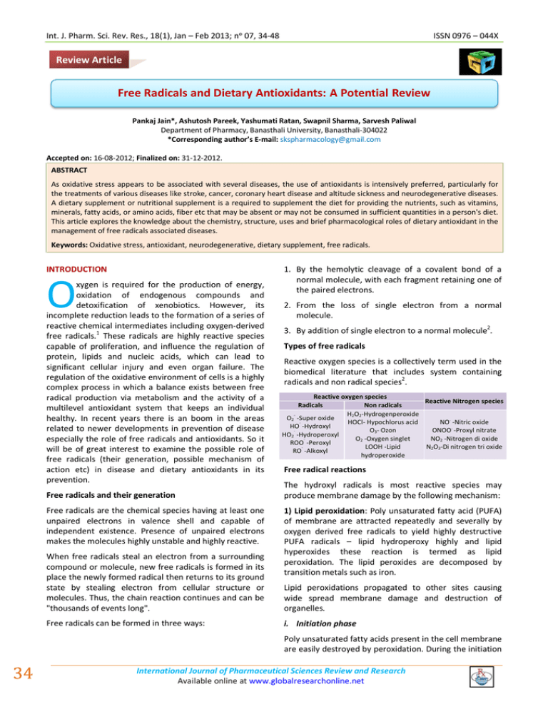
Int. J. Pharm. Sci. Rev. Res., 18(1), Jan – Feb 2013; nᵒ 07, 34-48 ISSN 0976 – 044X Review Article Free Radicals and Dietary Antioxidants: A Potential Review Pankaj Jain*, Ashutosh Pareek, Yashumati Ratan, Swapnil Sharma, Sarvesh Paliwal Department of Pharmacy, Banasthali University, Banasthali-304022 *Corresponding author’s E-mail: skspharmacology@gmail.com Accepted on: 16-08-2012; Finalized on: 31-12-2012. ABSTRACT As oxidative stress appears to be associated with several diseases, the use of antioxidants is intensively preferred, particularly for the treatments of various diseases like stroke, cancer, coronary heart disease and altitude sickness and neurodegenerative diseases. A dietary supplement or nutritional supplement is a required to supplement the diet for providing the nutrients, such as vitamins, minerals, fatty acids, or amino acids, fiber etc that may be absent or may not be consumed in sufficient quantities in a person's diet. This article explores the knowledge about the chemistry, structure, uses and brief pharmacological roles of dietary antioxidant in the management of free radicals associated diseases. Keywords: Oxidative stress, antioxidant, neurodegenerative, dietary supplement, free radicals. INTRODUCTION O xygen is required for the production of energy, oxidation of endogenous compounds and detoxification of xenobiotics. However, its incomplete reduction leads to the formation of a series of reactive chemical intermediates including oxygen-derived free radicals.1 These radicals are highly reactive species capable of proliferation, and influence the regulation of protein, lipids and nucleic acids, which can lead to significant cellular injury and even organ failure. The regulation of the oxidative environment of cells is a highly complex process in which a balance exists between free radical production via metabolism and the activity of a multilevel antioxidant system that keeps an individual healthy. In recent years there is an boom in the areas related to newer developments in prevention of disease especially the role of free radicals and antioxidants. So it will be of great interest to examine the possible role of free radicals (their generation, possible mechanism of action etc) in disease and dietary antioxidants in its prevention. Free radicals and their generation Free radicals are the chemical species having at least one unpaired electrons in valence shell and capable of independent existence. Presence of unpaired electrons makes the molecules highly unstable and highly reactive. When free radicals steal an electron from a surrounding compound or molecule, new free radicals is formed in its place the newly formed radical then returns to its ground state by stealing electron from cellular structure or molecules. Thus, the chain reaction continues and can be "thousands of events long". Free radicals can be formed in three ways: 1. By the hemolytic cleavage of a covalent bond of a normal molecule, with each fragment retaining one of the paired electrons. 2. From the loss of single electron from a normal molecule. 3. By addition of single electron to a normal molecule2. Types of free radicals Reactive oxygen species is a collectively term used in the biomedical literature that includes system containing radicals and non radical species2. Reactive oxygen species Reactive Nitrogen species Radicals Non radicals H2O2-Hydrogenperoxide O2 -Super oxide . HOCl- Hypochlorus acid NO -Nitric oxide · HO -Hydroxyl . O3- Ozon ONOO -Proxyl nitrate · HO2 -Hydroperoxyl O -Oxygen singlet NO -Nitrogen di oxide · 2 2 ROO -Peroxyl LOOH -Lipid N2O3-Di nitrogen tri oxide · RO -Alkoxyl hydroperoxide Free radical reactions The hydroxyl radicals is most reactive species may produce membrane damage by the following mechanism: 1) Lipid peroxidation: Poly unsaturated fatty acid (PUFA) of membrane are attracted repeatedly and severally by oxygen derived free radicals to yield highly destructive PUFA radicals – lipid hydroperoxy highly and lipid hyperoxides these reaction is termed as lipid peroxidation. The lipid peroxides are decomposed by transition metals such as iron. Lipid peroxidations propagated to other sites causing wide spread membrane damage and destruction of organelles. i. Initiation phase Poly unsaturated fatty acids present in the cell membrane are easily destroyed by peroxidation. During the initiation 34 International Journal of Pharmaceutical Sciences Review and Research Available online at www.globalresearchonline.net a Int. J. Pharm. Sci. Rev. Res., 18(1), Jan – Feb 2013; nᵒ 07, 34-48 ' phase, the primary events is the production of R (carbon centered radical) (PUFA radical) or ROO' (Lipid peroxide radical) by the interaction of PUFA molecule with free radicals generated by other means RH + OH’ stress that can result in serious cell damage if the stress is massive or prolonged4. Severe oxidative stress ' R + H2O (Reaction 1-A) ’ ROOH ISSN 0976 – 044X Excess ROS and low oxidant defence + ROO + H (Reaction1-B) metal ion Damage to biomolecule (Lipid, DNA, Protein) ii. Propagation phase The carbon centered radical (R') rapidly react with molecular oxygen forming a peroxyl radical (ROO') which can attack another polyunsaturated lipid molecule. R’ + O2 ROO' + RH ROOH + R’ (Reaction No.3) The reaction would proceed unchecked till peroxyl radicals react with another peroxyl radical to form inactive products.3 R R ’ ROO + R RO-OR + O2 (Reaction 4-A) R—R (Reaction 4-B) ' Ion channel, Ion transporters) Protein damage ( Damage to receptor, base modification) Enzyme, ion channel Raised intracellular iii. Termination phase ' (Strand breakage Raised intracellular Ca++) The net result of reaction 2 and 3 is the conversion of R to ROOH. But there is simultaneous conversion of a carbon centered radical to a peroxyl radical, ROO'. This would lead to continuous production of hydro peroxide with consumption of equimolecular quantities of PUFA. One free radical generates another free radicals in the neighbouring molecule; A chain reaction or "propagation" is initiated. ’+ DNA damage (Damage to membrane, ROO' (Reaction No. 2) ' ROO’ + ROO' Lipid peroxidation RO—OR (Reaction 4-C 2) Oxidation of protein: Oxygen derived free radical cause cell injury by oxidation of protein macromolecules of the cells, cross linking of labile amino acids as fragmentation of polypeptide directly. The end result is degradation of cytosolic neutral proteases and cell destruction. Ca++ Cellular damage with release of more radicals Cell death & tissue damage Carcinogenesis, Atherosclerosis, Ageing etc. ANTIOXIDANTS Antioxidant is any substance that when present in low concentrations compared to those of oxidisable substrate significantly delays or prevent the oxidation of substrate. These can safely interact with free radicals and terminate the chain reaction before vital molecules are damaged. The antioxidants themselves do not become free radicals by donating an electron because they are stable in either form. They act as scavengers, helping to prevent cell and tissue damage that could lead to cellular damage & disease.5 Dietary antioxidant Dietary antioxidants are a substance present in food that significantly decreases the adverse effect of reactive species, such as reactive oxygen and nitrogen on normal physiological function in humans. 3) DNA damage: Free radical cause breaking in the single strands of nuclear and mitochondrial DNA this result in cell injury. It may also cause malignant transformation of cells. Classification of dietary antioxidants: 4) Cytoskeletal damage: Reactive oxygen species are also known to interact with cytoskeletal elements and interfere in mitochondrial aerobic phosphorylation and thus cause ATP depletion. 1 Oxidative stress 2. C. Vitamin C (Ascorbic acid) Flavonoids Chemical compound and reactions capable of generating ROS are referred to as pro-oxidant and those disposing of these species, scavenging them, suppressing their reaction are called antioxidant. In normal cells, there is an appropriate pro-oxidant –antioxidant balance. When production of ROS is increased or when levels of antioxidant are diminished this state is called oxidative 3. 4. 5. Poly phenol antioxidants Vitamin cofactor and minerals Non-flavonoid phenolics 6. Other antioxidants I) Non enzymatic (Nutritional) and hormonal antioxidants Vitamins A. Carotenoids and vitamin A B. Vitamin E : Tocopherols and tocotrienols II) Enzymatic nutritional) 1. and hormonal antioxidants (Non- Superoxide dismutase (SOD) International Journal of Pharmaceutical Sciences Review and Research Available online at www.globalresearchonline.net 35 Int. J. Pharm. Sci. Rev. Res., 18(1), Jan – Feb 2013; nᵒ 07, 34-48 2. Catalase (CAT) 3. Glutathione peroxidase (GPx) 4. Melatonin are also present. Β-cryptoxanthin is a pro –VA xanthophylls. Carotenoids not containing oxygen molecules are classified as hydrocarbon carotenoids or carotenes. The major pro-VA carotenoids, β-carotene and α- carotene, as well as lycopene are found in this group. III) Food antioxidants NON ENZYMATIC AND HORMONAL ANTIOXIDANTS (NUTRITIONAL) Vitamins A. Carotenoids and vitamin A carotenoids are a group of over 600 naturally occurring plant pigments that provide the yellow, orange or red colors seen in many fruits and vegetables-carotene, the most well known carotenoids, can be converted with in the intestinal mucosal cell to two identical molecules of retinal, or vit A(VA), by the enzyme β-carotene 15,15' – dioxygenase. Most carotenoids are devoid of VA, activity. For example, lycopene, the carotenoids responsible for giving tomatoes their red colour, cannot be converted to 5 VA . H3C CH3 CH3 ISSN 0976 – 044X CH3 R CH3 Dietary Sources: Vitamin A is found in dark green and yellow vegetables and yellow fruits, such as broccoli spinach, turnip greens, carrots, squash, sweet potatoes, pumpkin, cantaloupe, and apricots, and in animal sources such as liver, milk, butter, cheese, and whole eggs. Spirulina species (algae) have been found to be a good source of vitamin A5. Role of carotenoids in disease prevention Carotenoids as Quenchers of Reactive species Carotenoids are the most potent biological quencher of singlet oxygen. Singlet oxygen is produced when a light – excited photo sensitizer passes its energy on to molecular oxygen. The efficiency with which carotenoids can quench singlet oxygen is related to their chemical structures because singlet oxygen quenching is directly related to polyene chain length; Lycopene appears to be the most efficient carotenoid at quenching singlet oxygen. Its isolated double bonds open chain, and lack of oxygen substituents apparently increase its activity. R = -CHO Retinal or vitamin A aldehyde The reaction of β-carotene with a lipid radical result in the formation of a carbon centered β-carotene radical intermediate. This intermediate structures his two possible fates; it can act as prooxidant by reacting with molecular oxygen or it can react with another lipid radical to form stable products. R= - COOH Retinoic acid or vitamin A acid Carotenoids and macular degeneration Vitamin A When R= -CH2OH retinol or vitamin A alcohol All three compounds contain as common structural unit Age related macular degeneration is the leading cause of irreversible blind ness in people over the age of 65. It is a result of blue light mediated free radical damage to the retina. the macular region of the eye has a distinct yellow coloration because of the presence of the two carotenoids lutein and zeaxanthin these two carotenoids are found within entire retina and macula and are thought to protect this area from light mediated damage via an antioxidant mechanism. A trimethyl cyclohexenyl ring (β-ionone) and Carotenoids and immune response An all Trans’ configurated polyene chain, (isoprenoid chain) with four double bonds Vitamin A is a derivative of certain carotenoids, which are hydrocarbon (polyene) pigments (yellow, red). These are widely distributed in nature. These are called as "provitamin A" and are α, β and γ carotenes. Carotenes are C40H56 hydrocarbons 18' 17 16 H3C CH3 1 2 7 6 3 CH3 18 20 CH3 CH3 9 8 5 4 19 11 10 13 12 H3C 15 14 14' 15' 12' 13' CH3 20' 10' 11' 8' 9' CH3 19' 4' 5' 3' 6' 7' 2' 1' H3C CH3 16' 17' β-Carotene Carotenoids can be classified into two major groups based on structure. Xanthophylls are oxygenated carotenoid containing carboxyl and / or hydroxyl group(s) as end group substituents. Lutin is the major xanthophylls found in human serum, but zeaxanthin and cryptoxanthin 36 The immune response that carotenoids are thought to modulate include increasing natural killer cells activity in the elderly, increasing the lymphocyte response to mitogens, protection of immune cells from their own bactericidal production of reactive species, and increasing total white blood cells and CD4/ CD8 ratio in HIV infected humans. Carotenoids and Cardiovascular disease Carotenoids are primarily transported in low-density lipo protein (LDL) in serum and because they have good quencher of reactive species, their role in the prevention of LDL oxidation and hence reducing risk of CVS disease. Carotenoids are synthesized in plant via the HMG-CoA International Journal of Pharmaceutical Sciences Review and Research Available online at www.globalresearchonline.net a Int. J. Pharm. Sci. Rev. Res., 18(1), Jan – Feb 2013; nᵒ 07, 34-48 pathway, and may act via a feed mechanism to inhibit HMG-CoA reductase in humans. Carotenoids and cancer β-carotene has recently been shown to have anticancer action. β-carotene increase the number of receptor on white blood cells for a molecule known as "major histocompatibility complex I" (MCH I). Cancerous cells have different proteins on their surface protein to identify foreign invaders of cancer cells. Other immune system cells known as monocytes help direct the CD8 cells and they use MCH II to do this5. B. Vitamin E: Tocopherols and Tocotrienols Tocopherol consists of a chromane ring, and a long saturated phytyl ring chain. The four tocopherols designated as α, β, γ, and δ, differ only in the number and position of the methyl groups on the chromane ring α-tocopherol: 5,7,8 trimethyl tocol β-tocopherol:5,8 dimethyl tocol γ- tocopherol: 7,8 dimethyl tocol δ-- tocopherol: 8 methyl tocol The- -tocopherol is most active in vitamin E activity. The presence of phenolic –OH groups on 6th carbon of the chromane ring is the most important group for its antioxidant activity.5 ISSN 0976 – 044X Vitamin E and cardiovascular disease Oxidative stress may affect cardiovascular function in two ways; one involving the long-term development of atherosclerosis and the other involving immediate damage during a heart attack or stroke. Free radicals may contribute to atherogenesis by oxidizing low density lipoproteins (LDLs) which then damage arterial walls. The oxidation of LDL cholesterol is suspected to occur at the initial stages of atherosclerosis, and vitamin E has been shown to inhibit this oxidative reaction. Vitamin E intakes are associated with lowered risk of angina and mortality from ischaemic heart disease. Vitamin E is known to inhibit platelet aggregation, as well as prostaglandin synthesis which stimulates platelet aggregation and elevates HDL- cholesterol level by increasing the scavenging action. Vitamin E and eye disorders Oxidative processes have been implicated in the causation of both cataracts and the age-related disorder of the retina, maculopathy. Antioxidants and antioxidant enzymes inactivate harmful free radicals and the proteinsplitting enzymes (proteases) remove the damaged portion from the lens, nerve tissue, blood vessels, cartilage, and others. All three of the major dietary antioxidants (vitamin C, vitamin E and carotenoids) have been associated with decreased cataract risk through the retardation of lens opacity.5 Vitamin E and neurological disorders CH3 HO CH3 H3C CH3 O CH3 CH3 CH3 CH3 Vitamin-E (- Tocopherol) Dietary sources Cottonseed oil, Corn oil, sunflower oil, wheat germ oil and margarine are the richest sources of the vitamin E. It is also found in fair quantities in dry soybeans, cabbage, yeast, lettuce apple seeds, peanuts. Role of vitamin E in disease prevention Vitamin E and cancer Several functions of vitamin E are relevant in considering its role in cancer prevention and control. Besides being a free radical scavenger, vitamin E at high intakes enhances the body's immune responses. Vitamin E also inhibits the conversion of nitrites in the stomach to nitrosamines, which are cancer promoters. Tocotrienols suppress the HMG-CoA reductase and tumor growth for several cell lines. tocotrienols may protection against early neoplasia and also affecting both glutathione –s- transferase and glutathione peroxidase. Both GST and GPx decreased with tocotrienol supplementation suggesting less severe 5 hepatocarcinogenesis. The role of antioxidants in slowing the progression of certain neurological disorders has been suggested as oxidation may be a causative factor in several disorders of the nervous system. Supplementation with vitamin C and E might be of benefit in slowing the progression of Parkinson's disease. Vitamin E and aging Cellular damage by active oxygen species, including damage associated with lipid peroxidation, contributes to the pathological changes attributed to aging. Buttner and Burns reported a decrease in the rate of free radical medicated lipid peroxidation on supplementation with vitamin E. Improvement in mental well-being of the elderly has been reported with vitamin E and selenium supplements. Vitamin E and rheumatoid arthritis Anti-oxidant vitamin E helps to prevent bone and joint damage. It is found that vitamin E may stimulate the growth of cartilage cells in joints and may have antiinflammatory properties.5 Vitamin E and Blood clotting α-tocopheryl hydroquinone is an oxidized product of αtocopherol and an efficient antioxidant. Vitamin E quinine is a potent anticoagulant as inhibitor of vit.K dependent carboxylase that controls blood clotting. International Journal of Pharmaceutical Sciences Review and Research Available online at www.globalresearchonline.net 37 Int. J. Pharm. Sci. Rev. Res., 18(1), Jan – Feb 2013; nᵒ 07, 34-48 Vitamin E and other uses Racently, Vitamin E has been used in nocturnal muscle cramps (NMC), fibrocystic breast disease (FBD) and atherosclerosis. The tocopherol derivative tocopheranolactone may be involved in synthesis of coenzyme Q or ubiquinone and Vitamin E may have some role in nucleic acid synthesis5 C. Vitamin-C (Ascorbic Acid) Vitamin C (Ascorbic Acid) is a required nutrient for humans. Most animals capable of synthesizing ascorbic acid with the exception of humans. Lack of Ascorbate in the diet leads to the deficiency disease scurvy. Scurvy is characterized by small areas of bleeding under the skin, bleeding gums, hyperkeratosis, joint pain, shortness of breath, and lethargy. Fatigue is an important early symptom of vitamin C deficiency. Ascorbic acid is an "enadiol-lactone" of an acid with a configuration similar to that of sugars L-glucose. HO O O HO HO OH Vitamin-C It is comparatively strong acid, stronger than acetic acid, owing to dissociation of enolic H at C2 and C3. D-forms are generally inactive as anti-scorbutic agent. Naturally occurring vitamin C is L-Ascorbic acid. Strong reducing property: Depend on the liberation of the H-atom from the enadiol- OH groups, on C2 and C3: the ascorbic acid being oxidized to dehydro-ascorbic acid.5 ISSN 0976 – 044X and may also lower total cholesterol in the blood, thus reducing the risk of cardiovascular disease Vitamin C and cataracts Use of vitamin C supplements has been inversely associated with cataract risk. High intake of fruits and vegetables which are rich sources of ascorbic acid appear to be protective too. In several epidemiological studies, cataract patients were shown to have low vitamin C and E intakes and low plasma vitamin C levels and smoking induces free radical formation, they are at increased risk of developing cataracts. Vitamin C and thalassemia People suffering from the hereditary diseasae iodopathic hemochromatosis and thalassemia (Hemoglobin is defective) have excess unbound, Fe, which acts as a strong prooxidant. Vitamin C can accentuate this prooxidant effect by reducing Fe3+ to Fe2+ and increasing 3+ the unbound Fe overload by reducing Fe in the gut to 2+ more soluble Fe thus increasing absorption Vitamin C and other diseases Since oxidative processes are implicated in a variety of clinical disorders, antioxidants like vitamin C may play a significant role in their risk reduction. Supplementation with vitamin C and E, for example, has been reported to prove beneficial in Parkinson disease. Cigarette smoking is associated not only with reduced sperm count and poor sperm quality but also with lower blood vitamin C levels. Supplementation with vitamin C has been shown to improve sperm quality in heavy smokers. It has also been reported to reduce oxidative damage to sperm DNA. Further research, however, needs to be undertaken to examine the role of such antioxidants in reducing infertility in men who are heavy smokers or who are exposed to oxidative stress from other causes5. Poly phenol antioxidant Dietary sources These are chiefly vegetable sources are citrous fruitsorange/lemon/lime, etc; other fruits like papaya, pineapple, banana, strawberry. Amongst vegetables- leafy vegetables like cabbage and cauliflower, germinating seeds, green peas and beans, potatoes, and tomatoes. Amla is the richest source. Considerable amount of Vitamin C activity is lost during cooking, processing and storage, because of its water-solubility and its irreversible oxidative degradation to inactive compounds.5 Role of vitamin C in disease prevention Vitamin C and cardiovascular disease A major culprit in the development of atherosclerosis is oxidized LDL. Vitamin C protects against this oxidation. There is also evidence that vitamin C increases HDL levels 38 Polyphenol antioxidants are characterized by the presence of several phenol functions. Several hundreds of different polyphenols have been identified in foods. The two main types of polyphenols are flavonoids and phenolic acids. Flavonoids are themselves distributed among several classes: flavones, flavonols, flavanols, flavanones, isoflavones, proanthocyanidins, and anthocyanins. As antioxidants, polyphenols may protect cell constituents against oxidative damage and, therefore, limit the risk of various degenerative diseases associated to oxidative stress.5 Flavonoids Flavonoids are a group of naturally occurring, low molecular weight polyphenols of plant origin, which formally should be considered as Benzo-γ-pyrone derivatives. Flavonoids are widely dispersed in the human food supply in fruits and vegetables & several of these compounds have anticarcinogenic effects. International Journal of Pharmaceutical Sciences Review and Research Available online at www.globalresearchonline.net a Int. J. Pharm. Sci. Rev. Res., 18(1), Jan – Feb 2013; nᵒ 07, 34-48 Chemically, flavonoids shows a 15-Carbon skeleton (C6-C3C6), which consists of two phenyl rings connected by three carbon bridge. OH O HO OH O HO OH OH OH OH O Apigenin O Quercetin Isoflavones are structural isomers of the flavonoid. Flavonoids have been reported to exert multiple biological effects. It includes anti-inflammatory, antiallergic, antiviral & anticancer activity. Dietary Source Some of the most common flavonoids are quercetin, a flavonol abundant in onion, tea, and apple; catechin, a flavanol foundin tea and several fruits; hesperetin, a flavanone present in citrusfruits; cyanidin, an anthocyanin giving its color to manyred fruits (blackcurrant, raspberry, strawberry, etc.); daidzein,the main isoflavone in soybean; proanthocyanidins, common in many fruits, such as apple, grape, or cocoa and are responsiblefor their characteristic astringency or bitterness One of the most common phenolic acids is caffeic acid, present inmany fruits and vegetables, most often esterified with quinic acidas in chlorogenic acid, which is the major phenolic compound in coffee. Another common phenolic acid is ferulic acid, which is present in cereals and is esterified to hemicelluloses in the cell wall6. Role of flavonoids in disease prevention Flavonoids and interaction with free radicals & metal ions Flavonoids are good scavengers of free radicals due to high reactivates of their hydroxyl substituents in a hydrogen atom abstraction reaction: F1 (OH) +R------->F1 (o) +RH------------------------------------ (I) The rate constant of equation (I) depend on the dissociation energy of the (O-H) bond D (O-H) & the-one electron reduction potential of the (F1 OH/F1O) pair. Unfortunately the values of D (O-H) for Flavonoids are unknown. Scavengers are estimated on the basis of their reduction potential. Flavonoids from Ginkgo biloba extract possess superoxide dismuting activity. Flavonoids apparently exhibit a double effect on superoxide generating systems via the inhibition of the enzymes responsible for superoxide production such as xanthine oxidase & proteine kinase or by direct interaction with superoxide ions.7 Flavonoids and lipid Peroxidation The effects of Flavonoids on the in-vitro & in-vivo lipid peroxidation have been studied extensively. Flavonoid inhibits the invitro peroxidation processes such as auto oxidation of linoleic acid, oxidation of low density lipoproteins, peroxidation of phospholipids membrane, ISSN 0976 – 044X microsomal & mitochondrial lipid peroxidation etc. Some Flavonoids stimulates lipid peroxidation. Kaempferol inhibits lipid peroxidation in human erythrocytes, rutin has no effects & myricetin enhances it. It was found rutin is much stronger inhibitors in the case of NADPHdependent microsomal lipid peroxidation. Flavonoids and enzymatic activity Flavonoids have been shown to inhibit a wide range of enzymes, including ATP ase, Aldole reductase, phoshodiesterases, protein tyrosine kinase etc.The inhibitory activity may depend on their antioxidant & chelating properties. Many Flavonoids having catechol moiety in the ring inhibit the mitochondrial succinoxidase & NADH oxidase activities. Flavonoids and suppression of free radical induced cellular and tissue damage Antioxidant & chelatory activities of Flavonoids are probably the most important factors of their protective action against free radical medicated damage in cells & tissue. Flavonoids were found to suppress the cytotoxicity of superoxide ion & hydrogen peroxide ion on Chinese hamster v79 cells. Myricitin & quercetin the constituents of Ginkgobiloba extract suppressed oxidative processes in brain neurons, possibly explaining the beneficial action of their Ginkgo biloba on brain neurons objected to ischemia. Flavonoids and cytotoxic effects against tumor cells Hirano (1994) has regarded 28 naturally occurring & synthetic flavonoids as novel anti-leukemic compounds with potent cytostatic activity and low cytotoxicity against normal cells. They also found that flavonoids significantly inhibit the growth of the human promylocytic leukemia cell line HL60, with their antiproliferative efficacy being either equivalent or even higher than that for traditional anticancer agent such as etoposide, vincristine, methotrexate, etc. Quercetin may inhibit the proliferation of human ovarian cancer cells by the enhancement of transforming growth factor. In addition to direct anticancer activity, quercetin exhibits synergistic antiproliferative effects with some chemotherapeutic agents such as cisplatin and cytosine. Dietary rutin and quercetin significantly reduce the tumor incidence and multiplicity in azoxymethanol induced colonic neoplasia, being effective at the stage of tumor promotion7. Vitamin, cofactor and minerals Coenzyme Q It is also known as ubiquinone. The structure of CoQ consists of a quinine ring attached to an isoprene side chain. CoQ is fat-soluble but becomes amphiphilic in the process of translocating electrons and protons. International Journal of Pharmaceutical Sciences Review and Research Available online at www.globalresearchonline.net 39 Int. J. Pharm. Sci. Rev. Res., 18(1), Jan – Feb 2013; nᵒ 07, 34-48 ISSN 0976 – 044X Mineral antioxidants and disease prevention Selenium Heavy exercise causes increase the production of free radicals because it increases the energy consumption and oxidation to produce ATP. The CoQ increase the volume of oxygen uptake and reduces the free radical production, which may increase energy output and lengthen the time to reach exhaustion.9 Selenium is an important part of endogenous enzymes. It is an essential trace mineral present in body. Selenium is a natural antioxidant that protects against free radicals and appears to preserve elasticity of tissue that becomes less elastic with aging. This is accomplished by delaying the oxidation of polyunsaturated fatty acids, which deal with the change in hormone production receptors. All disease that is associated with aging the affected by the working of Selenium. Selenium can take the place of vitamin E in some antioxidant functions, such as the production of cell membranes. The mineral selenium performs many important and essential roles in the human body. One of the major bio-chemical functions of this chemical include anti-oxidation actions at the cellular level, taking part in several enzyme systems, and as a vital component in the maintenance of muscle cell and red blood cell integrity, it also plays a role in the synthesis of nucleic acids - DNA and RNA. This mineral is vital in the detoxification of poisonous metals from the body, it plays a role in cellular respiration and energy transfer reactions, it is also a major player in the production of sperm cells, it plays a part in fetal development and growth, it is also essential in the maintenance of the integrity of keratinous tissues including the skin, the hair and the nails, it maintains pancreatic functioning, it is involved in the synthesis of antibodies as well as in the production of compounds called the ubiquinones - these chemicals are believed to help in protecting the body against infectious diseases and malignancies, aiding the body deal with inflammatory diseases, chronic heart disease and high blood pressure disorders.14 CoQ and Heart disease Zinc Coenzyme Q10 helps to maintain a healthy cardiovascular system. CoQ10 plasma concentrations have been demonstrated as an independent predictor of mortality in chronic heart failure, CoQ10deficiency being detrimental to the long-term prognosis of chronic heart failure. Oxidation of the circulating LDL is thought to play a key role in the pathogenesis of atherosclerosis, which is the underlying disorder leading to heart attack and ischemic strokes and CHD and the content of Ubiquinol in human LDL affords protection against the oxidative modifications of LDL themselves, thus lowering their atherogenic potency.9-11 Zinc is a trace metal, which is essential in human and animal tissues. It acts as an antioxidant to defend the body against the free radicals and to prevent lipid peroxidation and other damage. Zinc is an antioxidant that acts in a number of ways. It is an integral part of the cytoplasm, the fluid part of the cell and the enzyme SOD. Zinc chelated to the amino acid methionine has been shown to be more effective than other sources of zinc in defending against free radical generation.15 Ubiquinone Dietary sources Meat like beef, pork and chicken heart, chicken liver and fish are the richest source of dietary CoQ10. Dairy products are much poorer sources of CoQ10 compared to animal tissues. Vegetable oils are also quite rich in CoQ10. Within vegetables, parsley and perilla are the richest CoQ10 sources Broccoli, grape, and cauliflower is modest sources of CoQ10. Most fruit and berries represent a poor to very poor source of CoQ10, with the exception of avocado, with a relatively high CoQ10 content.8 Role of CoQ in disease prevention CoQ and ageing The role for CoQ in maintaining the efficiency of cellular energy production. The potential role of CoQ in age related brain degenerative disease.9 CoQ and Exercise CoQ and Periodontal disease CoQ level is dramatically lower in disease gingival. Oral administration and especially topical application of CoQ appeared to reduce gingival inflammation and 12 periodontal pocket depth. CoQ and Immune function CoQ appears to stimulate the immune system in both normal and deficient people. It delayed the progression of ARC to AIDS.13 40 Copper This mineral is one of the most important blood antioxidants and prevent the rancidity of PUFA and helps cell membranes remain healthy. It protects against free radicals by preserving the structural strength of the membranes, where the reactions take place. It protects 16 the immune system in a positive way. Iron The antioxidant activity of Iron is reflected in its action as a co-factor for catalase, the enzyme that mops up toxic peroxide and converts this to water. While Iron in catalase function as an antioxidant, iron in high doses, International Journal of Pharmaceutical Sciences Review and Research Available online at www.globalresearchonline.net a Int. J. Pharm. Sci. Rev. Res., 18(1), Jan – Feb 2013; nᵒ 07, 34-48 unbound to proteins, act as a pro-oxidant and generates free radicals, in particular the highly reactive hydroxyl radical. These points to the importance of a healthy and adequate antioxidant pool to take care of such events and 17 to the fact that "more is not necessarily better". Manganese Manganese plays an important role as an antioxidant in the prevention of toxic oxygen formation. it may play a central part in the degenerative process called aging. In addition, Manganese is the co-factor in the oxidation enzyme, SOD.18 Non flavonoids phenolics a. Curcumin It is an active ingredient of the Indian curry spice turmeric. It is polyphenol i.e with a molecular formula C21H20O6 and has at least two tautomeric forms keto and enols. Turmeric is obtained from the plant known as Curcuma longa (zingiberaceae). O ISSN 0976 – 044X extracts of turmeric and its curcumin components exhibit strong antioxidant activities, compared to vitamins C and E. A study has showed curcumin to be eight times more powerful than vitamin E in the case of preventing lipid 22 peroxidation. Cucuminoids induced glutathione Stransferase and are potent inhibitors of cytochrome P450 and curcumin might inhibit the accumulation of destructive beta amyloid in the brain of patients of Alzheimer's disease and also Inhibit Lox and Cox-2 inflammation. b. Silybinin Silibinin (INN), also known as silybin, is the major active constituent of silymarin, a standardized extract of the Milk Thistle (Silybum marianum) seeds containing mixture of flavonolignans consisting of among others of silibinin, isosilibinin, silicristin and silidianin. O Silibinin OH HO Role of Silybinin in disease prevention O O CH3 CH3 Curcumins Curcumin and disease prevention Curcumin is a component with anti-inflammatory, antitumor and antioxidant properties. Curcumin is the main biologically active Phytochemical (of chemical reactions resulting from the influence of light or radiation) compound of Turmeric.19 More than one billion people consume curcumin regularly in their daily diet. Curcumin has long been used in some Eastern medicine and also used for protection against cancer and cardiovascular disease. Curcumin keeps the heart healthy by preventing a plaque build-up in the arteries, which can lead to atherosclerosis. In one study, participants who take 500 milligrams of curcumin each day significantly have their cholesterol levels reduced in simply 10 days. Preliminary research indicates that curcumin may also help lower blood pressure and prevent blood clots.20 Numerous research teams provide evidence that curcumin contributes to the inhibition of tumour formation and is promoted as the inhibition of cancer. This compound is also known to decrease and block the progression of tumours. Most of the antioxidants have either a phenolic functional group or a B-diketone group. Curcumin is a unique antioxidant which has a variety of functional groups including the carbon-carbon double bond, B-diketon group and phenyl rings that contain varying amounts of hydroxyl and methoxy substituents.21 curcuminoids, such as curcumin, bisdemethoxycurcumin and demethoxycurcumin, for their antioxidant activities with the in vitro model systems. Water and fat-soluble Silybinin and antioxidant Silymarin has been used as an excellent antioxidant, scavenging free radicals (reactive oxygen species) and inhibiting lipid peroxidation thereby protecting cells against oxidative stress. It augments the non-enzymatic and enzymatic antioxidant defense systems of cells involving reduced glutathione, superoxide dismutase and catalase. It can protect the liver, brain, heart and other vital organs from oxidative damage for its ability to prevent lipid peroxidation and replenishing the reduced glutathione levels. Silibinin exhibits membrane protective properties and it may protect blood constituents from oxidative damage.23 Silybinin and hepatoprotective Silymarin is used for the treatment of several liver diseases characterized by degenerative necrosis and functional impairment including chronic liver disorders. Silymarin is used for the treatment of liver diseases, including hepatitis A, alcoholic cirrhosis, and chemically induced hepatitis. Ethanol metabolism involves formation of free radicals leading to oxidative stress in liver. Silymarin successfully opposes alcoholic cirrhosis with its antioxidant and hepatoprotective mechanisms restoring the normal liver biochemical parameters. Silymarin also ameliorates cytolysis in active cirrhosis patients. However use of silymarin is inadvisable in decompensated cirrhosis.24 Silybinin and anti-inflammation Milk thistle seed and its active extract silymarin have antiinflammatory and anti-arthritic effects due to International Journal of Pharmaceutical Sciences Review and Research Available online at www.globalresearchonline.net 41 Int. J. Pharm. Sci. Rev. Res., 18(1), Jan – Feb 2013; nᵒ 07, 34-48 ISSN 0976 – 044X excellent antioxidant property, scavenging free radicals which act as pro-inflammatory agents. Silymarin was found to be more effective in cases of developing arthritis compared to developed arthritis. Silymarin and silibinin hinder inflammatory process by inhibiting neutrophil migration and Kuppfer cell inhibition. They also inhibit the formation of inflammatory mediators’ viz. prostaglandins and leukotrienes especially (by inhibiting 5-lipoxigenase pathway) and release of histamine from basophils. Therefore, milk thistle seed may possess antiallergic and anti-asthmatic activities.25 half of the uric acid produced in the body, which the other half produced from endogenous purines Silybinin and antitumor and anticarcinogenic effects Uric acid binds strong prooxidant transition metals such as Fe and Cu thus preventing their reaction with H2O2 to produce hydroxyl radical. In blood uric acid appears to stabilize ascorbate probably due to binding of Fe and Cu and it also reacts with a variety of reactive oxygen species such as ozone, nitrogen, dioxide, peroxynitrite, singlet oxygen, hypochlorus acid, and the hydroxyl radicals. It inhibits per oxidation of lipids probably indirectly by binding Fe and Cu. Allantoin is the major oxidation product of urate. The ratio of allantoin to urate is used as a measure of oxidative stress in aqueous compartments.29 Silymarin significantly inhibits tumor growth and also cause regression of established tumors. Silibinin significantly induces growth inhibition, a moderate cell cycle arrest and a strong apoptotic cell death in small cell and non-small cell human lung carcinoma cells. Silibinin inhibits the growth of human prostate cancer cells both in vitro and in vivo.26 Silybinin and neuroprotection Silymarin was found to be useful in prevention and treatment of neurodegenerative and neurotoxic processes due to its antioxidant effects. Silymarin can effectively protect dopaminergic neurons against lipopolysaccharide-induced neurotoxicity in brain.27 Silybinin and Cardioprotection During cancer therapy, the use of certain chemotherapeutic agents like doxorubicin is limited by cardiotoxicity that is known to be mediated by oxidative stress and apoptosis induction. Silybinin has such cardioprotective properties due to its antioxidant and membrane protective actions.28 Silybinin and miscellaneous effects Silymarin helps to maintain normal renal function and also reduces oxidative damage to kidney cells in vitro. In rats, silibinin prevents cisplatin induced nephrotoxicity. As an antioxidant, silymarin can protect the pancreas against certain forms of damage. In a controlled trial of human diabetics treated with silymarin, patients experienced decreases in blood glucose and insulin requirements. It exhibits anti-ulcer activity in rats. In one study of post parturient cattle given milk thistle seed meal, milk production was increased and ketonuria reduced, as compared to controls.27 O HN O NH N H N H O Uric acid Uric acid and antioxidant function 2. Alpha- lipoic acid (Thioctic acid) Lipoic acid (LA), also known as α-lipoic acid and alpha lipoic acid (ALA) is an sulphur containing fatty acid called 6, 8-dithio-octanoic acid. It contains eight carbons and two sulphur atoms. LA contains two sulfur atoms (at C6 and C8) connected by a disulfide bond and is thus considered to be oxidized (although either sulfur atom can exist in higher oxidation states). Lipoate or its reduced form, dihydrolipoate reacts with reactive oxygen species. Alpha lipoic acid also contains an asymmetric carbon, which means that there are two possible optical isomers that are mirror images of each other: R-lipoic acid and S-lipoic acid. R-lipoic acid is the natural form of alpha lipoic acid, while S-lipoic acid is the synthetic form. It was suggested that it interact with vitamin C and glutathione, which may in turn recycle vitamin E. It is a naturally occurring metabolite and has been used primarily in Germany, for treatment of Diabetes and neurologic disease.30 O S OH S Other antioxidants Lipoic Acid 1. Uric acid Uric acid is produced in our body from the oxidation of xanthine and hypoxanthine by the enzymes xanthine oxidase and dehydrogenase. A diet rich in purines such as anchovies, sardines, sweetbreads, kidney, or liver increase the concentration of uric acid. The diet however, accounts of approximately 42 Dietary Sources In healthy person, body makes enough alpha-lipoic acid and it is also found in red meat, organ meats (such as brain, liver, kidney, heart, sweetbreads, tripe and tongue) and yeast, particularly brewer's yeast. The vegetables like Broccoli, spinach are rich source of alpha-lipoic acid.31 International Journal of Pharmaceutical Sciences Review and Research Available online at www.globalresearchonline.net a Int. J. Pharm. Sci. Rev. Res., 18(1), Jan – Feb 2013; nᵒ 07, 34-48 Role of Alpha- lipoic acid in disease prevention Alpha- lipoic acid and antioxidant property Reduced form of Alpha- lipoic acid has been found to exert a number of antioxidant and neuro-protective actions that are not seen in alpha lipoic acid. While both forms are able to scavenge a number of free radicals, only R-dihydro-lipoic acid has been shown effective against superoxide and peroxyl reactive oxygen species. The oxidized and reduced forms of R-lipoic acid made up a redox couple. Oxidation reduction or “redox” reactions involve the transfer of an electron from a donor to an acceptor. This transfer changes the reduced form to the oxidized form when the donor loses an electron. When an acceptor gains an electron, it changes from its oxidized form to its reduced form. It is through this cycle of reduction to R-dihydro-lipoic acid and oxidation back to R-lipoic acid that beneficial processes take place, such as the ability to provide cysteine, an amino acid that is critical for glutathione production.32 Alpha lipoic acid has been found to reduce the formation of glycosylated end products, or AGEs. AGEs are formed when proteins react with sugars, and this process increases the risk of cardiovascular disease by oxidizing LDL cholesterol and making blood vessels tough and inflexible. Alpha lipoic acid can also affect the left ventricle of the heart, reducing its ability to pump blood into the circulatory system, thereby increasing blood pressure. Glycosylated proteins are also unable to bind to receptors on liver cells to signal the cessation of cholesterol manufacturing. This causes the body to continue to produce too much cholesterol. Alpha lipoic acid stops these processes from happening by inhibiting glycation at the starting point. Alpha lipoic acid may be of benefit in cardiovascular disease. The risk factors of atherosclerosis, high cholesterol, smoking, diabetes mellitus, high homocysteine levels and hypertension, have one thing in common: the generation of oxidative stress. Oxidative influences on LDL cholesterol cause an increase in atherogenicity by altering cell receptor uptake ofthese particles. The oxidized LDL is taken up by scavenger receptors on monocytes, smooth muscle cells and macrophages in a process leading to the accumulation of lipids and the formation of foam cells, an early feature of atherosclerotic plaques. Inflammatory events then occur within the newly formed lesion that further generate peroxides, superperoxides and hydroxyl radicals within the endothelium, causing a cycle that damages the vasculature. Because of its ability to recycle antioxidants, alpha lipoic acid may protect against free radical LDL cholesterol oxidation and decrease the risk of cardiovascular disease. Alpha lipoic acid might help to improve the healing of wounds in diabetics. In a study with humans undergoing hyperbaric oxygen therapy, alpha lipoic acid supplementation was able to accelerate wound repair in ISSN 0976 – 044X patients affected by chronic wounds. Alpha lipoic acid, in combination with hyperbaric oxygen therapy, was able to down-regulate inflammatory cytokines and growth 32 factors, promoting the healing process. Alpha lipoic acid may also be beneficial during an acute stroke. It is able to inhibit platelet and leukocyte activation and adhesion, reduce free-radical generation, and increase cerebral blood flow. Alpha lipoic acid increased the plasma vitamin C levels, total glutathione levels, total blood thiol groups, and T helper lymphocyte levels, and decreased the lipid peroxidation products. The results of this study show that alpha lipoic acid changes the blood redox state of HIV infected individuals. Alpha lipoic acid has been used to improve mental function and might be a successful therapy for Alzheimer’s disease and other related dementias. Mitochondria are known to lose efficiency with age due to the oxidation of proteins, lipids, DNA, and RNA. Agerelated decay of mitochondrial function can be partially reversed by the treatment with dihydro-lipoic acid.33 Alpha lipoic acid has been found to be useful for multiple sclerosis. Reactive oxygen species, or free radicals, play an important role in many of the events underlying multiple sclerosis. Reactive oxygen species are known to affect the migration of monocytes and cause dysfunction of the blood brain barrier. To infiltrate the central nervous system, monocytes must cross the blood brain barrier. They are then able to begin the process of demyelination and axonal damage. In a rat model for multiplesclerosis, acute experimental allergic encephalomyelitis, alpha lipoic acid prevented the development of the clinical signs of multiple sclerosis. Alpha lipoic acid inhibited the migratory capacity of the monocytes, and also stabilized the integrity of the blood brain barrier.34 Alpha- lipoic acid and diabetes Insulin resistance is a major characterstic in type II (non insulin- dependent) diabetes. Therapeutic agents that enhance glucose uptake by skeletal muscle are potentially useful in type II diabetes. Animal research suggests that alpha-lipoic acid enhances insulin – stimulated glucose transport activity and glucose oxidation in peripheral tissues. Alpha- lipoic acid and heavy metal poisoning Research in animal models has shown beneficial effect of alpha-lipoic acid in the treatment of heavy metal poisoning. In a rat model, alpha-lipoic acid treatment completely prevented cadmium induced lipid peroxidation in heart, brain, and testes and restores 35 activities of ATPase and catalase. 3. Emblicanin antioxidant Emblicanins are a type of polyphenolic antioxidant found in Amla aka Indian gooseberry. Amla is known as Emblica Officinalis in botanical terms. Emblicanin is different from International Journal of Pharmaceutical Sciences Review and Research Available online at www.globalresearchonline.net 43 Int. J. Pharm. Sci. Rev. Res., 18(1), Jan – Feb 2013; nᵒ 07, 34-48 most other antioxidants as it is a pro-oxidation free cascading antioxidant.36 ISSN 0976 – 044X of superoxide dismutase as an anti-aging treatment, since it is now known that SOD levels drop while free radical levels increase as we age. Superoxide Dismutase helps the body use zinc, copper, and manganese. There are two types of SOD: copper/zinc (Cu/Zn) SOD and manganese (Mn) SOD. Each type of SOD plays a different role in keeping cells healthy. Cu/Zn SOD protects the cells’ cytoplasm, and Mn SOD protects their mitochondria from free radical damage.38 Emblicanin A Emblicanin B Emblicanin A (one of the key compounds in Emblicanin) aggressively seeks and attacks free radicals. After it neutralizes a free radical, emblicanin A is transformed into emblicanin B. Emblicanin B in turn also attacks free radicals and is transformed into Emblicanin oligomers. This makes emblicanins one of the best free radical scavenging antioxidant.37 NON NUTRITIONAL ANTIOXIDANTS Super oxide dismutase (SOD) Superoxide Dismutase (SOD) is an enzyme that repairs cells and reduces the damage done to them by superoxide, the most common free radical in the body. SOD is found in both the dermis and the epidermis, and is key to the production of healthy fibroblasts (skin-building cells). There are four families of SOD: Cu-SOD, Cu-Zn-SOD, Mn-SOD and Fe-SOD. The transition metal of the enzyme react with O2`- abstracting its electron. SODS are a family of metallo enzymes that convert O2. - To H2O2 according to the following reaction. Human SOD, Cu-Zn-SOD, it reported to inhibit OH- production. As a potent antioxidant that can neutralize most common free radicals, Superoxide Dismutase (SOD) is an enzyme that revitalizes cells and reduces the rate of cell destruction. SOD is often thought of as the body's first line of defense. It neutralizes the most common free radical- superoxide radical- by converting it into hydrogen peroxide and water. Because it rejuvenates cells and tissues that have become hardened or fibrotic from age, people who suffer from disease or injury benefit from a good dose of SOD. SOD .+ 2O2 + 2H H2O2 + O2 Dietary Sources SOD is found in barley grass, broccoli, Brussels sprouts, cabbage, wheatgrass, and most green plants. The body needs plenty of vitamin C and copper to make this natural antioxidant, so be sure to get enough of these substances in your diet as well. Role of Super oxide dismutase (SOD in disease prevention Studies have shown that SOD acts as both an antioxidant and anti-inflammatory in the body, neutralizing the free radicals that can lead to wrinkles and precancerous cell changes. Researchers are currently studying the potential 44 Abnormalities in the copper- and zinc-dependent superoxide dismutase gene may contribute to the development of Amyotrophic Lateral Sclerosis (ALS), or Lou Gehrig’s disease, in some people. ALS is a fatal disease that causes deterioration of motor nerve cells in the brain and spinal cord. It has been theorized that low levels of superoxide dismutase in those with ALS leaves nerve cells unprotected from the free radicals that can kill them, so researchers have been studying the effect of vitamin E and other antioxidant supplements on the progression of this disease. It was hoped that regular doses of antioxidants could make up for the lack of SOD and help neutralize free radicals. Initial studies were promising, and indicated that vitamin E supplementation could potentially slow the progression of ALS, with some researchers claiming that the risk of death from ALS was as much as 62 percent lower in regular vitamin E users compared to nonusers. Superoxide Dismutase has also been used to treat arthritis, prostate problems, corneal ulcers, burn injuries, inflammatory diseases, inflammatory bowel disease, and long-term damage from exposure to smoke and radiation, and to prevent side effects of cancer drugs. In its topical form, it may help to reduce facial wrinkles, scar tissue, heal wounds and burns, lighten dark or hyperpigmentation and protect against harmful UV rays.39 Catalase (CAT) Catalase is a common enzyme found in nearly all living organisms exposed to oxygen. It catalyzes the decomposition of hydrogen peroxide to water and oxygen. It is a very important enzyme in reproductive reactions. Likewise, catalase has one of the highest turnover numbers of all enzymes; one catalase molecule can convert millions of molecules of hydrogen peroxide to water and oxygen each second. Catalase is a tetramer of four polypeptide chains, each over 500 amino acids long. It contains four porphyrin heme (iron) groups that allow the enzyme to react with the hydrogen peroxide. The optimum pH for human catalase is approximately 7 and has a fairly broad maximum (the rate of reaction does not change appreciably at pHs between 6.8 and 7.5) The pH optimum for other catalases varies between 4 and 11 depending on the species. The optimum temperature also varies by species. Catalases are produced by aerobic organisms ranging from bacteria to man. Catalases haem-containing proteins that catalyse the conversion of hydrogen peroxide (H2O2) to water and molecular oxygen, thereby International Journal of Pharmaceutical Sciences Review and Research Available online at www.globalresearchonline.net a Int. J. Pharm. Sci. Rev. Res., 18(1), Jan – Feb 2013; nᵒ 07, 34-48 protecting cells from the toxic effects of hydrogen peroxide.40 2H2O2 CAT H2O + O2 4 CAT is found to act 10 times faster than peroxidase. It is present in mast cells and is localized in mitochondria and subcellular respiratory organelles. Dietary Sources Leeks, onions, broccoli, parsnips, zucchini, spinach, kale, radishes, carrots, red peppers, turnips, cucumbers, celery and red cabbage are rich in catalase while zucchini has less. Potatoes are an inexpensive, versatile source of catalase, Avocadoes are one of the richest sources of catalase, but they are also high in fat. Role of Catalase in disease prevention Powerful antioxidant support Catalases are perhaps the single most efficient enzymes found in the cells of the human body. Catalase has been shown to create a speedy reaction against hydrogen peroxide free radicals, turning them into water and oxygen.) Possible Anti-Aging & Anti-Degenerative Effects Catalase is currently being studied for its applications on extending life span and vitality. Research scientists from the University of Washington in Seattle conducted a lab study on rats and the augmentation of natural catalase in their bodies. By supplementing with increased catalase, the life span of these laboratory rats increased by almost 20%. This is the equivalent of nearly 25 human years.41 Catalase may increase lifespan Dr. David Sinclair of Harvard Medical School stated in The Scientist Magazine that there is a direct link between the catalase enzyme, free radical damage and extending our life span. This also suggests that the catalase enzyme may help ward off degenerative diseases. Similarly, studies done in Russia and Spain also show a correlation between these types of enzymes and the prolongation of life. In 2005, Spanish scientists found that very high doses of apple polyphenols boosted the gene expression of natural catalase in the body. Studies from China on apple polyphenol also confirmed significantly increased catalase. ISSN 0976 – 044X in the cytosol and mitochondrial extracts from liver cells of rats. This study also found that dietary supplements for increasing the activity of catalase in the liver mitochondria in rats led to reduced mitochondrial dysfunction and slowed the process of aging in these animals.41 Glutathione peroxidase (GPx) Glutathione peroxidase (GPx) is the general name of an enzyme family with peroxidase activity whose main biological role is to protect the organism from oxidative damage. The biochemical function of glutathione peroxidase is to reduce lipid hydroperoxides to their corresponding alcohols and to reduce free hydrogen peroxide to water. Glutathione peroxidase 1 (GPx1) is the most abundant version, found in the cytoplasm of nearly all mammalian tissues, whose preferred substrate is hydrogen peroxide. Glutathione peroxidase 4 (GPx4) has a high preference for lipid hydroperoxides; it is expressed in nearly every mammalian cell, though at much lower levels. Glutathione peroxidase 2 is an intestinal and extracellular enzyme, while glutathione peroxidase 3 is extracellular, especially abundant in plasma. So far, eight different isoforms of glutathione peroxidase (GPx1-8) have been identified in humans. Mammalian GPx1, GPx2, GPx3, and GPx4 have been shown to be selenium-containing enzymes, whereas GPx6 is a selenoprotein in humans with cysteine-containing homologues in rodents. GPx1, GPx2, and GPx3 are homotetrameric proteins, whereas GPx4 has a monomeric structure. As the integrity of the cellular and subcellular membranes depends heavily on glutathione peroxidase, its antioxidative protective system itself depends heavily on the presence of selenium. GPx is well known first line defense against oxidative stress, which in turn requires glutathione as cofactors. This selection containing enzyme catalyses the reduction of H2O2 and lipid peroxidation to water. GPx catalyses the oxidation of GSH to GSSG at the expense of H2O2.43 GPx H2O2 H2O GSH GSSG Fat Reduction Dietary sources Very exciting research from Japan shows a link between catalase and lowered amounts of organ fat in lab rats. This study also showed a link between the enzyme and an increase in muscle strength.41 Asparagus is a leading source of glutathione. Raw eggs, garlic and fresh unprocessed meats contain high levels of sulphur-containing amino acids and help to maintain optimal glutathione levels. Fresh and frozen meats have the highest levels of glutathione peroxidase and other foods high in glutathione peroxidase include avocados, spinach, tomatoes, apples, carrots, grapefruit and purslane and honey is a very good source of glutathione peroxidase. Helps Prevent DNA Damage A 2006 study from the Institute of Cytology and Genetics found that oxidative stress, accumulation of protein and DNA damage could be reduced in the presence of antioxidant enzymes, including catalase and glutathione International Journal of Pharmaceutical Sciences Review and Research Available online at www.globalresearchonline.net 45 Int. J. Pharm. Sci. Rev. Res., 18(1), Jan – Feb 2013; nᵒ 07, 34-48 Glutathione peroxidase and antioxidant activity Glutathione peroxidase acts as an enzyme protecting hemoglobin from oxidative destruction by H2O2 and also acts as a contraction factor of mitochondria i.e. as a compound preventing loss of contractibility of mitochondria under special condition. It catalyses reduction of H2O2 and organic hyperoxides including those derived from the unsaturated lipid to alcohol. It protects biomembranes from oxidative attack. It prevents lipid per oxidation by scavenging H2O2 and showing down H2O2 dependent free radical attack on the lipids. Glutathione peroxidase and miscellaneous effects The enzyme glutathione peroxidase is actively involved in resisting several disease states in the human body. It is useful in the treatment of disorders like neonatal jaundice, incidences of alcoholic liver disease and blood clotting disorders. The compound glutathione is also actively involved in protecting cells from oxidation and mutation damage induced by different oxidative and mutagenic agents. Glutathione is also involved in the metabolism of carbohydrates and compounds called prostaglandins. The proper functioning of white blood cells and red blood cells is also dependent on the actions of glutathione peroxidase. Cardiovascular disease is known to be caused by a variety of factors which includes the peroxidation of fats; the remedial actions of glutathione peroxidase in breaking down peroxides found in the fats are helpful in the prevention of such disorders.43 ISSN 0976 – 044X tryptophan may help increase levels of melatonin. The highest levels of tryptophan are found in milk and dairy products, soybeans, seafood, meats, poultry, peanuts and eggs. Eating carbohydrates together with tryptophan increases the effect on melatonin production. Vitamin B5, or pantothenic acid, is part of a coenzyme that is essential for the synthesis of melatonin. Melatonin and antioxidant function Melatonin has direct antioxidant effect such as scavenging hydroxyl radicals and reducing peroxidation of neural membrane in induced by thiobarbituric acid. High dose of melatonin reduced paraquat- induced lipid peroxidation in lungs prevented formation of DNA adducts induced by carcinogens, and increased brain glutathione synthesis in rats. Melatonin and miscellaneous effects The studies had proved the anti aging properties of melatonin. Melatonin increases human growth hormone and helps protect your mitochondria. Melatonin is thought to play a protective role in the development of the neurologic diseases, muscle weakness. Parkinson’s, Alzheimer’s and Huntington’s disease. A number of studies support the use of melatonin not only as a sleep aid, but also as an immune booster in immune compromised patients. It acts as an anticancer agent through its anti oxidant and immune enhancing properties, and a new tool in the treatment of cardiovascular disease.44 FOOD ANTIOXIDANTS Melatonin Melatonin, N-acetyl –5- methoxytryptamine, is a mammalian harmone produced by the metabolism of serotonin(5-hydroxytryptamine) which is derived from tryptophan. CH3 NH O H3C O N H Synthetic and natural food antioxidants are used routinely in many foods especially those containing oils and fats. Natural tocopherol, and other phenolic antioxidants such as BHA (butylated hydroxyl anisole), BHT (butylated hydroxytoluene), PG(propyl gallate) and TBHQ (tertiary butyl hydroquinone) are effective chain breaking antioxidants and most commonly used commercially. Water soluble antioxidants such ascorbic acid and citric acids are also used extensively.45 OH Melatonin It is synthesized primarily in the pineal gland and, to a lesser extent, in the retina, the GIT, and several other tissues. Melatonin production is higher during darkness and plays and important role in the daily (circadian) and annual (circannual) biological rhythms, such as sleep and 44 hormone patterns. HO OH H3C CH3 OH CH3 H3C H3C H3C O O Propyl gallate CH3 CH3 BHT Dietary sources Food antioxidants in health and disease Melatonin is found in most plant sources, White and black mustard seeds mashed into a condiment are two of the highest food sources of melatonin Other foods included on the list are rice, red radishes, broccoli, sweet potatoes, mushrooms, lentils poppy seed, tomatoes and bananas. The amino acid tryptophan is converted into serotonin, and at night, the pineal gland converts serotonin into melatonin. Foods that contain a high amount of The BHA, BHT, TBHQ, and PG their indirect benefits to health from inhibition of lipid oxidation in foods. BHA and BHT inhibit the carcinogenic effects of afflatoxin and the BHT inhibits photocarcinogenesis in animal model and 46 protects against UV induced erythema. 46 International Journal of Pharmaceutical Sciences Review and Research Available online at www.globalresearchonline.net a Int. J. Pharm. Sci. Rev. Res., 18(1), Jan – Feb 2013; nᵒ 07, 34-48 ISSN 0976 – 044X Green tea CONCLUSION Green tea is made solely from the leaves of Camellia sinensis. Tea has been cultivated for centuries, beginning in India and China. Today, tea is the most widely consumed beverage in the world, second only to water. Hundreds of millions of people drink tea, and studies suggest that green tea in particular has many health benefits. There are three main varieties of tea -- green, black, and oolong. Green tea is made from unfermented leaves and reportedly contains the highest concentration of powerful antioxidants called polyphenols. Antioxidants are substances that fight free radicals -- damaging compounds in the body that change cells, damage DNA, and even cause cell death. Many scientists believe that free radicals contribute to the aging process as well as the development of a number of health problems, including cancer and heart disease. Antioxidants such as polyphenols in green tea can neutralize free radicals and may reduce or even help prevent some of the damage they cause. In traditional Chinese and Indian medicine, practitioners used green tea as a stimulant, a diuretic (to help rid the body of excess fluid), an astringent (to control bleeding and help heal wounds), and to improve heart health. Other traditional uses of green tea include treating gas, regulating body temperature and blood sugar, promoting digestion, and improving mental processes.47 The present review provides a on oxidative stress mediated cellular damages and role of dietary antioxidants as functional foods in the management of various ailments. Free radicals reactive oxygen species and reactive nitrogen species are generated by our body by various endogenous systems, exposure to different physiochemical conditions or pathological states. Free radicals have been implicated in the etiology of large number of major diseases. They can adversely alter many crucial biological molecules leading to loss of form and function. Green tea and disease prevention: Green tea is the source for providing the most antioxidant polyphenols, notably a catechin called epigallocatechin-3-gallate (EGCG), responsible for most of the health benefits.48 It not just inhibits the growth of cancer cells but also helps in killing those dangerous cells without harming any healthy cell. Green tea lowers total cholesterol levels, as well as improves the ratio of good cholesterol to bad cholesterol. Moreover, it inhibits the abnormal formation of blood clots which is the leading cause of heart attacks and stroke. Green tea represses angiotensin II which leads to high blood pressure & reduces the possibilities of heart attacks. Green tea can even help in preventing tooth decay. The antibacterial abilities of green tea helps in preventing food poisoning, Green tea drinkers have lower risk for a wide range of diseases, from simple bacterial or viral infections to chronic disease including cardiovascular disease, stroke, periodontal disease, and delays the onset of osteoporosis. Green tea helps a lot in lowering down the sugar level in a body so it’s very good for a person who is suffering from diabetes. The polyphenols present in green tea extract reduces the amount of amylase produced by conversion of starch into sugar and hence the levels of sugar in the blood also decrease. As green tea helps in burning the fat & increasing metabolism naturally due to the presence of catechin polyphenols. It lowers down the cholesterol & enhances fat oxidation. Green tea catechin helps in preventing obesity by 49 inhibiting the movement of glucose in fat cells. 1. Halliwell B, Gutteridge JMC, “Cross CE. Free radicals, antioxidants,and human disease: where are we now I Lab” Clin Med 119, 1992, 598-620. 2. Devasagayam TPA, Tilak JC, Boloor KK, Sane KS, Ghaskadbi SS, Lele RD, “Free Radicals and Antioxidants in Huma Health:Current Status and Future Prospects” JAPI, 52, 2004, 794-804 3. Muller F L., Lustgarten, MS., Jang Y., Richardson A, Van Remmen, H, “Trends in oxidative aging theories” Free Radic. Biol. Med. 43, 477-503. 4. Nancy C, Joyce,Cheng C, Zhu, Deshea L, Harris “Relationship among Oxidative Stress, DNA Damage, and Proliferative Capacity in Human Corneal Endothelium” IOVS, 50, 5, 2009, 2116-2122. 5. Andreas M. Papas “Antioxidant status, Diet, Nutrition and health” CRC press, 89,107,133-251,497. 6. Velioglu, YS, Mazza G., Gao L, Oomah, BD, Antioxidant activity and total phenolics in selected fruits, vegetables, and grain products.J. Agric. Food Chem., 46, 1998, 4113–4117. 7. Ascalbert A., Manach C, Morand C, Remesy C “Dietary Polyphenols and the Prevention of Diseases Critical Reviews in Food Science and Nutrition” 45, 2005, 287–306. 8. http://heal-thyself.ning.com/profiles/blogs/coenzyme-q10-foodsources. 9. http://www.mbschachter.com/coenzyme_q10.htm Many novel approaches are made and significant findings have come to light in the last few years. The traditional Indian diet, spices and medicinal plants are rich sources of natural antioxidants. Antioxidants can protect against the damage induced by free radicals acting at various levels. Dietary and other components of plants form major sources of antioxidants. Higher intake of foods with functional attributes including high level of antioxidants in functional foods is one strategy that is gaining importance in advanced countries including India. This article explores the knowledge about the role of free radicals in various diseases and the management of these diseases by different dietary antioxidant therapies. REFERENCES 10. Witztum JL, "The oxidation hypothesis of atherosclerosis". Lancet 344, 1994, 793–5. 11. Mohr D, Bowry VW, Stocker R "Dietary supplementation with coenzyme Q10 results in increased levels of ubiquinol-10 within circulating lipoproteins and increased resistance of human lowdensity lipoprotein to the initiation of lipid peroxidation". Biochim. Biophys. Acta 1126 (3), 1992, 247–54. 12. Watts TLP, "Coënzyme Q10 and periodontal treatment: is there any beneficial effect?” British Dental Journal 178, 1995, 209–213 International Journal of Pharmaceutical Sciences Review and Research Available online at www.globalresearchonline.net 47 Int. J. Pharm. Sci. Rev. Res., 18(1), Jan – Feb 2013; nᵒ 07, 34-48 13. http://www.immunesupport.com/95sum096.htm 14. Fairweather-Tait SJ, BaoY, Broadley MR, Collings R, Ford D, Hesketh JE, Hurst R, “Selenium in human health and disease” Antioxid Redox Signal.,1;14, (7), 2011, 1337-83. 15. Hajo H, Overbeck S, Rink L, “Zinc supplementation for the treatment or prevention of disease: Current status and future perspectives; Experimental Gerontology” 43, 2008, 394–408 16. http://www.diet.com/g/copper 17. http://www.nutri-facts.org/Health-Functions.405+M54a708de802 18. http://www.manganese-health.org/about_us/healtheffects 19. Fahey JW, Talalay, P, “Antioxidant Functions of Sulforaphane: a PotentInducer of Phase II Detoxication Enzymes” 37, 1999, 973979 20. Bryant R., Ryder J, Martino P, Kim J, Craig, B, “ Effect of vitamin E and Csupplementation either alone or incombination on exerciseinduced lipidperoxidation in trained xyclists” J Strength Cond res, 17(4),2003 792-800. 21. Wright JS. “Predicting the antioxidantactivity of curcumin and curcuminoids” Journal of Molecular Structure, 2002, 207–217. 22. Majeed, M. “Turmeric and the Healing Curcuminoids” 1999, McGraw-Hill Professional. 23. Das SK., Mukherjee S., Vasudevan D.M. Medicinal properties of milk thistle with special reference to silymarin: An overview. Nat. Prod. Rad. 2008, 7: 182-192. 24. Jacobs B., Dennehy C., Ramirez G., Sapp J., “Lawrence V. Milk thistle for the treatment of liver disease; a systematic review and meta-analysis” Am. J. Med, 113, 2002, 506-515. 25. Fiebrich F, Koch H, “Silymarin, an inhibitor of lipoxygenase” Experentia, 35, 1979, 150-152. 26. Katiyar S.K., Korman N.J., Mukhtar H., Agarwal R, “Protective effects of silymarin against photo carcinogenesis in a mouse skin model” J. Nat. Cancer Inst; 89, 1997, 556-566. 27. Wang MJ., Lin WW., Chen YH., Ou H.C., Kuo JS, “Silymarin protectsdopaminergic neurons against lipopolysaccharide-induced neurotoxicity byinhibiting microglia activation” Eur. J. Neurosci. 16, 2002, 2103-2112. 28. Chlopeikova A., Psotova J., Miketova P., Simanek V, “Chemopreventive effect of plant phenolics against anthracyclineinduced toxicity on rat cardiomyocytes. Part I.Silymarin and its flavonolignans” Phytother. Res, 18, 2004, 107-110. 29. Mc Crudden, Francis H. (2008). Uric Acid. Biblio Bazaar. 30. Raddatz G, Bisswanger,H, "Receptor site and stereo specifity of dihydrolipoamide dehydrogenase for R- and S-lipoamide: a molecular modeling study". Journal of Biotechnology, 58 (2), 1997, 89–100. ISSN 0976 – 044X 34. Schreibelt G, Musters RJ, Reijerkerk A, “Lipoic acid affects cellular migration into the central nervous system and stabilizes bloodbrain barrier integrity” J Immunol. 15; 177(4), 2006, 2630-7. 35. Grunert RR. “The effect of dl-alpha-lipoic acid on heavy metal intoxication in mice and dogs” Arch Biochem Biophys, 86, 1960, 190-4. 36. Bhutan K K, “International Conference on Newer Developments in Drug Discovery From Natural Products and Traditional MedicinesAn Overview” 2008, 16. 37. Bhattacharya, A, Ghosal, S, Bhattacharya, SK, "Antioxidant activity of tannoid principles of Emblica officinalis (amla) in chronic stress induced changes in rat brain". Indian journal of experimental biology 38 (9): 2000, 877–880. 38. Borgstahl GE, Parge HE, Hickey MJ, Johnson MJ, Boissinot M, Hallewell RA, Lepock JR, Cabelli DE, Tainer JA "Human mitochondrial manganese superoxide dismutase polymorphic variant Ile58Thr reduces activity by destabilizing the tetrameric interface". Biochemistryv35, 14, 1996, 4287–97. 39. Gagliardi S, Cova E, Davin A, Guareschi S, Abel K, Alvisi E, Laforenza U, Ghidoni R, Cashman JR, Ceroni M, Cereda C "SOD1 mRNA expression in sporadic amyotrophic lateral sclerosis".Neurobiol. Dis. 39 (2): 2010, 198–203. 40. Chelikani P, Fita I, Loewen PC “Diversity of structures and properties among catalases". Cell. Mol. Life Sci. 61 (2): 2004, 192– 208. 41. Goodsell DS "Catalase". Molecule of the Month. RCSB Protein Data Bank. Retrieved 2007. 42. Muller FL, Lustgarten MS, Jang Y, Richardson A, Van Remmen H "Trends in oxidative aging theories". Free Radic. Biol. Med. 43 (4), 2007, 477–503. 43. Newburger PE, Malawista SE, Dinauer MC, Gelbart T, Woodman RC, Chada S, Shen, Q G van Blaricom, Quie PG, Curnutte JT, “Chronic granulomatous disease and glutathione peroxidase deficiency” revisited Blood, 84, 11,1994, 3861-3869. 44. Tan DX, Manchester LC, Terron MP, Flores LJ, Reiter RJ "One molecule, many derivatives: a never-ending interaction of melatonin with reactive oxygen and nitrogen species?" J. Pineal Res. 42 (1), 2007, 28–42. 45. Wang XC, Zhang J, Yu X, Han L, Zhou ZT, Zhang Y, Wang JZ "Prevention of isoproterenol-induced tau hyperphosphorylation by melatonin in the rat". Sheng Li Xue Bao 57 (1), 2005, 7–12. 46. Burton, G. W.; Ingold, K. U., "Autoxidation of biological molecules. Antioxidant activity of vitamin E and related chain-breaking phenolic antioxidants in vitro", Journal of the American Chemical Society, 103, 1981, 6472 - 6477. 47. The Tea Guardian. "Quality Basics 1: Various Plants, Various Qualities". Retrieved 20 December 2010 31. Pershandsingh HA. “Alpha-lipoic acid: physiologic mechanisms and indications for the treatment of metabolic syndrome” Expert Opin Investig Drugs. 16(3), 2007, 291-302. 48. Rodríguez-Caso C, Rodríguez-Agudo D, Sánchez-Jiménez F, Medina MA "Green tea epigallocatechin-3-gallate is an inhibitor of mammalian histidine decarboxylase". Cell. Mol. Life Sci. 60 (8), 2003, 1760–3. 32. Alleva R, Tomasetti M, Sartini D, Emanuelli M, “Alpha-lipoic acid modulates extracellular matrix and angiogenesis gene expression in non-healing wounds treated with hyperbaric oxygen therapy” Mol Med, 14(3-4), 2008, 175-83. 49. Nagao T, Komine Y, Soga S. "Ingestion of a tea rich in catechins leads to a reduction in body fat and malondialdehyde-modified LDL in men". Am. J. Clin. Nutr. 81 (1), 2005, 122–9 33. Mitsui Y, Schmelzer JD, Zollman PJ “Alpha-lipoic acid provides neuroprotection from ischemiareperfusion injury of peripheral nerve” J Neurol Sci. 1;163 (1), 1999, 11-16. Source of Support: Nil, Conflict of Interest: None. 48 International Journal of Pharmaceutical Sciences Review and Research Available online at www.globalresearchonline.net a
