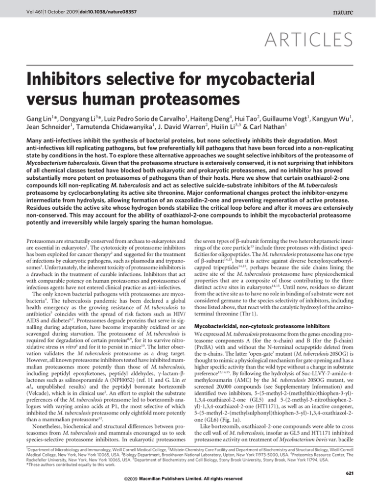
Vol 461 | 1 October 2009 | doi:10.1038/nature08357
ARTICLES
Inhibitors selective for mycobacterial
versus human proteasomes
Gang Lin1*, Dongyang Li3*, Luiz Pedro Sorio de Carvalho1, Haiteng Deng4, Hui Tao2, Guillaume Vogt1, Kangyun Wu1,
Jean Schneider1, Tamutenda Chidawanyika1, J. David Warren2, Huilin Li3,5 & Carl Nathan1
Many anti-infectives inhibit the synthesis of bacterial proteins, but none selectively inhibits their degradation. Most
anti-infectives kill replicating pathogens, but few preferentially kill pathogens that have been forced into a non-replicating
state by conditions in the host. To explore these alternative approaches we sought selective inhibitors of the proteasome of
Mycobacterium tuberculosis. Given that the proteasome structure is extensively conserved, it is not surprising that inhibitors
of all chemical classes tested have blocked both eukaryotic and prokaryotic proteasomes, and no inhibitor has proved
substantially more potent on proteasomes of pathogens than of their hosts. Here we show that certain oxathiazol-2-one
compounds kill non-replicating M. tuberculosis and act as selective suicide-substrate inhibitors of the M. tuberculosis
proteasome by cyclocarbonylating its active site threonine. Major conformational changes protect the inhibitor-enzyme
intermediate from hydrolysis, allowing formation of an oxazolidin-2-one and preventing regeneration of active protease.
Residues outside the active site whose hydrogen bonds stabilize the critical loop before and after it moves are extensively
non-conserved. This may account for the ability of oxathiazol-2-one compounds to inhibit the mycobacterial proteasome
potently and irreversibly while largely sparing the human homologue.
Proteasomes are structurally conserved from archaea to eukaryotes and
are essential in eukaryotes1. The cytotoxicity of proteasome inhibitors
has been exploited for cancer therapy2 and suggested for the treatment
of infections by eukaryotic pathogens, such as plasmodia and trypanosomes3. Unfortunately, the inherent toxicity of proteasome inhibitors is
a drawback in the treatment of curable infections. Inhibitors that act
with comparable potency on human proteasomes and proteasomes of
infectious agents have not entered clinical practice as anti-infectives.
The only known bacterial pathogens with proteasomes are mycobacteria4. The tuberculosis pandemic has been declared a global
health emergency as the growing resistance of M. tuberculosis to
antibiotics5 coincides with the spread of risk factors such as HIV/
AIDS and diabetes6,7. Proteasomes degrade proteins that serve in signalling during adaptation, have become irreparably oxidized or are
scavenged during starvation. The proteasome of M. tuberculosis is
required for degradation of certain proteins8,9, for it to survive nitroxidative stress in vitro4 and for it to persist in mice10. The latter observation validates the M. tuberculosis proteasome as a drug target.
However, all known proteasome inhibitors tested have inhibited mammalian proteasomes more potently than those of M. tuberculosis,
including peptidyl epoxyketones, peptidyl aldehydes, c-lactam-blactones such as salinosporamide A (NPI0052) (ref. 11 and G. Lin et
al., unpublished results) and the peptidyl boronate bortezomib
(Velcade), which is in clinical use2. An effort to exploit the substrate
preferences of the M. tuberculosis proteasome led to bortezomib analogues with varying amino acids at P1, the most selective of which
inhibited the M. tuberculosis proteasome only eightfold more potently
than a mammalian proteasome12.
Nonetheless, biochemical and structural differences between proteasomes from M. tuberculosis and mammals encouraged us to seek
species-selective proteasome inhibitors. In eukaryotic proteasomes
the seven types of b-subunit forming the two heteroheptameric inner
rings of the core particle13 include three proteases with distinct specificities for oligopeptides. The M. tuberculosis proteasome has one type
of b-subunit14,15, but it is active against diverse benzyloxycarbonylcapped tripeptides14,15, perhaps because the side chains lining the
active site of the M. tuberculosis proteasome have physicochemical
properties that are a composite of those contributing to the three
distinct active sites in eukaryotes14,15. Until now, residues so distant
from the active site as to have no role in binding of substrate were not
considered germane to the species selectivity of inhibitors, including
those listed above, that react with the catalytic hydroxyl of the aminoterminal threonine (Thr 1).
Mycobactericidal, non-cytotoxic proteasome inhibitors
We expressed M. tuberculosis proteasome from the genes encoding proteasome components A (for the a-chain) and B (for the b-chain)
(PrcBA) with and without the N-terminal octapeptide deleted from
the a-chains. The latter ‘open-gate’ mutant (M. tuberculosis 20SOG) is
thought to mimic a physiological mechanism for gate opening and has a
higher specific activity than the wild type without a change in substrate
preference12,14,15. By following the hydrolysis of Suc-LLVY-7-amido-4methylcoumarin (AMC) by the M. tuberculosis 20SOG mutant, we
screened 20,000 compounds (see Supplementary Information) and
identified two inhibitors, 5-(5-methyl-2-(methylthio)thiophen-3-yl)1,3,4-oxathiazol-2-one (GL5) and 5-(2-methyl-3-nitrothiophen-2yl)-1,3,4-oxathiazol-2-one (HT1171), as well as an inactive congener,
5-(5-methyl-2-(methylsulphonyl)thiophen-3-yl)-1,3,4-oxathiazol-2one (GL6) (Fig. 1a).
Like bortezomib, oxathiazol-2-one compounds were able to cross
the cell wall of M. tuberculosis, insofar as GL5 and HT1171 inhibited
proteasome activity on treatment of Mycobacterium bovis var. bacille
1
Department of Microbiology and Immunology, Weill Cornell Medical College, 2Milstein Chemistry Core Facility and Department of Biochemistry and Structural Biology, Weill Cornell
Medical College, New York, New York 10065, USA. 3Biology Department, Brookhaven National Laboratory, Upton, New York 11973-5000, USA. 4Proteomics Resource Center, The
Rockefeller University, New York, New York 10065, USA. 5Department of Biochemistry and Cell Biology, Stony Brook University, Stony Brook, New York 11794, USA.
*These authors contributed equally to this work.
621
©2009 Macmillan Publishers Limited. All rights reserved
ARTICLES
NATURE | Vol 461 | 1 October 2009
R
O2N
N S
O
b
HT1171
S
O
N S
S
GL6
c
GL5
d
SO
5
µM
10
µ
25 M
µM
50
µM
U
nt
re
at
DM ed
S
0 O
m
15 in
m
30 in
m
45 in
m
60 in
m
in
DM
DM
SO
G
H L5
Bo T1
rte 171
zo
m
ib
Proteasome
activity (%)
100
80
60
40
20
0
O
N S
O S
O
N S
S
1,3,4-oxathiazol-2-one
S
O
f
2
12.5 µM
25 µM
50 µM
µ
10 M
0
µM
3
µM
4
75
5
HT1171
140
120
100
80
60
40
20
0
µM
6
Viability (%)
log10 [c.f.u. ml–1]
7
50
GL5
25
Bortezomib
5 0
n
10 M
nM
e
Figure 1 | Oxathiazol-2-one compounds inhibit mycobacterial
proteasomes and kill non-replicating M. tuberculosis. a, Compound
structures. b, Inhibition of proteasomes in intact BCG (A580 0.6–1) by GL5,
HT1171 or bortezomib (each 50 mM) after 4 h. Proteasome activity in lysates
was tested with Ac-YQW-AMC (50 mM) as substrate.
c, Concentration–response for GL5 after 1 h exposure as in b. Control,
DMSO (,1% v/v) alone. d, Time course for effect of GL5 (50 mM) as in
b, except that washing began at the times indicated. ‘Untreated’ and ‘DMSO’
cells were handled in the same manner but received nothing or DMSO at
time 0 and were lysed at 60 min. ‘T0’ cells were treated and washed
immediately. e, Killing of M. tuberculosis Erdman in Sauton’s medium with
time 0 addition of 50 mM DETA-NO, a nitric oxide donor (t1/2, ,20 h),
mimicking the nitroxidative stress that limits the replication of
M. tuberculosis in mice22. Upper dashed line, c.f.u. ml21 after exposure to
DMSO control. Lower dashed line, limit of detection. Arrow, initial
inoculum. f, Monkey kidney epithelial cells (Vero76) were incubated with
compounds for 4 days before 3-(4,5-dimethylthiazol-2-yl)-5-(3carboxymethoxyphenyl)-2-(4-sulfophenyl)-2H-tetrazolium (MTS) assay
for viability; microscopy gave concordant results. Data are means 6 s.d. of
triplicates in one of at least two experiments. Some error bars fall within the
symbols.
Calmette–Guérin (BCG; Fig. 1b). At 50 mM, GL5 and HT1171 inhibited
,90% of mycobacterial proteasome activity, whereas bortezomib
(50 mM) inhibited 52%. Exposure of BCG to 25 mM GL5 for 4 h led
to .80% reduction of proteasome activity (Fig. 1c), whereas as little as
,15 min exposure to GL5 at 50 mM was sufficient to reduce activity by
,50% (Fig. 1d).
Moreover, GL5 killed BCG alone and in synergy with sub-bacteriostatic fluxes of nitric oxide arising from the decomposition of
2,2-(hydroxynitrosohydrazino)-bis-ethanamine (DETA-NO) (Supplementary Fig. 1). GL5 and HT1171 also dose-dependently killed
1.5–2.5 log10 M. tuberculosis over 4 days in synergy with sufficient
nitric oxide to induce a pathophysiologically relevant state of bacterial non-replication16 (Fig. 1e). Bortezomib was less mycobactericidal (Fig. 1e), and it was toxic to monkey epithelial cells (Fig. 1f) and
human macrophages (Supplementary Fig. 2). In contrast, GL5 and
HT1171 showed no apparent toxicity to mammalian cells (Fig. 1f and
Supplementary Fig. 2) at concentrations up to 75 mM, 3,000-fold
greater than those at which bortezomib killed the epithelial cells.
The oxathiazol-2-one compounds exerted no antibacterial activity
against Mycobacterium avium intracellulare, Staphylococcus aureus,
Salmonella enterica var. Typhimurium or Pseudomonas aeruginosa
(data not shown). Although some oxathiazol-2-one compounds
were reported to react with thiols17, those studied here did not inhibit
the thiol-dependent cathepsin B (data not shown). Moreover, 11 of
23 oxathiazol-2-one compounds tested were ,5% reactive with glutathione; the others reacted to a limited degree (Supplementary Table
1). Thus, at a functional level, the oxathiazol-2-one compounds
tested here seem to be relatively selective and nontoxic, although they
may have additional targets.
Selective inhibition of mycobacterial proteasomes
The different impact of oxathiazol-2-one compounds on
M. tuberculosis and mammalian cells prompted us to ask if these
compounds differentially inhibit isolated mycobacterial and human
proteasomes. In dialysis (Fig. 2a) and kinetic studies (Fig. 2b, c),
oxathiazol-2-one compounds inhibited M. tuberculosis proteasomes
irreversibly (Fig. 2a), whereas inhibition of human proteasome b1,
b2 and b5 sites was so minimal (Fig. 2b, c and Supplementary Table
1) as to preclude definition of a mode of inhibition. After establishing
that oxathiazol-2-one compounds spontaneously hydrolyse in tissue
culture medium to the corresponding amide with a half time (t1/2)
ranging from 7 to 180 min (Supplementary Table 2), we used partition
ratios (the ratios of rate constants for catalysis and inactivation) to
assess their relative potency18. By this measure, GL5 and HT1171 were
.1,000-fold more effective against M. tuberculosis proteasomes than
human proteasomes (Fig. 2d and Supplementary Fig. 3a, b). Inhibition was competitive with the substrate benzyloxycarbonyl-valylleucyl-arginyl-AMC (Z-VLR-AMC) (Supplementary Fig. 3c, d), indicating that the inhibitor binds at or near the active site, and was timedependent (Supplementary Fig. 4). Potencies of all 21 oxathiazol-2-one
compounds tested against wild-type M. tuberculosis proteasome correlated closely (r2 5 0.82) with their potencies against the open-gate
form (Supplementary Fig. 5). Oxathiazol-2-one-treated M. tuberculosis
proteasomes lost the ability to degrade not only oligopeptides, but
also a protein substrate, b-casein (Supplementary Fig. 6). In contrast,
oxathiazol-2-one compounds were inactive or very weak inhibitors of
trypsin (half-maximal inhibitory concentration (IC50) . 50 mM), cathepsin B (IC50 . 50 mM), matrix metalloproteinase-2 (IC50 . 100 mM)
a
b
100
kobs/l × 103 s–1
O
O
Activity recovered (%)
O
O
75
50
25
0
D
N MS
PI O
-0
05
Bo
2
rte GL
zo 5
m
ib
a
d
c
3.0
2.5
2.0
20SOG
Hu20S β5
1.5
1.0
0.5
0
0
25 50 75
GL5 (µM)
kobs/l M–1 s–1
Compound 20SOG
0
10 20 30
HT1171 (µM)
Partition ratio
Hu20S
β5
β2
β1
20SOG Hu20S
GL5
376.4
0.4
NI
0.03
11.5
16,170
HT1171
2,134
10.1
6.9
2.8
8.9
8,963
Figure 2 | Kinetic analysis of inactivation of M. tuberculosis 20SOG and
human proteasomes (Hu20S) by oxathiazol-2-one compounds.
a, M. tuberculosis 20SOG (0.23 nM) was pre-treated with DMSO, GL5
(5 mM), salinosporamide A (NPI-0052, 10 nM) or bortezomib (2.7 mM) for
3 h and assayed, then dialysed against assay buffer overnight and assayed
again. b, c, Plots of kobs as function of inhibitor concentration for GL5 (b) and
HT1171 (c). Values for kobs, derived from the data in Supplementary Fig. 4,
were plotted against inhibitor concentration I. d, Kinetic parameters and
partition ratios of GL5 and HT1171, determined as in Supplementary Figs 3
and 4. Kobs/I is second-order rate constant of inactivation. Partition ratios
for human proteasomes refer to the b5 subunit. Standard errors were ,10%.
NI, no inhibition.
622
©2009 Macmillan Publishers Limited. All rights reserved
ARTICLES
NATURE | Vol 461 | 1 October 2009
HT1171 increased the mass of the same peptide by only 68.06 Da
(Fig. 3a, (2)R(4) and Supplementary Fig. 7d), indicating that one
primary amine was no longer available for reductive alkylation. MS/
MS analysis (Supplementary Fig. 7d) of this mono-alkylated peptide
showed that only the lysine was modified, confirming that the
N-terminal Thr was modified on HT1171 treatment. The mass spectrometric results were the same when HT1171 was replaced by GL5 or
GL3 and when the proteasome was wild type or open-gate (not
shown). These findings support the mechanism of inhibition shown
in Fig. 3b: the attack of the oxathiazol-2-one by the OH of Thr 1 forms
a carbonated or carbonothioated enzyme intermediate on Thr 1 that
can either undergo hydrolysis to reactivate the enzyme, or donate a
carbonyl to Thr 1 that links its a-amino and c-hydroxyl groups to
form an oxazolidin-2-one, a chemically stable moiety20, consonant
with the ability of oxathiazol-2-one compounds to cyclocarbonylate
1,2-aminoalcohols21.
and mycobacterial blaC-encoded b-lactamase, a serine proteaselike enzyme (IC50 . 100 mM). Although GL5 reversibly inhibited
a-chymotrypsin with Ki 64 nM, antichymotryptic potency was minimal
in 13 other oxathiazol-2-one compounds tested (Supplementary Table
4). Supplementary Table 1 summarizes biochemical and mycobactericidal properties of 24 oxathiazol-2-one compounds, including 21 we
synthesized as described in the Supplementary Information.
Competitive, irreversible, mechanism-based inhibition indicated
that the oxathiazol-2-one compounds inactivate the M. tuberculosis
proteasome by covalent attack on the active site Thr 1. To test this, we
trypsinized M. tuberculosis proteasomes that had been treated or not
with HT1171. Results were identical for both wild-type and opengate forms. Liquid chromatography–tandem mass spectrometry
(LC–MS/MS) identified peptides representing 98% of the b-subunit.
Only one peptide ion was unique to the M. tuberculosis proteasome
b-subunit treated with HT1171 (Fig. 3a). Its mass was 26 Da higher
than that of the N-terminal heptapeptide (TTIVALK) identified only
in the untreated samples (Fig. 3a and Supplementary Fig. 7a, b), indicating the addition of a carbonyl at the expense of two hydrogen atoms
((1)R(2) in Fig. 3a). MS/MS analysis of this modified peptide
(Supplementary Fig. 7a) indicated that the modification was on
one of the first two residues. To determine whether the N-terminal
residue was modified, the tryptic peptides from untreated and treated
M. tuberculosis proteasome b-subunits were subjected to reductive
glutaraldehydation, which modifies amino groups of proteins
and peptides19. Subjecting the trypsinized oligopeptides to reductive
glutaraldehydation without prior exposure to oxathiazol-2-one
increased the mass of the N-terminal heptapeptide by 136.13 Da, consistent with modification of two primary amines, those of Thr 1 and
Lys 7 (Fig. 3a, (1)R(3) and Supplementary Fig. 7c). In contrast, applying the same procedure to proteasomes that had been pre-treated with
a
Structural basis of species selectivity
To determine the basis for species selectivity, we solved four crystal
structures: wild-type M. tuberculosis proteasome following exposure to
GL1 at 2.4 Å resolution and to HT1171 at 2.5 Å resolution, and the
open-gate variant (20SOG) alone at 2.5 Å resolution or following
exposure to HT1171 at 2.9 Å resolution. N-terminal octapeptide deletion in the a-subunit of 20SOG did not alter the overall structure of the
M. tuberculosis proteasome (Supplementary Fig. 8a). Furthermore,
wild type and open-gate proteasomes underwent the same conformational changes (described later) on inhibitor treatment (Supplementary Fig. 8b). The three structures (Fig. 4, Supplementary Figs 8, 9 and
Supplementary Table 5) each confirmed that oxathiazol-2-one compounds cyclocarbonylate Thr 1. Not only the oxazolidin-2-one ring,
but also its protruding methyl group and carbonyl oxygen, were
NH2
NH2
745.49
771.47
746.49
HO
740 750 760 770 780
m/z
H2N
OH
IVAL
O
772.47
O
HT1171
Mass shift: 26 Da
O
H
N
N
H
O
OH
(1)
773.47
740 750 760 770 780
m/z
IVAL
O
O
OH
O
H
N
O
(2)
Mass shift: Glutaraldehyde
68 Da
Na(CN)BH3
Mass shift: Glutaraldehyde
136 Da
Na(CN)BH3
N
N
839.52
881.61
HO
882.61
H
N
N
883.61
O
O
760 800 840 880
m/z
H
N
O
OH
IVAL
O
N
H
O
OH
(3)
b
840.53
OH
IVAL
O
O
OH
O
760
841.53
840
800
m/z
(4)
–
O
O
H
H O
R
OH
N S
O
CO2 + R
+S
H
NH2
H
O
O
R
R
O
N
O
c
a
O
S
H
Ö
H
H
O
Ö
R
PROT
H2Ö
N
O
O–
b
O
d
N
HS H2N
PROT
+ R
N
H
SH
+ CO2
O
c
PROT
O
PROT
H2N
O
O
b
N S
+
H3N
O
HO
c
a
OH
R
N S
O
H
c
O
O
R
H
O
d
d
PROT
H2Ö
N
O
O
O
O
Figure 3 | LC–MS/MS identification of the modified N terminus of the
M. tuberculosis proteasome treated with oxathiazol-2-one compounds.
a, Mass spectra of tryptic N-terminal heptapeptides from M. tuberculosis
proteasomes that were untreated (1), HT1171-treated (2), treated with
glutaraldehyde/Na(CN)BH3 after trypsin digestion (3), or treated with
glutaraldehyde/Na(CN)BH3 after HT1171 treatment and trypsin digestion
(4). All ions were confirmed by MS/MS analysis (Supplementary Fig. 7a–d).
Reaction equations illustrate proposed modification of active site Thr 1 by
NH2
d
N
H
PROT
+ R
N
H
SH
+ CO2
O
oxathiazol-2-one ((1)R(2)), and modification of primary amino groups at
Thr 1 and Lys 7 with glutaraldehyde and Na(CN)BH3 ((1)R(3) and
(2)R(4)). b, Proposed mechanism of proteasome inactivation by
oxathiazol-2-one. Paths marked by a, b and d lead to irreversible inhibition.
In paths marked by c, hydrolysis of the inhibitor-enzyme intermediate
allows the proteasome to degrade the oxathiazol-2-one without losing
activity.
623
©2009 Macmillan Publishers Limited. All rights reserved
ARTICLES
NATURE | Vol 461 | 1 October 2009
a
c
H2
S4–H1 loop
L99
I3
OXZ
L101
T48
S5 strand
A46
S4–H1 loop
D124
T2
S4 strand
V4
M. tuberculosis β
Human β2
L99- A100- L101
G95 - A96 - A97
T48 - G47 - A46
T48 - G47 - A46
M95 - A96 - G97
S96 - M97 - G98
S48 - G47 - S46
G48 - G47 - A46
Human β1
Human β5
S1 strand
OXZ
Neighbour β-subunit
b
d
H2
H1
E54
S4–H1 loop Neighbour β Bottom
S5
H1
S4
W129
8º
OXZ
S1
T48
S6′
D124
M. tuberculosis β T48A49E54
Human
β1 S48A49A54
β 2 T48A49M54
Human
Human
β5 G48A49F54
S4–H1 loop
A49
D124W129
Y114T119
D125P130
D125Y130
S20
T20
A20
A20
OXZ
S7′
S20
M. tuberculosis 20S
β-subunit
M. tuberculosis 20S
β-HT1171
Figure 4 | Crystal structure of the full-length M. tuberculosis 20S
proteasome after exposure to HT1171 shows cyclocarbonylation of active
site Thr 1 and conformational changes in the b-subunit. a, 2Fo2Fc electron
density map contoured at 1.2s and superimposed on the crystal structure of
HT1171-treated proteasome at the active site in the b-subunit. Map was
calculated by omitting oxazolidin-2-one and the N-terminal four amino
acids from the crystal structure. b, Superposition of HT1171-modified
b-subunit in green with native b-subunit in grey (Protein Data Bank 2FHG).
OXZ labels oxazolidin-2-one ring on Thr 1. The conformational changes can
be approximately described by an 8u tilt of H1 (dashed line) and a downward
shift of the S4–H1 loop (red arrow). c, Active site structure of HT1171modified b-subunit in green, in comparison with native b-subunit structure
in grey. In the native structure, Ala 46-Gly 47-Thr 48 is part of the S4 strand
forming a b-sheet with the S5 strand. In the HT1171-treated structure,
Ala 46-Gly 47-Thr 48 loses contacts with S5 and converts to the S4–H1 loop.
Panel at right specifies amino acid pairs that form S4–S5 b-sheet interaction
in this region in M. tuberculosis b-chain before oxathiazol-2-one treatment
and compares these with corresponding sequences in the proteolytically
active human proteasome b-chains. d, Active site structure of HT1171treated M. tuberculosis 20S oriented to view hydrogen bonds stabilizing the
new position of S4–H1 loop in the M. tuberculosis b-chain after oxathiazol-2one treatment. S4–H1 loop as upper surface of the constricted substratebinding pocket is stabilized by a hydrogen bond between Glu 54 and Trp 129
of the neighbouring b-subunit and by three pairs of water-mediated
hydrogen bonds with the neighbouring b-subunit and with Ser 20 at the
lower substrate-binding surface of the same subunit. Panel at right compares
the hydrogen bonding of the residues of M. tuberculosis with human
counterparts. The 2Fo2Fc electron density map contoured at 1.2s is
superimposed at the oxazolidin-2-one ring site in c and d. Thr 1 modification
and b-subunit conformational changes on exposure to GL1 or HT1171 in
the wild-type and the open-gate M. tuberculosis 20S are virtually the same.
resolved in the electron density (Fig. 4). The oxazolidin-2-one ring is
stabilized by a hydrogen-bond network involving Ala 180, Ser 141 and
Asn 24 of the neighbouring b-subunit, and a water molecule in the
substrate cavity (Supplementary Fig. 9). Use of HT1171 and GL1 in the
crystallographic studies and HT1171, GL5 and GL3 in the mass spectroscopic studies brought to four the number of oxathiazol-2-one
compounds for which the same suicide-substrate inhibition mechanism was confirmed.
Surprisingly, the substrate-binding pocket of the M. tuberculosis
proteasome underwent a major conformational change on cyclocarbonylation of Thr 1 by HT1171 or GL1. Such a change is unprecedented among the dozens of crystal structures of proteasomes in
complex with inhibitors11. An ,8u downward tilt of the H1 helix in
the b-subunit moved the N-terminal end of H1 (Ala 49–Phe 55) downward by as much as 4.2 Å (Fig. 4b). The H1 shift brought with it a
3-amino-acid stretch (Ala 46–Thr 48) of the S4 b-strand, converting
this segment into a short loop (S4–H1). As a consequence, another
short loop (Met 95-Gln 96-Gly 97) between H2 and S5 lost stabilizing
contacts with the shifted components and became disordered, as illustrated by a black dashed curve in Fig. 4b. The S4–H1 loop region
comprises the upper surface of the substrate-binding pocket11,14. Its
downward shift constricted the pocket to the point that it could not
accommodate a peptide substrate, affording an additional mechanism
of inhibition over and above incorporation of the active site hydroxyl
into an oxazolidin-2-one.
In the native M. tuberculosis b-subunit, the three S4–H1 loop
amino acids Ala 46, Gly 47 and Thr 48 are in b-strand configuration
and form a b-sheet interaction with Leu 101, Ala 100 and Leu 99,
respectively, in the S5 b-strand (Fig. 4c). None of these residue pairs
is conserved in the human proteasome b5 subunit and only one of
them (Gly 47–Ala 96) in the human b1 and b2 subunits. One direct
hydrogen bond and three pairs of water-mediated hydrogen bonds
624
©2009 Macmillan Publishers Limited. All rights reserved
ARTICLES
NATURE | Vol 461 | 1 October 2009
stabilized the new position of the S4–H1 loop (Fig. 4d): between
Glu 54 and Trp 129 of the neighbouring b-subunit (3.0 Å); between
Thr 48, a water molecule and Asp 124 in the S6–S7 loop of the neighbouring b-subunit (2.8 and 3.1 Å, respectively); between Ala 50, a
water molecule and the carbonyl O of Trp 129 in the short S7
b-strand of the neighbouring b-subunit (3.0 and 3.1 Å, respectively);
and between Ala 49, a water molecule and Ser 20 in the same bsubunit (3.2 and 3.1 Å, respectively). This last pair of hydrogen bonds
crosslinked the upper and the lower substrate-binding surfaces, sealing the entrance of the substrate pocket. The residue pairs involved in
the post-shift hydrogen bonds are not well conserved in the human
proteasome (Fig. 4d). We speculate that the upper substrate-binding
surfaces (S4–H1 loops) in the three catalytic b-subunits of the human
proteasome might have difficulty breaking off from their corresponding b-sheet cores on initial modification of Thr 1 by oxathiazol2-one compounds.
Thus, selectivity of oxathiazol-2-one compounds seems to be
imparted in three ways: by the presence of a 1,2-aminoalcohol at the
active site of the target, which, among enzymes, is likely to be limited
to the N-terminal Thr hydrolase family; by the ability of the inhibitor
to bind rapidly (before its spontaneous decay) and precisely adjacent
to the 1,2-aminoalcohol; and by the degree to which the amino group
of the 1,2-aminoalcohol has better access to the inhibitor’s carbonyl
(now attached to the alcohol) than water has. The protein landscape
near the 1,2-aminoalcohol can thus determine species selectivity, both
in how it binds the R group on the oxathiazol-2-one and in the conformations it adopts, that may either permit or limit access of water to
the intermediate formed during reaction of the oxathiazol-2-one with
the active site. Detailed understanding of the sequence of steps by
which oxathiazol-2-one compounds cause a major conformational
shift in the substrate-binding domain could guide the design of the
next generation of inhibitors selective for the M. tuberculosis proteasome over the human proteasome.
Discussion
Non-replicating M. tuberculosis displays ‘phenotypic tolerance’, that
is, relative resistance to conventional anti-infectives, imposing the
need to treat tuberculosis longer than almost any other infectious
disease. Prolonged treatment leads to interruption of therapy and
emergence of hereditable drug resistance. Hence agents are needed
that can kill M. tuberculosis when its replication is halted by conditions encountered in the host, such as those imposed by inducible
nitric oxide synthase22. Oxathiazol-2-one compounds identified here
phenocopy genetic deletion of the M. tuberculosis proteasome10 by
killing M. tuberculosis rendered non-replicative by exposure to sublethal nitric oxide. Along with thioxothiazolidines that inhibit the
dihydrolipoamide acyltransferase of M. tuberculosis16, oxathiazol-2one compounds are only the second class of compounds, to our
knowledge, that are selectively bactericidal for a non-replicating
pathogen.
Certain oxathiazol-2-one compounds are selective in other ways as
well. They inhibit the M. tuberculosis proteasome, but not the human
proteasome N-terminal threonine b1, b2 or b5 proteases, cysteine
proteases, serine proteases, or metalloproteases. They do not kill
monkey epithelial cells, human macrophages or any bacteria we
tested other than mycobacteria. The oxathiazol-2-one compounds
react with proteasomes at their active site; however, residues distant
from the active site, with which oxathiazol-2-one compounds do not
interact, seem to impart species selectivity.
Inhibition of protein synthesis at the stages of transcription (for
example, by rifamycins) or translation (for example, by aminoglycosides
and capreomycin) are among the best validated antibiotic strategies23. It
may prove synergistic to interfere at the same time with bacterial protein
degradation. The ability of a brief exposure to oxathiazol-2-one compounds to inhibit M. tuberculosis proteasomes permanently is a potential advantage. M. tuberculosis may have difficulty replacing irreversibly
inactivated proteasomes, not only when the protein synthesis of
M. tuberculosis is markedly diminished in the non-replicative state24,
but also when its protein synthesis is impaired by antibiotics.
Much of biology can be viewed as the interplay of two principles:
conservation and diversity. The conservation of proteasomes across
vast evolutionary distances is striking. The present work demonstrates that functionally exploitable diversity exists between the proteasomes of M. tuberculosis and its obligate human host.
METHODS SUMMARY
High-throughput screening was performed in 384-well plates with 33 mM test
compounds. To test inhibition of proteasomes in intact mycobacteria, treated
cells were centrifuged, washed twice and lysed mechanically, and aliquots (10 mg
protein) of the supernatants assayed with Suc-LLVY-AMC (50 mM). Killing of
DETANO-treated, non-replicating M. tuberculosis Erdman was tested in
Sauton’s medium, pH 7.4. After 4 days, samples were plated for enumeration
of colony-forming units (c.f.u.) 3 weeks later. Monkey kidney cells were cultured
in Dulbecco’s Eagle’s medium with 10% fetal bovine serum for cytotoxicity
assessment after 48 h by tetrazolium reduction and microscopy. Recombinant
M. tuberculosis proteasomes were purified as reported15. Human proteasomes
were activated with PA28 (Boston Biochem). Kinetic studies were conducted in a
Molecular Devices fluorescent plate reader with AMC-derivatized peptide substrates according to which proteasomal protease was being studied
(M. tuberculosis or human b1, b2 or b5). Values of kobs were derived from the
fit of data to equation (1) (ref. 25) in Prism (GraphPad Software)
P~vs tz
(v0 {vs )
½1{e ({kobs t) kobs
ð1Þ
where P 5 product, vs 5 steady-state (final) velocity, t 5 time, v0 5 initial velocity
and kobs 5 apparent first-order rate constant for interconversion between v0 and vs.
LC–MS/MS analysis used Thermo LTQ Orbitrap and Applied Biosystems
QSTAR mass spectrometers. Matrix-assisted laser desorption/ionization–time
of flight (MALDI–TOF) analysis was performed on a PerSeptive DE-STR instrument. Crystallization of the M. tuberculosis proteasome, diffraction data collection and structure solution and refinement are included along with other details
in the Methods.
Full Methods and any associated references are available in the online version of
the paper at www.nature.com/nature.
Received 9 June; accepted 4 August 2009.
Published online 16 September 2009.
1.
2.
3.
4.
5.
6.
7.
8.
9.
10.
11.
12.
13.
14.
15.
16.
Baumeister, W., Walz, J., Zuhl, F. & Seemuller, E. The proteasome: paradigm of a
self-compartmentalizing protease. Cell 92, 367–380 (1998).
Kropff, M. et al. Bortezomib in combination with intermediate-dose
dexamethasone and continuous low-dose oral cyclophosphamide for relapsed
multiple myeloma. Br. J. Haematol. 138, 330–337 (2007).
Glenn, R. J. et al. Trypanocidal effect of a9, b9-epoxyketones indicates that
trypanosomes are particularly sensitive to inhibitors of proteasome trypsin-like
activity. Int. J. Antimicrob. Agents 24, 286–289 (2004).
Darwin, K. H. et al. The proteasome of Mycobacterium tuberculosis is required for
resistance to nitric oxide. Science 302, 1963–1966 (2003).
Raviglione, M. C. & Smith, I. M. XDR tuberculosis–implications for global public
health. N. Engl. J. Med. 356, 656–659 (2007).
Corbett, E. L. et al. The growing burden of tuberculosis: global trends and
interactions with the HIV epidemic. Arch. Intern. Med. 163, 1009–1021 (2003).
Restrepo, B. I. Convergence of the tuberculosis and diabetes epidemics: renewal
of old acquaintances. Clin. Infect. Dis. 45, 436–438 (2007).
Pearce, M. J. et al. Ubiquitin-like protein involved in the proteasome pathway of
Mycobacterium tuberculosis. Science 322, 1104–1107 (2008).
Burns, K. E. et al. Proteasomal protein degradation in mycobacteria is dependent
upon a prokaryotic ubiquitin-like protein. J. Biol. Chem. 284, 3069–3075 (2009).
Gandotra, S. et al. In vivo gene silencing identifies the Mycobacterium tuberculosis
proteasome as essential for the bacteria to persist in mice. Nature Med. 13,
1515–1520 (2007).
Borissenko, L. & Groll, M. 20S proteasome and its inhibitors: crystallographic
knowledge for drug development. Chem. Rev. 107, 687–717 (2007).
Lin, G. et al. Distinct specificities of Mycobacterium tuberculosis and mammalian
proteasomes for N-acetyl tripeptide substrates. J. Biol. Chem. 283, 34423–34431
(2008).
Kisselev, A. F. & Goldberg, A. L. Proteasome inhibitors: from research tools to drug
candidates. Chem. Biol. 8, 739–758 (2001).
Hu, G. et al. Structure of the Mycobacterium tuberculosis proteasome and mechanism
of inhibition by a peptidyl boronate. Mol. Microbiol. 59, 1417–1428 (2006).
Lin, G. et al. Mycobacterium tuberculosis prcBA genes encode a gated proteasome
with broad oligopeptide specificity. Mol. Microbiol. 59, 1405–1416 (2006).
Bryk, R. et al. Selective killing of nonreplicating mycobacteria. Cell Host Microbe 3,
137–145 (2008).
625
©2009 Macmillan Publishers Limited. All rights reserved
ARTICLES
NATURE | Vol 461 | 1 October 2009
17. Huth, J. R. et al. Toxicological evaluation of thiol-reactive compounds identified
using a La assay to detect reactive molecules by nuclear magnetic resonance.
Chem. Res. Toxicol. 20, 1752–1759 (2007).
18. Copp, L. J. in Enzyme Kinetics: A Modern Approach (ed. Marangoni, A. G.) Ch. 13
158–173 (Wiley, 2003).
19. Russo, A., Chandramouli, N., Zhang, L. & Deng, H. T. Reductive glutaraldehydation
of amine groups for identification of protein N-termini. J. Proteome Res. 7,
4178–4182 (2008).
20. Lu, L. Q. et al. A new entry to cascade organocatalysis: reactions of stable sulfur
ylides and nitroolefins sequentially catalyzed by thiourea and DMAP. J. Am. Chem.
Soc. 130, 6946–6948 (2008).
21. Rajca, A., Grobelny, D., Witek, S. & Zbirovsky, M. 5-Aryl-2-oxo-1,2,4-oxathiazoles
as cyclocarbonylating agents for 2-aminoalcohols and 1,2-diamines. Synthesis 12,
1032–1033 (1983).
22. MacMicking, J. D. et al. Identification of nitric oxide synthase as a protective locus
against tuberculosis. Proc. Natl Acad. Sci. USA 94, 5243–5248 (1997).
23. Walsh, C. Where will new antibiotics come from? Nature Rev. Microbiol. 1, 65–70
(2003).
24. Hu, Y. M. et al. Protein synthesis is shutdown in dormant Mycobacterium
tuberculosis and is reversed by oxygen or heat shock. FEMS Microbiol. Lett. 158,
139–145 (1998).
25. Copeland, R. A. in Enzymes: a Practical Introduction to Structure, Mechanism, and
Data Analysis Chs 9–10, 305–349 (Wiley, 2000).
26. Otwinoswki, Z. & Minor, W. in Methods in Enzymology (eds Carter, C. W. Jr &
Sweet, R. M.) 307–326 (Academic, 1997).
27. McCoy, A. J. et al. Phaser crystallographic software. J. Appl. Cryst. 40, 658–674
(2007).
28. Brünger, A. T. et al. Crystallography & NMR system: a new software suite for
macromolecular structure determination. Acta Crystallogr. D 54, 905–921 (1998).
29. Emsley, P. & Cowtan, K. Coot: model-building tools for molecular graphics. Acta
Crystallogr. D 60, 2126–2132 (2004).
30. Adams, P. D. et al. PHENIX: building new software for automated crystallographic
structure determination. Acta Crystallogr. D 58, 1948–1954 (2002).
Supplementary Information is linked to the online version of the paper at
www.nature.com/nature.
Acknowledgements S. Eswaramoorthy helped with crystallography software,
C. Lipinski, C. Walsh and M. Fischbach proposed reaction mechanisms, and
C. Karan assisted with screening. S. Ehrt and S. Gandotra performed some
bactericidal assays, C. Tsu and L. Dick donated a fluorimeter and J. Blanchard
provided BlaC. Supported by NIH PO1-AI056293, NIH R01AI070285 and the
Milstein Program in Chemical Biology of Infectious Diseases. X-ray diffraction data
were collected at beamline X6A, X25 and X29 in the National Synchrotron Light
Source, a facility supported by the US DOE and NIH. The Department of
Microbiology and Immunology is supported by the William Randolph Hearst
Foundation.
Author Contributions G.L. purified recombinant proteasome, conducted the
screen, designed new oxathiazol-2-one compounds and performed most of the
assays. D.L. and H.L. purified and crystallized recombinant proteasomes and solved
their structures. L.P.S.C. helped analyse kinetics. H.D. performed mass
spectrometry. H.T. synthesized oxathiazol-2-one compounds under the
supervision of J.D.W.; G.V. studied human macrophages. K.W. conducted studies
with viable M. tuberculosis. J.S. and T.C. performed kinetic, bactericidal and
cytotoxicity experiments. C.N. organized the effort and helped design and interpret
experiments. C.N., G.L. and H.L. wrote the paper.
Author Information Coordinates have been deposited in the Protein Data Bank
under ID codes 3H6F, 3H6I, 3HFA and 3HF9. Reprints and permissions information
is available at www.nature.com/reprints. Correspondence and requests for
materials should be addressed to C.N. (cnathan@med.cornell.edu), G.L.
(gal2005@med.cornell.edu) and H.L. (hli@bnl.gov).
626
©2009 Macmillan Publishers Limited. All rights reserved
doi:10.1038/nature08357
METHODS
High-throughput screen. Compounds from ChemDiv, ChemBridge, Spectrum,
Prestwick Chemicals and Cerep were dissolved in DMSO and robotically
dispensed to Falcon Microtest 384-well plates, with DMSO as vehicle control.
After a 45-min preincubation with M. tuberculosis 20SOG (10 ml) at 37 uC, reaction buffer containing substrate (5 ml) was added. Final concentrations were as
follows: test compounds, 33 mM; Suc-LLVY-AMC, 50 mM; M. tuberculosis
20SOG, 1 nM; HEPES, 20 mM; EDTA, 0.5 mM; pH 7.5. Plates were placed on
an orbital shaker in a humidified incubator at 37 uC for 45 min. Fluorescence was
recorded at excitation 360 nm, emission 460 nm. Z9 values were .0.5. We
screened 20,400 compounds and picked 72 (0.35%) that gave inhibition
$65%. Of these, inhibition was confirmed with 26 compounds; concentrationdependent activity was most potent for HT1171 and GL5. A search of the library
with the core structure (1,3,4-oxathiazol-2-one) identified GL6. GL1 and GL3
were identified in commercial collections using SCIFINDER. GL1, GL3 and GL5
were purchased from TimTec and GL6 was purchased from ChemDiv. HT1171
was resynthesized and the other compounds listed in Supplementary Table 1 were
synthesized at the Abby and Howard P. Milstein Chemistry Core Facility of Weill
Cornell Medical College.
Mycobactericidal activity. Initial experiments showed that GL5 killed M. bovis
var. BCG in synergy with nitric oxide, which was provided from the decomposition of 2,2-(hydroxynitrosohydrazino)-bis-ethanamine (DETA-NO) at 50 mM
(Supplementary Fig. 1). In the experiment illustrated in Supplementary Fig. 1,
DETA-NO alone reduced but did not completely prevent replication of BCG,
whereas in the experiments with M. tuberculosis illustrated in Fig. 1e, DETA-NO
prevented replication without reducing viability as assessed by plating.
M. tuberculosis Erdman was cultivated in Sauton’s medium pH 7.4 with 0.4%
L-asparagine, 0.2% glycerol and 0.02% Tween 80. Mid-log phase cultures (A580
0.8–1.0) were diluted to A580 0.05–0.1 and quantified by c.f.u. Mycobacteria were
incubated with bortezomib, GL5 or HT1171 at indicated concentrations in the
presence of DETA-NO (50 mM) in 96-well plates in 200 ml for 4 days, then serially
diluted in PBS with 0.02% Tween 80, pH 7.2 and plated for c.f.u. determination
on Middlebrook 7H11 agar plates with 10% Middlebrook OADC enrichment
(Fig. 1e).
Impact of oxathiazol-2-one compounds on eukaryotic cells. Vero76 African
green monkey kidney cells (ATCC CRL-1587) were cultured in DMEM containing 4.5 g l21 glucose, 0.584 g l21 L-glutamine, 1 mM pyruvate, 100 IU ml21 penicillin, 100 mg ml21 streptomycin and 10% fetal bovine serum. Fresh medium was
supplied containing test compounds at 25 or 50 mM. At indicated intervals of
1–25 h, this medium was removed and fresh medium added without test compounds. After exposure (48 h) to GL5, HT1171 or bortezomib at indicated concentrations, viability was assessed by the CellTiter96 Aqueous Assay (Promega)
for tetrazolium reduction according to the manufacturer’s protocol. Human
monocyte-derived macrophages were allowed to differentiate in vitro for 2 weeks.
GL5 or bortezomib were added in DMSO at indicated concentrations (,1%).
Photomicrographs were taken 1 week later.
Titration of M. tuberculosis and human proteasome by GL5 or HT1171. GL5
or HT1171 at concentration 0.2–50 mM were incubated with 3 nM
M. tuberculosis 20SOG or 0.5 nM Hu20S (from human red blood cells, purchased
from Boston Biochem), 2 nM PA28 (eukaryotic proteasome activator, Boston
Biochem) in assay buffer overnight (19 h). The remaining activity of proteasomes was assayed with 50 mM Suc-LLVY-AMC at 37 uC. The plot of vi/v0 versus
I/Et was used to estimate the partition ratios18. Here vi is the velocity of enzymatic
reaction in the presence of inhibitor, v0 is the velocity of enzymatic reaction in the
absence of inhibitor, Et is the total concentration of the active sites of the
enzymes. In this experiment, Et of M. tuberculosis 20SOG is 42 nM counting
the concentration of active sites (14 active sites for each M. tuberculosis proteasome core particle), and Et of Hu20S is 1 nM counting the concentration of active
sites (two b5 active sites for each human proteasome core particle). Because GL5
did not completely inhibit the Hu20S activity in our experiment (Supplementary
Fig. 3a, b), its partition ratio as listed in Fig. 2b was significantly underestimated.
Standard errors were less than 10%.
Determination of mode of inhibition. Assays were conducted on a Spectra
MAX Gemini plate-reader from Molecular Devices. Assay buffer containing
1 nM M. tuberculosis 20SOG was added to the GL5 (5 mM, final concentration)
and Z-VLR-AMC (indicated concentrations ranging from 15 to 60 mM). The
reaction progress was recorded by monitoring fluorescence at 460 nm
(lex 5 360 nm) for 60 min at 37 uC (Supplementary Fig. 3c). Values of kobs were
derived from the fit of data to equation (1) (ref. 25) in Prism (GraphPad
Software), and then were plotted against substrate concentration (Supplementary Fig. 3d). The diminution in kobs with increasing concentration
of substrate indicated that GL5 was a competitive inhibitor. The line was the
fit of data to equation (2) (ref. 25), where kinact is the maximal rate of enzyme
inactivation, I is the concentration of inhibitor, Ki is the dissociation constant for
the inhibitor, S is the concentration of substrate and Km is the substrate concentration at which the reaction reaches half-maximal velocity.
P~vs tz
kobs ~
(v0 {vs )
½1{e ({kobs t) kobs
ð1Þ
kinact I
Ki (1zS=Km )zI
ð2Þ
Kinetics. Kinetic measurements were performed at 37 uC on a 96-well plate assay
on a Spectra MAX Gemini plate-reader from Molecular Devices with 1.5 nM
M. tuberculosis 20SOG, 5 nM M. tuberculosis 20SWT or 0.5 nM Hu20S plus 2 nM
PA28 in 20 mM HEPES, 0.5 mM EDTA, pH 7.5, and 25 mM Suc-LLVY-AMC (for
Hu20S b5 activity), 25 mM Z-LLE-AMC (for Hu20S b1 activity) or 25 mM
Z-VLR-AMC (for h20S b2 activity). The release of AMC from substrate cleavage
was monitored (lex 5 360 nm, lem 5 460 nm) for 120 min. The kobs values were
determined by fitting the raw data to equation (1). The slopes of the plots of kobs
versus I gave an apparent value of kobs/I, also referred to as kinact/Ki for an
irreversible inhibitor, which was then corrected by equation (3) to compensate
for the effect of substrate competition, where app is the apparent value at different inhibitor concentrations (Supplementary Figs 4, 5 and Supplementary
Table 1).
kobs/I 5 (kobs/I)app(1 1 S/Km)
(3)
Inhibition of degradation of b-casein by M. tuberculosis 20SOG.
M. tuberculosis 20SOG (final concentration 12 nM) was pre-incubated in the
assay buffer with 10 mM GL5/GL6 at 37 uC for 1 h. b-Casein (Sigma) was added
at a final concentration of 0.2 mg ml21. The mixtures were incubated at room
temperature. Aliquots were withdrawn at 1, 2, 3 and 4 h, mixed with SDS– PAGE
loading buffer and analysed by SDS–PAGE (Supplementary Fig. 6).
Stability of oxathiazol-2-one compounds in RPMI medium. Oxathiazol-2-one
compounds in DMSO (final concentration 75 mM) were added to RPMI medium,
which mimics human extracellular fluid, using pyrazinamide (75 mM) as an internal
control. The solutions were incubated at 37 uC and aliquots were withdrawn at
designed time points for analysis by ultra performance liquid chromatography/MS
(Waters). Normalized data were fitted to an exponential decay equation to yield the
half time for each compound.
LC–MS/MS analysis. M. tuberculosis 20SOG or M. tuberculosis 20SWT
(415 mg ml21; 8.2 mM active sites) in 20 mM HEPES, 0.5 mM EDTA, pH 7.5,
was incubated with 500 mM HT1171, GL5 or GL3 at room temperature until
the activity assay demonstrated that inactivation was complete. A control sample
was incubated for the same time with an equivalent volume of DMSO. The
samples were then run on a SDS–PAGE gel to separate a- and b-subunits. The
gel bands corresponding to untreated and inhibitor-treated PrcB were excised
from the gel, reduced with 10 mM dithiothreitol, alkylated with 55 mM iodoacetamide and digested with sequencing grade modified trypsin (Promega) in
ammonium bicarbonate buffer at 37 uC overnight. The digestion products were
analysed by LC–MS/MS and LC–MS with Thermo LTQ Orbitrap and Applied
Biosystems QSTAR mass spectrometers, respectively. One tenth of the digestion
products for each sample was also analysed by MALDI–TOF with a PerSeptive
MALDI–TOF DE-STR mass spectrometer. For LC–MS/MS analysis, each digestion product was separated by gradient elution with a Dionex capillary/nanoHPLC system that is directly interfaced with the mass spectrometer. MS/MS data
were searched using the MASCOT search engine for identifying proteins and
modifications (Fig. 3a and Supplementary Fig. 7a–d).
For in-gel modification of primary amine groups, gel slices of untreated and
treated proteins were incubated with 500 mM sodium cyanoborohydride and
2.5% glutaraldehyde at 37 uC for 1 h. Reactions were stopped by addition of 1 M
Tris-HCl. Note: sodium cyanoborohydride is toxic, glutaraldehyde is carcinogenic and mutagenic. All reactions were carried out in a fume hood.
Protease selectivity of oxathiazol-2-one compounds. Although the oxathiazol2-one GL5 displayed marked selectivity for the M. tuberculosis proteasome over
the three human proteasome subunits, the question remained whether oxathiazol-2-one compounds might inhibit proteases of other classes, given that many
heterocyclic compounds are irreversible protease inhibitors. Therefore, we tested
oxathiazol-2-one compounds on bovine spleen cathepsin B, a cysteine protease,
human matrix metalloproteinase 2 (gelatinase), a metalloprotease, and bovine
a-chymotrypsin, a serine protease. Most oxathiazol-2-one compounds were not
active or weak inhibitors of Cat B and MMP-2 with IC50 values greater than
50 mM.
Inhibition of a-chymotrypsin by GL5 and HT1171. GL6, which was inactive on
the M. tuberculosis proteasome, inhibited a-chymotrypsin more potently than
GL5, with IC50 values of 0.1 and 0.4 mM, respectively. We further determined that
©2009 Macmillan Publishers Limited. All rights reserved
doi:10.1038/nature08357
inhibition of a-chymotrypsin by GL5 was not time-dependent (data not shown),
indicating that it was reversible. Double-reciprocal analysis of inhibition at
different concentrations of GL5 versus different concentrations of substrate
demonstrated that GL5 was a competitive inhibitor of a-chymotrypsin, with
Ki 5 64 nM (not shown). However, other oxathiazol-2-one compounds showed
only weak activity against bovine a-chymotrypsin (Supplementary Table 4). For
example, the IC50 for HT1171 was 18.9 mM.
Glutathione reactivity of oxathiazol-2-one compounds. Oxathiazol-2-one compounds (10 mM) were spotted in a 96-well plate in triplicate. N-ethylmaleimide
(NEM) (10 mM) and DMSO were used as positive and negative controls, respectively. An identical 96-well plate was prepared to serve as the blank. The working
buffer (100 mM potassium phosphate, 2 mM EDTA, pH 7.0) with glutathione
(25 mM) was added to each well on one plate and working buffer without glutathione was added to the blank plate. The plates were incubated for 30 min at
room temperature, at which point 5,59-dithio-bis(2-nitrobenzoic acid) (DTNB;
100 mM) was added to each well. The plates were shaken and absorbance read at
405 nm. Percentage of reacted glutathione was calculated as % GSH reacted 5
(negative control 2 sample)/(negative control 2 positive control) 3 100.
Purification and crystallization of the M. tuberculosis 20S proteasome. The
wild-type M. tuberculosis proteasome was purified as described14. In our current
work, we improved the sample preparation and crystallization conditions in
order to obtain M. tuberculosis 20S proteasome crystals that diffracted to better
than 3 Å resolution14. Briefly, the purified and concentrated M. tuberculosis proteasome was dialysed against 10 mM HEPES buffer at pH 7.5 for 12 h (with 0.2 M
NaCl in the case of M. tuberculosis 20SOG). Then the sample was set up in
crystallization plates with the sitting drop method at 4 uC. The crystallization
droplet contained 3 ml of 20S proteasome at a concentration of 10 mg ml21,
mixed with 3 ml of well solution containing 60 mM Na citrate (pH 5.8) and
13% PEG 6000 as the precipitant. The crystals with the approximate dimensions
of 200 3 60 3 50 mm grew after 5–7 days. For cryo-crystallography, the crystal
solution was replaced in several concentration steps with a cryo-protecting solution containing the original mother liquid and 35% dimethylformamide. For
inhibitor soaking, the crystals were moved into the cryo-protecting solution
containing 1 mM HT1171 or 1 mM GL1, and incubated at 4 uC for 10 to 14 h
before the crystals were flash frozen.
Diffraction data collection and structure solution. X-ray diffraction data were
collected at National Synchrotron Light Source in Brookhaven National
Laboratory. The diffraction data were processed and scaled with the HKL2000
package26. The crystals of the M. tuberculosis 20S proteasome soaked with various
inhibitors belonged to the same space group P21 (See Supplementary Table 5 for
the statistics of the crystals). For crystals with unit cells different from our
previously reported structure, molecular replacement was carried out using
the program PHASER27 with the published structure as search model (Protein
Data Bank ID 2FGH)14 and the top solution was subjected to rigid body refinement in CNS28. For crystals with the same unit cell parameters as our published
structure, the native wild-type 20S structure was used directly to calculate the
difference density maps.
Structure refinement. The following is the refinement procedure for the structure of wild-type 20S soaked with HT1171; the structure of GL1 was refined in
the same way. The two loop regions, Thr 48 to Val 53 and Gly 89 to Leu 101, were
rebuilt according to the electron density in program Coot29. The refinement was
carried out using CNS with simulated annealing protocol. Fourteen noncrystallographic symmetry (NCS) restraints were applied during the refinements. A composite omit map was calculated to verify the conformation of
the S4–H1 loop region. Several rounds of manual rebuilding and refinement
improved the R factor to a value of 24.2% for Rwork and 25.6% for Rfree. At this
point, the oxazolidin-2-one structure covalently linked to Thr 1 was built into
the model. The topology and parameter files were generated for oxazolidin-2one-modified Thr 1 with idealized geometry. The energy minimization and
individual B-factor refinement were carried out with NCS restraints in CNS.
Water molecules were then added. Certain residues had different conformations
in different NCS-related subunits, and these side chains were fixed. The structure
was further refined with relaxed NCS restraints. For the open-gated structures,
further refinement of the model against the crystallographic data was achieved
using the refinement program of PHENIX30, which included anisotropic scaling,
bulk solvent correction, rigid body refinement, simulated annealing, NCS and
TLS (Translation/Libration/Screw, determined by TLSMD server) refinement.
In the final models, .99% of the residues are in the core and allowed regions of
the Ramachandran plot. Refinement and model statistics for all structures are
given in Supplementary Table 5.
©2009 Macmillan Publishers Limited. All rights reserved


