→ P3 Jesse J. Chen , Xin Cai
advertisement
N2′′→P3′′ phosphoramidate glycerol nucleic acid as a potential alternative genetic
system
Jesse J. Chen†, Xin Cai†, and Jack W. Szostak†,*
†
Howard Hughes Medical Institute, and Department of Molecular Biology and Center for
Computational and Integrative Biology, Massachusetts General Hospital, 185 Cambridge
Street, Boston, Massachusetts 02114
*
E-mail: szostak@molbio.mgh.harvard.edu
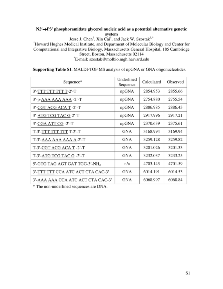
Supporting Table S1. MALDI-TOF MS analysis of npGNA or GNA oligonucleotides.
Underlined
Sequence
Calculated
Observed
3′-TTT TTT TTT T-2′-T
npGNA
2854.953
2855.66
3′-p-AAA AAA AAA -2′-T
npGNA
2754.880
2755.54
3′-CGT ACG ACA T -2′-T
npGNA
2886.985
2886.43
3′-ATG TCG TAC G-2′-T
npGNA
2917.996
2917.21
3′-CGA ATT CG -2′-T
npGNA
2370.639
2375.61
T-3′-TTT TTT TTT T-2′-T
GNA
3168.994
3169.94
T-3′-AAA AAA AAA A-2′-T
GNA
3259.128
3259.82
T-3′-CGT ACG ACA T -2′-T
GNA
3201.026
3201.33
T-3′-ATG TCG TAC G -2′-T
GNA
3232.037
3233.25
n/a
4703.143
4701.59
3′-TTT TTT CCA ATC ACT CTA CAC-3′
GNA
6014.191
6014.53
3′-AAA AAA CCA ATC ACT CTA CAC-3′
* The non-underlined sequences are DNA.
GNA
6068.997
6068.84
Sequence*
5′-GTG TAG AGT GAT TGG-3′-NH2
S1
Supporting Figures
HO
HO
T
H
B
H
*
31
*
(B= C, A, or G)
NH
NH
O P O−
O P O−
O
O
HO
5t
G
O
O
HO
5c-g
T
5t
5c
5g
5a
P NMR δ:
(ppm)
7.69
8.00
7.51
7.56
Figure S1. Structures and 31P-NMR chemical shifts of dinucleotide model compounds
containing N2′→P3′ phosphoramidate bonds.
Figure S2. Anion-exchange HPLC analysis of the crude product of npGNA synthesis.
Sequence: 3′-TTT TTT TTT T-2′-T, the underlined denotes the npGNA sequence. The
two truncated by-products were identified by MALDI-TOF MS analysis. For the 9mer,
[M+H]+ calcd: 2593.782, obsd:2594.17. For the 8mer, [M+H]+ calcd: 2332.610,
obsd:2332.14.
S2
Figure S3. Mixing curve showing 1:1 stoichiometry in the 3′-AAA AAA AAA-2′-T/3′TTT TTT TTT T-2′-T npGNA complex (the underlined portion of the sequence is
npGNA, the non-underlined bases are DNA). In each sample, the sum of the
concentrations of two npGNA oligomers was 4 µM.
S3
Figure S4. Circular dichroism studies on an npGNA duplex with the sequence 3′-CGT
ACG ACA T-2′-T / T-2′-GCA TGC TGT A-3′ (the underlined portion of the sequence is
npGNA, the non-underlined bases are DNA). The concentration of the npGNA duplex
was 10 µM. A. CD spectra obtained in the temperature range of 10-80°C at 10°C
intervals. B. Temperature-dependent CD signal change monitored at 278 nm. C. Plot of
derivative of temperature-dependent CD signal change in B.
S4
Figure S5. Circular dichroism studies showing structural similarity among various GNA
and npGNA duplexes. The core sequences are the same for the three duplexes with a
single thymidine at the 2′-terminus of npGNA or at 3′- and 2′-termini of GNA. A. GNA
homoduplex with the sequence: T-3′-CGT ACG ACA T-2′-T / T-2′-GCA TGC TGT A3′-T. B. GNA:npGNA heteroduplex with the sequence: T-3′-CGT ACG ACA T-2′-T
(GNA) / T-2′-GCA TGC TGT A-3′ (npGNA). C. npGNA homoduplex with the
sequence: T-3′-CGT ACG ACA T-2′-T / T-2′-GCA TGC TGT A-3′-T. (the underlined
denotes the GNA or npGNA sequences). The concentration of each GNA or npGNA
strand was 10 µM. The red traces are the sum of signals from single-stranded
components. The blue traces are from duplexes.
S5
Figure S6. CD studies on T-3′-AAA AAA AAA A-2′-T(GNA): T-2′-TTT TTT TTT T3′(npGNA) showing heteroduplex formation. (The underlined denotes the GNA or
npGNA sequences). The concentration of each GNA or npGNA strand was 10 µM. A.
CD signals of single-stranded (ss) A10 GNA (black trace) and T10 npGNA (red trace). B.
CD signal of A10:T10 GNA:npGNA heteroduplex (blue trace) compared with the sum of
CD signals of each strand (magenta trace).
S6
Figure S7. Temperature-dependent CD studies on the T-3′-AAA AAA AAA A-2′T(GNA) /T-2′-TTT TTT TTT T-3′(npGNA) heteroduplex. (The underlined denotes GNA
or npGNA sequences). The concentration of the heteroduplex was 10 µM. A. CD spectra
obtained in the temperature range of 5-50°C at 5 °C intervals. B. Temperature-dependent
CD signal change monitored at 273 nm. C. Plot of derivative of temperature-dependent
CD signal change in B.
S7
Figure S8. Stability of 14a monitored by reverse-phase HPLC. A. Proposed degradation
pathways of 14a. B. HPLC profiles of time-dependent decomposition of 14a at pH 8.4
and 4 °C. The breakdown products were identified by ESI-MS analysis. [M−H]− for 14a1 calcd, 558.1128, obsd, 558.1; for 14a-2, calcd, 540.1022, obsd, 540.1. C. Kinetic
analysis of degradation of 14a at pH 8.4 (solid circle, k: 1.3×10⁻² h-1, half-life: 53.7 h) and
pH 10 (solid square, k: 1.1×10⁻³ h-1, half-life: 615.6 h). Data were fit to a single
exponential decay.
S8
Materials and methods
Reagents and solvents were purchased from Sigma-Aldrich. Pyridine, triethylamine,
and diisopropylethylamine were distilled from CaH2. Compounds 2t, 2c, 2g, and 2a were
prepared as described (Zhang, L.; Peritz, A. E.; Carrikk, P. J.; Meggers, E. Synthesis
2005, 4, 645-653). Flash column chromatography was performed using silica gel from
Sigma-Aldrich (Grade 9385, 230-400 mesh) with solvents indicated below. 1H-, 31P-,
and 13C-NMR experiments were performed on a Varian 400MHz spectrometer. Chemical
shifts are reported in ppm with reference to tetramethylsilane (TMS) or trisilyl propionic
acid (TSP) (0.00 ppm) for 1H, phosphoric acid (0.00 ppm) for 31P, or CDCl3 (77.16 ppm)
for 13C. Coupling constants are reported in Hz. Low-resolution mass spectrometry
(LSMS) analysis was performed on a Bruker Daltonics Esquire 6000 mass spectrometer.
Photolysis was performed using a Rayonet RPR-100 photo-reactor (400W, Southern New
England Ultraviolet Company, Branford, Connecticut).
Matrix-Assisted Laser Desorption Ionization-Time of Flight (MALDI-TOF) Mass
Spectrometry (MS) A sample of ~200 pmol oligonucleotide was adsorbed on a C18 Zip
Tip. Samples were eluted with 1.5 µL of a matrix solution containing a 2:1 mixture of
52.5 mg/mL 3-hydroxypicolinic acid (or 10 mg/mL 2′,4′,6′-trihydroxyacetophenone for
oligonucleotides with MW < 2000) in 50% acetontrile and 0.1 M ammonium citrate in
water. Eluents were directly spotted onto a stainless steel MALDI-TOF plate and were
analyzed in positive mode on a MALDI-TOF mass spectrometer (PerSeptive Biosystems,
Model Voyager DE).
Tm determination by thermal denaturation Thermal denaturation experiments were
performed on a Varian Cary 1E spectrophotometer equipped with a programmable
S9
temperature control. Optical absorbance was monitored at 260nm (1 nm width) with a
heating rate of 1 °C/min. Samples (200 µL) contained 2.0 µM of each strand in 100mM
NaCl, 10 mM sodium phosphate, pH 7.0. Melting temperatures (Tm) were calculated
from first derivatives of melting curves.
Circular dichroism (CD) spectroscopy CD spectroscopic studies were carried out on an
Aviv CD spectrometer (Model 202) at 25 °C. The samples (30 µL) contained 10 µM of
each strand in 100mM NaCl, 10 mM sodium phosphate, pH 7.0, path length 1.0 mm. The
samples were scanned from 200 to 350 nm with a 1-nm increment. The signal was
recorded from the average of 10 measurements for each wavelength. A sample containing
only buffer was used as the control for all measurements.
Solid-phase synthesis of npGNA oligonucleotides npGNA oligonucleotides were
synthesized by oxidative amination coupling using controlled-pore glass solid support
modified with T (0.5 µmol, Glen Research, Sterling, VA) (Chen, J. K.; Schultz, R. G.;
Lloyd, D. H.; Gryaznov, S. M. Nucleic Acids Research 1995, 23, 2661-8). The following
procedure was developed for manually delivering reagents and washing solution using
syringes during each step: 1. Detritylation with 1 mL of 3% dichloroacetic
acid/dichloromethane for 1 min; 2. Phosphitylation with 0.4 M 2-cyanoethyl-N,N′diisopropylchlorophosphite/0.4 M diisopropylethylamine in dichloromethane (0.5 mL for
10 min); 3. Hydrolysis with 0.4 M tetrazole in 9:1 acetonitrile/water (1 mL for 5 min). 4.
Coupling with 150 µL of 0.2 M 1t, 1c, 1a, or 1g and 0.2 M triethylamine in 1:1
CCl4/acetonitrile for 20 min (C, or T) or 40 min (A or G). Between each step, the solid
support was washed with acetonitrile (5 mL). The npGNA oligonucleotides were cleaved
off the solid support and deprotected by concentrated ammonia at 55 °C overnight. The
S10
average coupling yields were 40-80% based on the trityl assay, which limits this method
to synthesizing short npGNAs. All npGNA, GNA or DNA oligonucleotides used in this
study were purified by polyacrylamide gel electrophoresis and characterized by MALDITOF MS (See Supporting Table S1).
Stability studies of 14a A solution of 100 µL containing 50 µM 14a in 0.2 M NaCl, 20
mM MgCl2, and 100 mM HEPBS (N-(2-hydroxyethyl)piperazine-N′-(4-butanesulfonic
acid), pH 8.4 (or 10 mM NaOH, pH 10) was incubated at 4 °C. At each time point, a 20µL aliquot was removed and analyzed by reverse-phase HPLC using a Varian Microsorb100 (4.6 mm × 250 mm) column. Conditions: solution A: 25 mM triethylammonium
bicarbonate, 2.5 % acetonitrile, pH 7.0; solution B: 100% acetonitrile; Gradient: 0-2 min,
0% B; 2-22 min, 0-40% B. Compound, retention time: 14a, 9.4 min, 14a-1, 8.1 min, 14a2, 8.4 min.
Polymerization of in situ generated 14a on the T-GNA-(T)10-T template The reaction
mixture of 10 µL contained 200 µM T-GNA-(T)10-T template, 2 mM 13a, 20 mM
MgCl2, 0.2 M NaCl, 0.1 M HEPBS, pH 8.4. The reaction was initiated by photolysis at 4
°C for 6 h and was then incubated for 3 d at 4 °C. Oligonucleotides were then
precipitated from the reaction mixture by adding 90 µL of water, 50 µL of 3 M sodium
acetate, pH 5.3, and 400 µL of ethanol. The pellet was washed with 80% ethanol once,
air-dried, and analyzed by MALDI-TOF MS as described above. For templateindependent polymerization, the reaction conditions were the same as described above
except that no template was included. After ethanol precipitation of the reaction mixture,
the template was co-spotted with the sample on the MALDI plate as an internal control.
S11
Polymerization of in situ generated 14a in primer-extension experiments The reaction
mixture of 10 µL contained 1 µM 5′-[32P]-primer/template, 2 mM 13a, 20 mM MgCl2,
0.2 M NaCl, 0.1 M HEPBS, pH 8.4. The reaction was initiated by photolysis at 4 °C for 6
h and, after another 12 h incubation at 4 °C, was analyzed by 20% denaturing
polyacrylamide gel electrophoresis.
S12
Synthesis of npGNA intermediates
O
DMT O
T
H
DAST
Pyridine/CH2Cl2
NaN3
DMF/HMPA, 110 oC
N
DMT O
not purified
N
O
42% Yield
OH
H
2t
DMT O
DMT O
T
H2, Pd/C
78% Yield
H
3t
T
H
N3
NH2
4t
1t
Scheme S1. Synthesis of 1t.
Synthesis of 1t (Scheme S1)
Synthesis of (R)-1′-(thymidine-1-yl)-3′-O-(4,4′-dimethoxytrityl)-2,2′-anhydro-2′,3′propanediol (3t) Compound 2t (4.6 g, 9.2 mmol) was dissolved in 70 mL of anhydrous
CH2Cl2. (Diethylamino)sulfur trifluoride (DAST) (2 mL, 15.3 mmol) and 0.8 mL
anhydrous pyridine were added to the solution simultaneously at room temperature while
stirring. The reaction mixture was incubated at room temperature for 15 min and was
quenched by adding 10 mL of saturated NaHCO3 at 0 °C. The reaction mixture was then
extracted twice with saturated NaHCO3 (100 mL ×2) and once with brine (100 mL), dried
over Na2SO4, and evaporated in vacuo to afford 3t as light yellow foam (4.4 g, 9.1
mmol), which was used for subsequent synthesis without further purification. 1H NMR
(400 MHz, CDCl3) δ:7.18-7.32 (9H, m), 7.11 (1H, d, J=1.0), 6.78-6.82 (4H, m), 4.98
(1H, m), 4.21 (1H, dd, J=9.2, 9.2), 3.97 (1H, dd, 9.4, 5.2), 3.77 (6H, s), 3.64 (1H, dd,
J=3.6, 10.8), 3.15 (1H, dd, J=3.2, 10.8), 1.98 (3H, d, J=1.0). 13C NMR (100.5 MHz,
CDCl3) δ: 172.8, 160.7, 158.7, 144.1, 135.2, 135.0, 132.0, 130.0, 128.1, 127.9, 127.1,
118.4, 113.43, 113.38, 86.6, 77.4, 63.5, 55.3, 48.4, 14.1; LRMS calcd for [M+H]+
(C29H29N2O5): 485.2076, obsd, 485.2.
S13
Synthesis of (S)-1′-(thymidine-1-yl)-2′-azido-3′-O-(4,4′-dimethoxytrityl)-3′-propanol (4t)
Compound 3t (4.4 g, 9.1 mmol) and NaN3 (3 g, 460 mmol) was dissolved in 20 mL of
anhydrous dimethylformamide and 20 mL of hexamethylphosphoramide and was stirred
at 100 °C for 18 h. The reaction mixture was then poured into 200 mL of ethyl acetate.
The organic layer was washed twice with water (200 mL ×2), dried over Na2SO4, and
evaporated in vacuo. The crude pellet was further purified by silica column
chromatography (1% to 2% MeOH/CH2Cl2) to afford 4t as white foam (2.0 g, 3.8 mmol,
42%).1H NMR (400 MHz, CDCl3) δ: 8.72 (1H, br), 7.45 (1H, s), 7.43 (1H, s), 7.23-7.33
(7H, m), 6.97 (1H, s), 6.85(4H, d, J=8), 3.96 (1H, dd, J=5.6, 13.6), 3.89 (1H, m), 3.80
(6H, s), 3.43 (1H, dd, J= 8.4, 13.6), 3.37 (1H, dd, J= 2.4, 9.4), 3.19 (1H, dd, J=5.6, 9.4),
1.87 (3H, s). 13C NMR (100.5 MHz, CDCl3) δ:164.2, 158.8, 150.9, 144.4, 141.3, 135.44,
135.38, 130.08, 130.05, 128.14, 128.06, 127.2, 113.42, 113.40, 110.6, 87.0, 63.5, 60.4,
55.4, 49.5, 12.5; LRMS calcd for [M+Na]+ (C29H29N5NaO5): 550.2066, obsd, 550.2.
Synthesis of (S)-1′-(thymidine-1-yl)-2′-amino-3′-O-(4,4′-dimethoxytrityl)-3′-propanol (1t)
Compound 4t (4.6 g, 8.7 mmol) was dissolved in 100 mL of anhydrous methanol and
10% Pd on activated carbon (750 mg) was added. The reaction mixture was then shaken
vigorously under H2 (30 psi) using a hydrogenator at room temperature for 20 h. The
reaction mixture was then filtered through Celite, evaporated in vacuo, and purified by
silica gel chromatography (1%-4% MeOH/ CH2Cl2) to afford 1t as white foam (3.4 g, 6.8
mmol, 78%). 1H NMR (400 MHz, CDCl3) δ: 7.42 (1H, s), 7.40 (1H, s), 7.22-7.31 (7H,
m), 6.96 (1H, d, J=0.4), 6.83 (4H, d, J=7.2), 3.87 (1H, dd, J= 4.8, 14.0), 3.79 (6H, s), 3.64
(1H, dd, J= 7.6, 13.6), 3.28 (1H, m), 3.13 (2H, m), 1.85 (3H, s). 13C NMR (100.5 MHz,
CDCl3) δ: 164.4, 158.7, 151.5, 144.7, 141.6, 135.8, 130.1, 128.1, 128.0, 127.1, 113.3,
S14
110.1, 86.3, 65.6, 55.3, 52.2, 50.9, 12.4. LRMS calcd for [M+Na]+ (C29H31N3NaO5):
524.2161, obsd, 524.2.
NBz
CNBz
DMT O
DAST
Pyridine/CH2Cl2
H
NaN3
DMF/HMPA, 110 oC
N
DMT O
54% Yield
N
O
46% Yield
OH
H
2c
3c
CNBz
DMT O
H
N3
4c
H2S
Et3N/Pyridine, 0 oC
83% Yield
CNBz
DMT O
H
NH2
1c
Scheme S2. Synthesis of 1c.
Synthesis of 1c (Scheme S2)
Synthesis of (R)-1′-(4-N-benzoylcytosine-1-yl)-3′-O-(4,4′-dimethoxytrityl)-2,2′-anhydro2′,3′-propanediol (3c) Compound 2c (2.8 g, 4.7 mmol) was dissolved in 50 mL of
anhydrous CH2Cl2. (Diethylamino)sulfur trifluoride (DAST) (1.0 mL, 7.7 mmol) and 0.4
mL anhydrous pyridine were added to the solution simultaneously at room temperature
while stirring. The reaction mixture was incubated at room temperature for 15 min and
was quenched by adding 6 mL of saturated NaHCO3 at 0 °C. The reaction mixture was
then extracted twice with saturated NaHCO3 (100 mL ×2) and once with brine (100 mL),
dried over Na2SO4, and evaporated in vacuo. Compount 3c was further purified by silica
gel chromatography (2% MeOH/ CH2Cl2) as light yellow foam (1.4 g, 2.5 mmol, 54%).
Samples of 3c contained ~5% impurity due to partial decomposition during purification.
However, the impurity did not seem to affect the next step of synthesis. 1H NMR (400
MHz, CDCl3) δ: 8.17 (2H, d, J= 6.4), 7.2-7.5 (10H, m), 7.17 (1H, d, J=7.2), 6.80(4H, d,
J=7.2), 5.02 (1H, m), 4.23 (1H, dd, J=9.2,9.2), 3.99 (1H, dd, J=5.2, 8.4), 3.78 (6H,s), 3.66
S15
(1H, dd, J=3.6, 11.2), 3.18 (1H, dd, J= 3.2, 11.2); 13C NMR (100.5 MHz, CDCl3) δ:179.3,
163.3, 159.8, 158.8, 158.7, 144.1, 136.3, 135.7, 135.2, 134.9, 131.9, 130.0, 129.7, 128.2,
128.1, 127.9, 127.2, 113.5, 113.4, 107.2, 86.6, 77.9, 63.3, 55.3, 48.3, LRMS calcd for
[M+H]+ (C35H32N3O5): 574.2342, obsd, 574.1.
Synthesis of (S)-1′-(4-N-benzoylcytosine-1-yl)-2′-azido-3′-O-(4,4′-dimethoxytrityl)-3′propanol (4c) Compound 3c (2.0 g, 3.5 mmol) and NaN3 (1.2 g, 17.5 mmol) was
dissolved in 10 mL of dimethylformamide/hexamethylphosphoramide (1:1) and was
stirred at 110 °C for 4 h. The reaction mixture was then poured into 100 mL of ethyl
acetate. The organic layer was washed twice with water (200 mL ×2), dried over
Na2SO4, and evaporated in vacuo. The crude pellet was further purified by silica column
chromatography (1% to 2% MeOH/CH2Cl2) to afford 4c as light yellow foam (1.0 g, 1.6
mmol, 46%).1H NMR (400 MHz, CDCl3) δ: 8.80 (1H, br), 7.89 (2H, d, J=7.6), 7.21-7.63
(14H, m), 6.85(4H, d, J=8.8), 4.21 (1H, dd, J=4, 13.2), 4.12 (1H, m), 3.79 (6H, s), 3.58
(1H, dd, J= 8.8, 13.2), 3.45 (1H, dd, J= 2.8, 10), 3.18 (1H, dd, J=5.6, 10). 13C NMR
(100.5 MHz, CDCl3) δ: 162.7, 158.8, 150.0, 144.3, 135.4, 135.3, 133.4, 133.0, 130.1,
129.2, 128.1, 128.0, 127.6, 127.1, 113.4, 96.5, 86.9, 63.4, 59.7, 55.3, 51.8. LRMS calcd
for [M+H]+ (C35H33N6O5): 617.2512, obsd, 617.3.
Synthesis of (S)-1′-(4-N-benzoylcytosine-1-yl)-2′-amino-3′-O-(4,4′-dimethoxytrityl)-3′propanol (1c) Compound 4c (1.4g, 2.3 mmol) and triethylamine (0.6 mL) were dissolved
in 3.4 mL of anhydrous pyridine. The solution was then cooled to 0 °C on ice and
hydrogen sulfide was bubbled through the reaction mixture for 15 min. The solvent was
then evaporated in vacuo. The crude product was redissolved in 100 mL of CH2Cl2,
extracted twice with saturated NaHCO3 (100 mL ×2) and once with brine (100 mL), and
S16
evaporated. Compound 1c was further purified by silica gel chromatography (1%-3%
MeOH/CH2Cl2) as light yellow foam (1.1 g, 1.9 mmol, 83%). 1H NMR (400 MHz,
CDCl3) δ: 7.89 (2H, d, J=7.2), 7.22-7.59 (14H, m), 6.84 (4H, d, J=8.8), 4.10 (1H, dd,
J=2.8, 13.2), 3.80 (1H, J= 3.6, 13.2), 3.78 (6H, s), 3.42 (1H, m), 3.22 (1H, dd, J= 4.4,
9.6), 3.10 (1H, dd, J=4, 9.6). 13C NMR (100.5 MHz, CDCl3) δ: 162.2, 158.7, 150.2,
144.6, 135.7, 135.6, 133.2, 130.1, 129.1, 128.11, 128.06, 127.7, 127.1, 113.3, 96.1, 86.2,
64.9, 55.3, 54.6, 50.1. LRMS calcd for [M+H]+ (C35H35N4O5): 591.2607, obsd, 591.2.
NHBz
N
H
N
DMT O
R *
OH
NHBz
N
1. MesCl, Et3N, CH2Cl2
2. NaN3, HMPA/DMF, 100 oC
N
71% Yield
2a
DMT O
S H
NHBz
N
H2S
Et3N/Pyridine, 0 oC
N
DMT O
N
N
N
N
*
N3
4a
N
N
H
91% Yield
NH2
1a
Scheme S3. Synthesis of 1a.
Synthesis of 1a (Scheme S3)
Synthesis of (S)-1′-(6-N-benzoyladenine-9-yl)-2′-azido-3′-O-(4,4′-dimethoxytrityl)-3′propanol (4a) Compound 2a (4.3 g, 7.0 mmol) was dissolved in 70 mL of anhydrous
pyridine. The solution was cooled to 0 °C and methanesulfonyl chloride (1.1 mL, 14.1
mmol) was added drop-wise. The reaction mixture was then warmed up to room
temperature and stirred for 4 h. The reaction was quenched by adding 5 mL of methanol
at 0 °C and the solvent was then evaporated in vacuo. The crude product was dissolved in
200 mL of CH2Cl2, washed twice with saturated NaHCO3 (100 mL ×2) and once with
brine (100 mL), dried over Na2SO4, and evaporated in vacuo. The resulting pellet was
S17
dissolved in 20 mL of dimethylformamide/hexamethylphosphoramide (1:1). Sodium
azide (2.3 g, 35 mmol) was added to the solution and the reaction mixture was incubated
at 100 °C for 16 h. The reaction mixture was then poured into 200 mL of ethyl acetate.
The organic layer was washed twice with water (200 mL ×2), dried over Na2SO4, and
evaporated in vacuo. The crude product was further purified by silica column
chromatography (0.5% to 1% MeOH/CH2Cl2) to afford 4a as light yellow foam (3.2 g,
5.0 mmol, 71%). 1H NMR (400 MHz, CDCl3) δ: 9.06 (1H, s), 8.77 (1H, s), 8.02 (2H, d,
J= 7.2), 7.99 (1H, s), 7.23-7.61 (12H, m), 6.84 (4H, d, J= 8.8), 4.40 (1H, dd, J=3.6, 14.0),
4.18 (1H, dd, J= 8.4, 14.0), 4.00 (1H, m), 3.79 (6H, s), 3.44 (1H, dd, J= 4.0, 10.0), 3.29
(1H, dd, J=6.4, 10.0). 13C NMR (100.5 MHz, CDCl3) δ: 164.7, 158.8, 152.8, 152.1,
149.6, 144.3, 143.6, 135.34, 135.31, 133.7, 132.9, 130.1, 129.0, 128.2, 128.0, 127.9,
127.2, 122.9, 113.4, 87.2, 63.8, 60.9, 55.4, 44.7. LRMS calcd for [M+H]+ (C36H33N8O4):
641.2625, obsd, 641.2.
Synthesis of (S)-1′-(6-N-benzoyladenine-9-yl)-2′-amino-3′-O-(4,4′-dimethoxytrityl)-3′propanol (1a) Compound 4a (1.0 g, 1.6 mmol) and triethylamine (0.6 mL) were
dissolved in 3.4 mL of anhydrous pyridine at 0 °C. Hydrogen sulfide was bubbled
through the solution at 0 °C for 20 min. The reaction mixture was then evaporated, redissolved in 50 mL of CH2Cl2, extracted twice with saturated NaHCO3 (100 mL ×2) and
once with brine (100 mL), dried over Na2SO4, and evaporated in vacuo. The crude
product was further purified by silica gel chromatography to afford 1a as white foam
(0.9g, 1.46 mmol, 91%). 1H NMR (400 MHz, CDCl3) δ: 9.12 (1H, br), 8.75 (1H, br),
8.00-8.02(3H, m), 7.20-7.61(12H, m), 6.83 (4H, d, J=9.2), 4.43 (1H, dd, J= 4.4, 14.0),
4.22 (1H, dd, J=7.6, 14.0), 3.78 (6H, s), 3.45 (1H, m), 3.16 (2H, m); 13C NMR (100.5
S18
MHz, CDCl3) δ: 164.7, 158.7, 152.6, 152.5, 149.4, 144.7, 144.1, 135.73, 135.70, 133.8,
132.8, 130.1, 128.9, 128.1, 128.0, 127.9, 127.1, 122.9, 113.3, 86.5, 65.6, 55.3, 51.4, 48.1 .
LRMS calcd for [M+H]+ (C36H35N6O4): 615.2720, obsd, 615.3.
O
N
O
N
DMT O
H
N
NH
N
1. MesCl, Pyridine
2. NaN3, HMPA/DMF, 100 oC
O
N
H
96% Yield
NH
N
DMT O
H
N
O
N
H
N3
OH
4g
2g
O
N
H2, Pd/C
77 % Yield
DMT O
H
N
NH
N
O
N
H
NH2
1g
Scheme S4. Synthesis of 1g.
Synthesis of 1g (Scheme S4)
Synthesis of (S)-1′-(2-N-isobutyrylguanine-9-yl)-2′-azido-3′-O-(4,4′-dimethoxytrityl)-3′propanol (4g) Compound 2g (3.6 g, 6 mmol) was dissolved in 40 mL of anhydrous
pyridine. The solution was cooled to 0 °C and methanesulfonyl chloride (1.1 mL, 14.1
mmol) was added drop-wise. The reaction mixture was then warmed up to room
temperature and stirred for 4 h. The reaction was quenched by adding 5 mL of methanol
at 0 °C. The solvent was then evaporated and the crude product was redissolved in 100
mL of CH2Cl2. The solution was then washed twice with saturated NaHCO3 (100 mL ×2)
and once with brine (100 mL), dried over Na2SO4, and evaporated in vacuo. The resulting
pellet was dissolved in 20 mL of dimethylformamide/hexamethylphosphoramide (1:1).
Sodium azide (2 g, 30 mmol) was added to the solution and the reaction mixture was
incubated at 100 °C for 2 h. The reaction mixture was then poured into 200 mL of ethyl
acetate. The organic layer was washed twice with water (200 mL ×2), dried over
S19
Na2SO4, and evaporated in vacuo. The crude product was further purified by silica
column chromatography (0.5% to 1% MeOH/CH2Cl2) to afford 4g as white foam (3.6 g,
5.8 mmol, 96%). 1H NMR (400 MHz, CDCl3) δ: 11.98 (1H, br), 8.63 (1H, br), 7.53 (1H,
s), 7.42 (2H, d, J= 7.6), 7.21-7.31 (7H, m), 6.81 (4H, d, J= 8.8), 4.10 (1H, dd, J=4.8,
14.0), 3.98 (1H, dd, J=8.0, 14.0), 3.81(1H, m), 3.78 (6H, s), 3.28 (1H, dd, J= 4.0, 10.0),
3.20 (1H, dd, J= 6.0, 10.0), 2.68 (1H, sept, J= 7.0), 1.26 (6H, d, J= 7.0); 13C NMR (100.5
MHz, CDCl3) δ: 178.6, 158.8, 155.6, 148.4, 147.5, 144.3, 139.4, 135.4, 135.3, 130.07,
130.05, 128.1, 128.0, 127.2, 121.1, 113.4, 87.2, 63.5, 60.9, 55.4, 44.2, 36.6, 19.13, 19.11.
LRMS calcd for [M+Na]+ (C33H34N8NaO5): 645.2250, obsd, 645.2.
Synthesis of (S)-1′-(2-N-isobutyrylguanine-9-yl)-2′-amino-3′-O-(4,4′-dimethoxytrityl)-3′propanol (1g) Compound 4g (1.9 g, 3.0 mmol) was dissolved in 100 mL of anhydrous
methanol and 10% Pd on activated carbon (750 mg) was added. The reaction mixture was
then shaken vigorously under H2 (30 psi) using a hydrogenator at room temperature for
20 h. The reaction mixture was then filtered through Celite and evaporated in vacuo to
afford 1g as white foam (1.4 g, 2.3 mmol, 77%). 1H NMR (400 MHz, CDCl3) δ: 7.51
(1H, s), 7.39 (1H, s), 7.38 (1H, s), 7.22-7.31 (7H, m), 6.77 (4H, d, J=7.2), 4.18 (1H, dd,
J= 4.4, 14.0), 4.01 (1H, dd, J= 7.2, 14.0), 3.75 (6H, s), 3.40 (1H, m), 3.15 (1H, dd, J= 4.8,
9.2), 3.04 (1H, dd, J= 6.4, 9.0), 2.64 (1H, sept, J=6.8), 1.17 (6H, d, J= 6.8). 13C NMR
(100.5 MHz, CDCl3) δ:179.1, 158.7, 155.7, 148.8, 147.7, 144.6, 139.7, 135.6, 135.5,
130.0, 128.0, 127.1, 120.8, 113.3, 86.5, 65.2, 55.4, 51.5, 47.9, 46.0, 36.3, 19.1. LRMS
calcd for [M+H]+ (C33H37N6NO5): 597.2820, obsd, 597.3.
S20
HO
HO
T
B
H
H
(B= C, A, or G)
NH
NH
O P O−
O P O−
O
O
HO
5t
G
O
O
T
HO
5c-g
Scheme S5. Structures of dinucleotide model compounds 5t, 5c, 5a, and 5g.
Synthesis and characterization of 5t-5g (Scheme S5)
Dinucleotides 5t, 5c, 5a, and 5g were synthesized via oxidative amination coupling as
described above using controlled pore glass solid support modified with dG or T (2 µmol,
Glen Research, Sterling, VA) as the starting material. The dinucleotides were cleaved off
the solid support and deprotected by concentrated ammonia treatment at 55 °C overnight.
The dinucleotides were further purified by reverse phase HPLC (Column: Varian C18
Microsorb 100, 250 × 21.4 mm; Solution A: 10 mM NH4HCO3, pH 6.8, Solution B:
100% acetonitrile; Gradient: 10-25% B over 15 min; Flow rate: 15 mL/min). Compound,
retention time, yield: 5t, 5.5 min, 520 nmol; 5c, 5.1 min, 652 nmol, 5a, 6.2 min, 390
nmol, and 5g, 5.1 min, 190 nmol.
Compound 5t: 1H NMR (400 MHz, D2O) δ: 8.39 (1H, s), 8.03 (1H, s), 7.32 (1H, s), 6.21
(1H, t, d=6.8), 4.63 (1H, m), 4.06 (1H, m), 3.87 (2H, m), 3.69 (1H, m), 3.59 (1H, dd, J=
7.6, 14), 3.48 (2H, m), 3.23 (1H, m), 2.75 (1H, m), 2.48 (1H, m), 1.67 (3H, s); 31P NMR
(160.84 MHz, D2O) δ: 7.69 ppm; LRMS calcd for [M-H]−:527.1404, obsd, 527.1.
Compound 5c: 1H NMR (400 MHz, D2O) δ: 7.70 (1H, s), 7.58 (1H, d, J=7.3), 6.31 (1H, t,
J=6.4), 5.87 (1H, d, J= 7.2), 4.48 (1H, m), 4.07 (1H, m), 4.01 (1H, m), 3.87 (2H, m),
S21
3.56-3.68 (3H, m), 3.38 (1H, m), 2.20-2.36 (2H, m), 1.90 (3H, s); 31P NMR (160.84
MHz, D2O) δ: 8.00 ; LRMS calcd for [M-H]−:487.1342, obsd, 487.1.
Compound 5g: 1H NMR (400 MHz, D2O) δ: 7.84 (1H, s), 7.62 (1H, s), 6.15 (1H, t, J=
6.4), 4.39 (1H, m), 4.27 (1H, m), 3.95 (1H, dd, J=10.4, 14.4), 3.77 (2H, m), 3.63-3.69
(2H, m), 3.34 (1H, m), 3.05 (1H, m), 2.12-2.28 (2H, m), 1.90 (3H, s); 31P NMR (160.84
MHz, D2O) δ: 7.51; LRMS calcd for [M-H]−:527.1404, obsd, 527.1.
Compound 5a: 1H NMR (400 MHz, D2O) δ: 8.20 (1H, s), 7.98 (1H, s), 7.29 (1H, s), 6.09
(1H, t, J= 6.8), 4.43 (1H, m), 4.33 (1H, m), 4.10 (1H, dd, J= 10.4, 14.4), 3.80 (2H, m),
3.66-3.73 (2H, m), 3.39 (2H, m), 2.18 (1H, m), 2.04 (1H, m); 1.81 (3H, s); 31P NMR
(160.84 MHz, D2O) δ: 7.56; LRMS calcd for [M-H]−:511.1455, obsd, 511.1.
NHBz
N
N
DMT O
N
H
NHBz
N
80% HOAc/H2O
73% Yield
N
N
H
N3
N3
P
OH
N
N
O
N
H
65 % Yield
NH2
NH2
O
N
1. POCl3 /(EtO)3PO
2. NH3-H2O
6a
4a
-O
N
HO
N
O
-O
H2S, Py/TEA
96% Yield
P
OH
N
N
O
N
H
N
H2N
N3
7a
8a
Scheme S6. Synthesis of 8a.
Synthesis of 8a (Scheme S6)
Synthesis of (S)-1′-(6-N-benzoyladenine-9-yl)-2′-azido-3′-propanol (6a) Compound 4a
(0.49g, 0.77 mmol) was dissolved in 10 mL 3% dichloroacetic acid in dichloromethane
and the reaction mixture was stirred at room temperature for 15 min. The reaction was
quenched by adding 1 mL of methanol and solvent was evaporated in vacuo. The crude
S22
product was then purified by silica gel chromatography (2.5-4% methanol in
dichloromethane) to afford 6a as white solid (Yield: 190 mg, 0.56 mmol, 73%). 1H NMR
(400 MHz, CDCl3) δ: 9.34 (1H, s), 8.76 (1H, s), 8.06 (1H, s), 8.03 (2H, d, J=7.2), 7.61
(1H, dd, J= 7.6, 7.6), 7.52 (2H, dd, J= 8.0, 8.0), 4.67 (1H, br), 4.46 (2H, m), 4.00 (1H, m),
3.74 (1H, dd, J=4.8, 12), 3.55 (1H, dd, J=6.8, 12); 13C NMR (100.5 MHz, CDCl3) δ:
164.9, 152.7, 152.3, 149.9, 144.0, 133.5, 133.1, 129.0, 128.0, 122.7, 61.3, 60.8, 43.7;
LRMS calcd for [M+H]+ (C15H15N8O2) : 339.1318, obsd, 339.0.
Synthesis of (S)-1′-(adenine-9-yl)-2′-azido-3′-phosphopropanol (7a) Compound 6a (91
mg, 0.268 mmol) was rendered anhydrous by co-evaporating with dry
dimethylformamide (1 mL ×3) and was dissolved in 2 mL of freshly distilled
triethylphosphate at 0 °C. Freshly distilled POCl3 (123 µL, 1.34 mmol, 5 eq.) was added
to the reaction mixture. The solution was incubated at 0 °C for 3 h and was quenched by
adding 50 mL H2O at 0 °C. The pH of the solution was adjusted to ~ 7 by adding 1 M
NaOH solution. The solvent was then evaporated in vacuo and the resulting pellet was
dissolved in concentrated ammonia to remove the benzoyl protection group on the
nucleobase. Crude compound 7a (161 µmol) was purified by anion-exchange liquid
chromatography using DEAE A-25 resin with a linear gradient of 0-0.5 M
triethylammonium bicarbonate (final yield: 105 µmol, 65%). 1H NMR (400 MHz, D2O)
δ: 8.01 (1H, s), 7.99 (1H, s), 4.27 (1H, dd, J=3.2, 14.4), 4.12 (1H, dd, J=8.8, 14.4), 4.02
(1H, m), 3.87 (1H, m), 3.75 (1H, m); 31P NMR (160.84 MHz, D2O) δ: 1.39; LRMS calcd
for [M-H]− (C8H10N8O4P): 313.0563, obsd, 312.9.
Synthesis of (S)-1′-(adenine-9-yl)-2′-amino-3′-phosphopropanol (8a) Compound 7a (105
µmol) was dissolved in a mixture of triethylamine (1.5 mL) and pyridine (8.5 mL).
S23
Hydrogen sulfide was then bubbled through the mixture at 0 °C for 1 h. The solvent was
then evaporated in vacuo and the crude product 8a was purified by reverse-phase
preparative HPLC (Column: Varian C18 Microsorb 100, 250 × 21.4 mm; Solution A: 10
mM triethylammonium acetate, pH 7.0, Solution B: 100% acetonitrile; Gradient: 0-50%
B over 20 min; Flow rate: 15 mL/min). Compound, retention time, yield: 8a, 6.4 min,
101 µmol (96%). 1H NMR (400 MHz, D2O) δ: 8.23 (1H, s), 8.19 (1H, s), 4.57 (2H, m),
4.03 (1H, m), 3.97 (1H, m), 3.87 (1H, m); 31P NMR (160.84 MHz, D2O) δ: 4.73; LRMS
calcd for [M-H]− (C8H12N6O4P): 287.0658, obsd, 286.8.
NH2
NH2
O
-O
P
O
N
N
O
OH
N
H
N
Nvoc-Cl, Na2CO3
dioxane/H2O (1:9)
-O
N
P
OH
65 % Yield
N
H
9a
NH2
NH2
O
Im
CDI, DMF
54%
8a
P
N
HN Nvoc
H2N
-O
N
O
O
N
N
O
N
H
hv
N
-O
P
Im
N
N
O
N
H
N
Im
H2N
HN Nvoc
10a
11a
NH2
N
N
O
O
P
-O
H
N
Nvoc:
N
OCH3
O
N
H
12a
O
O2N
OCH3
Scheme S7. Synthesis of 10a and its photolytic conversion to 11a and 12a
Synthesis of 10a and its photolytic conversion (Scheme S7)
Synthesis of 9a Compound 8a (20 µmol) was dissolved in 2 mL of 0.5 M Na2CO3
solution. Nvoc-Cl (100 µmol, 5 eq.) in 1 mL of dioxane was then added. The reaction
mixture was incubated at room temperature overnight and was then subjected to reversephase HPLC purification as described above for 8a. Compound, retention time, yield: 9a,
S24
15.2 min, 13 µmol (65%) 1H NMR (400 MHz, D2O) δ: 8.01 (1H, s), 7.75 (1H, s), 7.64
(1H, s), 6.57 (1H, s), 4.89 (1H, d, J=14.0), 4.59 (1H, d, J=14.0), 4.41 (1H, dd, J= < 1,
14.4), 4.24 (1H, dd, J=13.6, 14.2), 4.12 (1H, m), 4.02 (2H, m), 3.96 (3H, s), 3.89 (3H, s);
P NMR (160.84 MHz, D2O) δ: 4.86; LRMS calcd for [M-H]− (C18H21N7O10P) :
31
526.1088, obsd, 525.9.
Synthesis of 10a Compound 9a (25 µmol) was rendered anhydrous by evaporating with
dry dimethylformamide (1 mL ×3) and was then re-dissolved in 1 mL of
dimethylformamide. Carbonyl diimidazole (25 mg, 125 µmol, 5 eq.) in 1 mL of
dimethylformamide was added to the mixture. The solution was then incubated at room
temperature overnight. The product, 10a, was precipitated from the reaction by adding
acetone (20 mL), ether (15 mL), and 0.2 g NaClO4 followed by incubation at -20 °C for 1
h. The pellet was washed once was 20 mL of 1:1 acetone/ether and was air-dried to
afford 10a as light yellow solid (13.5 µmol, 54%). 31P NMR (160.84 MHz, D2O) δ: -7.0;
LRMS calcd for [M-H]− (C21H23N9O9P) : 576.1356, obsd, 575.9.
Conversion of 10a to 11a or 12a by photolysis Compound 10a (1 µmol) was dissolved in
500 µL of 10 % D2O/H2O and the pH of the solution was adjusted either to 13 by 1M
NaOH solution or to 7 by adding 50 µL 0.5 M sodium phosphate (pH 7.0). The solutions
were UV radiated for 5 h at 4 °C. The resulting mixtures were analyzed by 31P NMR and
by ESI-MS. At pH 13, 10a was quantitatively converted to the desired product, 11a. 31P
NMR (160.84 MHz, D2O) δ: -8.2; LRMS calcd for [M-H]− (C11H14N8O3P) : 337.0926,
obsd, 336.9. At pH 7, 11a was not detected in the reaction mixture. Instead, 10a was
quantitatively converted to the cyclic nucleotide 12a. 31P NMR (160.84 MHz, D2O) δ:
31.6. The unusually large 31P chemical shift was consistent with the previously reported
S25
value for a phosphoramidate group in a cyclic, 5-membered ring (28 ppm for 2′-aminouridine-2′,3′-cyclic phosphoramidate) (Thomson, J. B.; Patel, B. K.; Jimenez, V.; Eckart,
K.; Eckstein, F. J. Org. Chem. 1996, 61, 6273-6281). LRMS calcd for [MH]− (C8H10N6O3P) : 269.0552, obsd, 269.0.
−O
O−
P O
A
O
H
−O
O
−O
−O
A
O
P O
O
A
H
80% AcOH/H2O
71%
H
NH
-O
O−
P O
O
−O
P O
O
O
O
T
NH2
14a-1
O−
Im P O
A
O
Nvoc-Cl, Na2CO3
dioxane/H2O (1:9)
60%
A
H
P O
O−
P O
H
H
−O
A
H
NH Nvoc
16a
H
O
95%
P O
O
H
A
O
hv, pH 10
O
89%
P O
O
A
O
CDI, DMF
O
−O
O−
Im P O
−O
A
P O
O
H
NH Nvoc
13a
A
NH2
14a
HO
15a
Scheme S8. Synthesis of 13a and its photolytic conversion to 14a
Synthesis of 13a and its conversion to 14a (Scheme S8)
Solid-phase synthesis of trinucleotide 15a Compound 15a was prepared using a
combination of solid-phase oxidative amination and phosphoramidite chemistry.
Controlled-pore glass beads (110µmol/g, Glen Research) pre-charged with thymidine
(total loading, 20 µmol, 180 mg) were first coupled with 1a (100 µmol, 5 eq.) using
oxidative coupling as described above. The dinucleotide with a phosphoramidate linkage
was then further extended with S-adenosine-glyceronucleotide by standard
phosphoramidite chemistry as described before {Zhang, 2005 #55}. The resulting
trinucleotide was then phosphorylated using the chemical phosphorylation reagent from
Glen Research (3-(4,4'-dimethoxytrityloxy)-2,2-dicarboxyethyl-propyl-(2-cyanoethyl)(N,N′-diisopropyl)-phosphoramidite, 100 µmol, 5 eq.). The resin was then deprotected by
incubating with concentrated ammonia at 55 °C overnight. The crude product (6.5 µmol
S26
based on calculated ε260: 38.5 mM-1cm-1) was further purified by reverse-phase
preparative HPLC (Column: Varian C18 Microsorb 100, 250 × 21.4 mm; Solution A: 10
mM NH4HCO3, pH 6.8, Solution B: 100% acetonitrile; Gradient: 0-40% B over 20 min;
Flow rate: 15 mL/min). Compound, retention time, yield: 15a, 6.6 min, 2.8 µmol (43%).
31
P NMR (160.84 MHz, D2O) δ: 6.81, 4.76, 0.49; LRMS calcd for [M-
H]− (C26H35N13O15P3) : 862.1588, obsd, 862.2.
Conversion of 15a to dinucleotide 14a-1 by acid hydrolysis Compound 15a (2.8 µmol)
was treated with 5 mL of 80% acetic acid in water at room temperature. The progress of
hydrolysis was monitored by 31P NMR and ~80% of the starting material was hydrolyzed
in 3 d. The reaction mixture was then evaporated to remove solvent. The crude product
was purified by reverse-phase HPLC as described for 15a. Compound, retention time,
yield: 14a-1, 5.5 min, 2.0 µmol (71%, based on calculated ε260: 30 mM-1cm-1). 31P NMR
(160.84 MHz, D2O) δ: 4.61, 0.39; LRMS calcd for [M-H]− (C16H22N11O8P2) : 558.1128,
obsd, 558.1.
Synthesis of 2′-NH2 protected dinucleotide 16a from 14a-1 Compound 14a-1 (2 µmol)
was dissolved in 1.5 mL of 0.5 M Na2CO3 solution. Nvoc-Cl (10 µmol, 5 eq.) in 0.5 mL
of dioxane was then added. The reaction mixture was incubated at room temperature
overnight and was then subjected to reverse-phase HPLC purification (Column: Varian
C18 Microsorb 100, 250 × 21.4 mm; Solution A: 25 mM triethylammonium bicarbonate,
2.5 % acetonitrile, pH 7.0, Solution B: 100% acetonitrile; Gradient: 0-40% B over 20
min; Flow rate: 15 mL/min). Compound, retention time, yield: 16a, 12 min, 1.2 µmol (60
%, based on calculated ε260: 32.2 mM-1cm-1). 31P NMR (160.84 MHz, D2O) δ: 4.41, 0.45;
LRMS calcd for [M-H]− (C26H31N12O14P2) : 797.1558, obsd, 797.1.
S27
Synthesis of 3′-imidazole activated dinucleotide 13a Compound 16a (0.76 µmol) was
rendered anhydrous by evaporating with dry dimethylformamide (1mL ×3) and was then
re-dissolved in 0.5 mL of dimethylformamide. A solution of 1M carbonyl diimidazole
(10 µL, 13 eq.) in dimethylformamide was added to the mixture. The solution was then
incubated at room temperature overnight. The solvent was evaporated and the crude
product was purified by reverse-phase HPLC as described for 16a. Compound, retention
time, yield: 13a, 14 min, 0.68 µmol (89%, based on calculated ε260: 32.2 mM-1cm-1).
LRMS calcd for [M-H]− (C29H33N14O13P2) : 847.1827, obsd, 847.2.
Photolytic conversion of 13a to 14a Compound 13a (2 nmol) was dissolved in 20 µL of
10 mM NaOH, pH 10. The mixture was then transferred to a microcapillary pipette (100
µL, Drummond Scientific) and was UV radiated for 5 h at 4 °C. The reaction mixture
was then analyzed by analytical reverse-phase HPLC (Column: Varian C18 Microsorb
100, 250 × 4.6 mm; Solution A: 25 mM triethylammonium bicarbonate, 2.5 %
acetonitrile, pH 7.0, Solution B: 100% acetonitrile; Gradient: 0-40% B over 20 min; Flow
rate: 15 mL/min). Compound, retention time, yield: 13a, 9.4 min, 1.9 nmol (95%). LRMS
calcd for [M-H]− (C19H24N13O7P2) : 608.1397, obsd, 608.1.
S28
