Integral Geometry Descriptors for Characterizing Emphysema and Lung Fibrosis in... Michal Charemza , Elke Th¨ onnes
advertisement
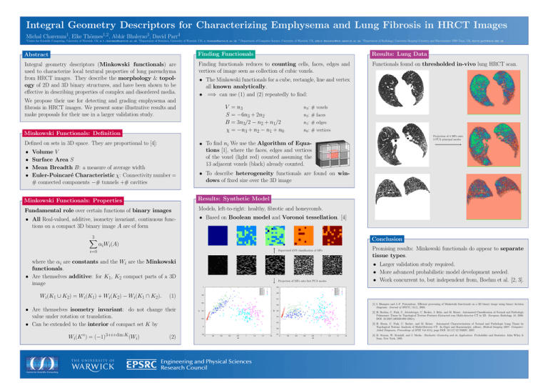
Integral Geometry Descriptors for Characterizing Emphysema and Lung Fibrosis in HRCT Images Michal Charemza1, Elke Thönnes1,2, Abhir Bhalerao3, David Parr4 1 Centre for Scientific Computing, University of Warwick, UK, m.t.charemza@warwick.ac.uk. 2 Department of Statistics, University of Warwick, UK, e.thonnes@warwick.ac.uk. 3 Department of Computer Science, University of Warwick, UK, abhir.bhalerao@dcs.warwick.ac.uk. 4 Department of Radiology, University Hospital Coventry and Warwickshire NHS Trust, UK, david.parr@uhcw.nhs.uk. Abstract Finding Functionals Results: Lung Data Integral geometry descriptors (Minkowski functionals) are used to characterize local textural properties of lung parenchyma from HRCT images. They describe the morphology & topology of 2D and 3D binary structures, and have been shown to be effective in describing properties of complex and disordered media. Finding functionals reduces to counting cells, faces, edges and vertices of image seen as collection of cubic voxels. Functionals found on thresholded in-vivo lung HRCT scan. We propose their use for detecting and grading emphysema and fibrosis in HRCT images. We present some illustrative results and make proposals for their use in a larger validation study. Minkowski Functionals: Definition V = n3 S = −6n3 + 2n2 B = 3n3/2 − n2 + n1/2 χ = −n3 + n2 − n1 + n0 n3: # voxels n2: # faces n1: # edges n0: # vertices Projection of 4 MFs onto 3 PCA principal modes Defined on sets in 3D space. They are proportional to [4]: • • • • • The Minkowski functionals for a cube, rectangle, line and vertex all known analytically. • =⇒ can use (1) and (2) repeatedly to find: Volume V Surface Area S Mean Breadth B: a measure of average width Euler-Poincaré Characteristic χ: Connectivity number = # connected components −# tunnels +# cavities • To find ni We use the Algorithm of Equations [1], where the faces, edges and vertices of the voxel (light red) counted assuming the 13 adjacent voxels (black) already counted. • To describe heterogeneity functionals are found on windows of fixed size over the 3D image Minkowski Functionals: Properties Results: Synthetic Model Fundamental role over certain functions of binary images Models, left-to-right: healthy, fibrotic and honeycomb. • All Real-valued, additive, isometry invariant, continuous functions on a compact 3D binary image A are of form • Based on Boolean model and Voronoi tessellation. [4] 3 X Conclusion αiWi(A) Supervised kNN classification of MFs i=0 where the αi are constants and the Wi are the Minkowski functionals. • Are themselves additive: for K1, K2 compact parts of a 3D image Wi(K1 ∪ K2) = Wi(K1) + Wi(K2) − Wi(K1 ∩ K2). • Larger validation study required. • More advanced probabilistic model development needed. • Work concurrent to, but independent from, Boehm et al. [2, 3]. (1) • Are themselves isometry invariant: do not change their value under rotation or translation. • Can be extended to the interior of compact set K by Wi(K ◦) = (−1)3+i+dim K (Wi) Projection of MFs onto first PCA modes Promising results: Minkowski functionals do appear to separate tissue types. (2) [1] I. Blasquez and J.-F. Poiraudeau. Efficient processing of Minkowski functionals on a 3D binary image using binary decision diagrams. Journal of MSCG, 11(1), 2003. [2] H. Boehm, C. Fink, U. Attenberger, C. Becker, J. Behr, and M. Reiser. Automated Classification of Normal and Pathologic Pulmonary Tissue by Topological Texture Features Extracted rom Multi-detector CT in 3D. European Radiology, 18, 2008. DOI: 10.1007/s00330-008-1082-y. [3] H. Boem, C. Fink, C. Becker, and M. Reiser. Automated Characterization of Normal and Pathologic Lung Tissue by Topological Texture Analysis of Multi-Detector CT. In Giger and Karssemeijer, editors, Medical Imaging 2007: ComputerAided Diagnosis, Proceedings of SPIE Vol 6514, page DOI: 10.117/12.702697, 2007. [4] D. Stoyan, W. Kendall, and J. Mecke. Stochastic Geometry and its Application. Probability and Statistics. John Wiley & Sons, New York, 1995.
