Non-covalent delivery of proteins into mammalian cells† PAPER www.rsc.org/obc
advertisement
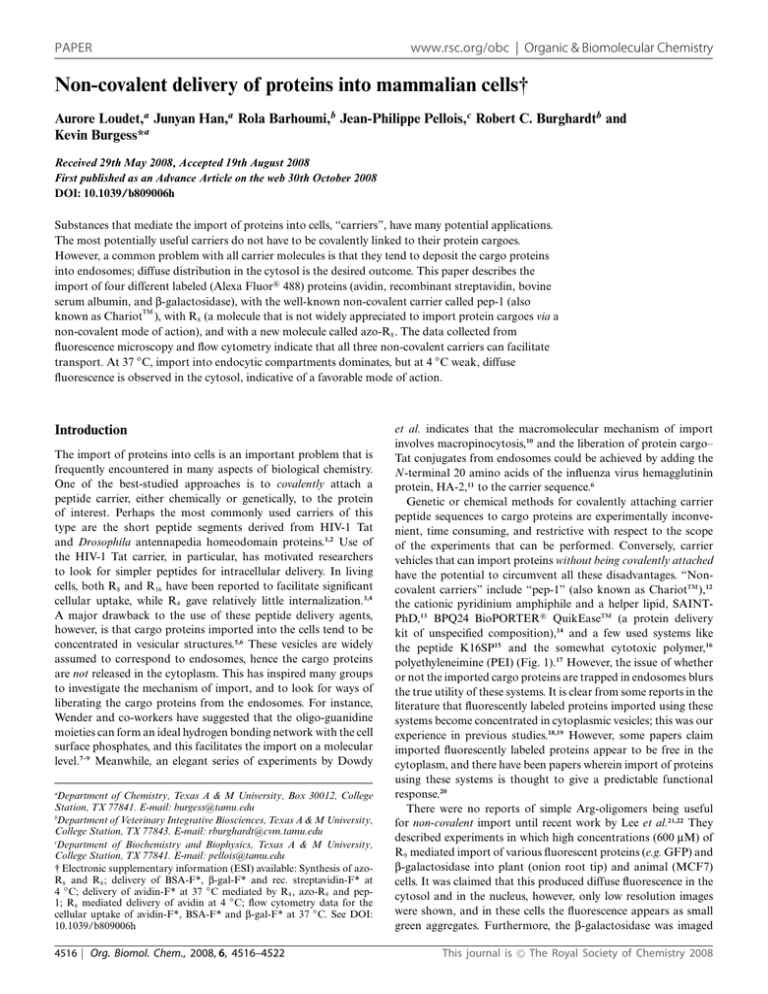
PAPER www.rsc.org/obc | Organic & Biomolecular Chemistry Non-covalent delivery of proteins into mammalian cells† Aurore Loudet,a Junyan Han,a Rola Barhoumi,b Jean-Philippe Pellois,c Robert C. Burghardtb and Kevin Burgess*a Received 29th May 2008, Accepted 19th August 2008 First published as an Advance Article on the web 30th October 2008 DOI: 10.1039/b809006h Substances that mediate the import of proteins into cells, “carriers”, have many potential applications. The most potentially useful carriers do not have to be covalently linked to their protein cargoes. However, a common problem with all carrier molecules is that they tend to deposit the cargo proteins into endosomes; diffuse distribution in the cytosol is the desired outcome. This paper describes the import of four different labeled (Alexa Fluor!R 488) proteins (avidin, recombinant streptavidin, bovine serum albumin, and b-galactosidase), with the well-known non-covalent carrier called pep-1 (also TM known as Chariot ), with R8 (a molecule that is not widely appreciated to import protein cargoes via a non-covalent mode of action), and with a new molecule called azo-R8 . The data collected from fluorescence microscopy and flow cytometry indicate that all three non-covalent carriers can facilitate transport. At 37 ◦ C, import into endocytic compartments dominates, but at 4 ◦ C weak, diffuse fluorescence is observed in the cytosol, indicative of a favorable mode of action. Introduction The import of proteins into cells is an important problem that is frequently encountered in many aspects of biological chemistry. One of the best-studied approaches is to covalently attach a peptide carrier, either chemically or genetically, to the protein of interest. Perhaps the most commonly used carriers of this type are the short peptide segments derived from HIV-1 Tat and Drosophila antennapedia homeodomain proteins.1,2 Use of the HIV-1 Tat carrier, in particular, has motivated researchers to look for simpler peptides for intracellular delivery. In living cells, both R8 and R16 have been reported to facilitate significant cellular uptake, while R4 gave relatively little internalization.3,4 A major drawback to the use of these peptide delivery agents, however, is that cargo proteins imported into the cells tend to be concentrated in vesicular structures.5,6 These vesicles are widely assumed to correspond to endosomes, hence the cargo proteins are not released in the cytoplasm. This has inspired many groups to investigate the mechanism of import, and to look for ways of liberating the cargo proteins from the endosomes. For instance, Wender and co-workers have suggested that the oligo-guanidine moieties can form an ideal hydrogen bonding network with the cell surface phosphates, and this facilitates the import on a molecular level.7–9 Meanwhile, an elegant series of experiments by Dowdy a Department of Chemistry, Texas A & M University, Box 30012, College Station, TX 77841. E-mail: burgess@tamu.edu b Department of Veterinary Integrative Biosciences, Texas A & M University, College Station, TX 77843. E-mail: rburghardt@cvm.tamu.edu c Department of Biochemistry and Biophysics, Texas A & M University, College Station, TX 77841. E-mail: pellois@tamu.edu † Electronic supplementary information (ESI) available: Synthesis of azoR8 and R8 ; delivery of BSA-F*, b-gal-F* and rec. streptavidin-F* at 4 ◦ C; delivery of avidin-F* at 37 ◦ C mediated by R8 , azo-R8 and pep1; R8 mediated delivery of avidin at 4 ◦ C; flow cytometry data for the cellular uptake of avidin-F*, BSA-F* and b-gal-F* at 37 ◦ C. See DOI: 10.1039/b809006h 4516 | Org. Biomol. Chem., 2008, 6, 4516–4522 et al. indicates that the macromolecular mechanism of import involves macropinocytosis,10 and the liberation of protein cargo– Tat conjugates from endosomes could be achieved by adding the N-terminal 20 amino acids of the influenza virus hemagglutinin protein, HA-2,11 to the carrier sequence.6 Genetic or chemical methods for covalently attaching carrier peptide sequences to cargo proteins are experimentally inconvenient, time consuming, and restrictive with respect to the scope of the experiments that can be performed. Conversely, carrier vehicles that can import proteins without being covalently attached have the potential to circumvent all these disadvantages. “Noncovalent carriers” include “pep-1” (also known as ChariotTM ),12 the cationic pyridinium amphiphile and a helper lipid, SAINTPhD,13 BPQ24 BioPORTER!R QuikEaseTM (a protein delivery kit of unspecified composition),14 and a few used systems like the peptide K16SP15 and the somewhat cytotoxic polymer,16 polyethyleneimine (PEI) (Fig. 1).17 However, the issue of whether or not the imported cargo proteins are trapped in endosomes blurs the true utility of these systems. It is clear from some reports in the literature that fluorescently labeled proteins imported using these systems become concentrated in cytoplasmic vesicles; this was our experience in previous studies.18,19 However, some papers claim imported fluorescently labeled proteins appear to be free in the cytoplasm, and there have been papers wherein import of proteins using these systems is thought to give a predictable functional response.20 There were no reports of simple Arg-oligomers being useful for non-covalent import until recent work by Lee et al.21,22 They described experiments in which high concentrations (600 mM) of R9 mediated import of various fluorescent proteins (e.g. GFP) and b-galactosidase into plant (onion root tip) and animal (MCF7) cells. It was claimed that this produced diffuse fluorescence in the cytosol and in the nucleus, however, only low resolution images were shown, and in these cells the fluorescence appears as small green aggregates. Furthermore, the b-galactosidase was imaged This journal is © The Royal Society of Chemistry 2008 Results and discussion Azo-R8 : design and synthesis At the onset of this project, we had hypothesized that mimics of pep-1 could be made by fusing well-known “promiscuous binders” (molecules that seem to bind many proteins in high throughput screens for drug leads)24,25 to a cell-penetrating warhead. The idea was that the promiscuous binder parts of several pep-1 analogs might non-covalently attach to the protein, coating it with entities that promote cell penetration. Thus, an azo-compound was chosen for the promiscuous binder part, and R8 for the cell-penetrating unit; these were synthetically joined to give azo-R8 (Fig. 2). However, azo-R8 was found to behave in much the same way as R8 , but with some minor differences as indicated below. Fig. 1 Structures of the “non-covalent carriers” (A) pep-1 (also known as ChariotTM ), (B) polyethyleneimine (PEI) and (C) K16SP. after fixing the cells, and this is known to give different results relative to live cells.21,22 Data presented in this paper deal with the import of four proteins labeled with Alexa Fluor!R 488 (F*):23 specifically, avidin, bovine serum albumin (BSA), b-galactosidase (b-gal), and a recombinant streptavidin. These cargoes were chosen to represent proteins with different pI values and sizes (Table 1). The potential carriers examined were pep-1, R8 , and a novel system that was synthesized “in house”, azo-R8 , all of which were not covalently attached to the protein. The key observations are that: (i) at 37 ◦ C, avidin was imported by all three carriers (pep-1, R8 , and azo-R8 ) but in each case the labeled protein primarily accumulated in vesicles that co-localized with the endosomal marker FM 4–64; however, (ii) at 4 ◦ C weak, diffuse fluorescence was observed within the cytoplasm with little evidence of punctate vesicle formation for all four proteins. These observations indicate a temperature dependence of carrier-mediated protein delivery that was similar for three chemically different carriers. Fig. 2 Structure of azo-R8 . Formation of carrier : cargo complexes Pep-1 and azo-R8 are amphipathic peptides, while R8 is cationic. Pep-1 is known to associate with protein cargoes through noncovalent electrostatic or hydrophobic interactions and form stable complexes.12,26–28 The formation of the carrier : cargo complex for azo-R8 was easily monitored by fluorescence spectroscopy. The azo compound can act as a quencher of the label on the protein, proving that the two are in close contact. Fig. 3 shows that the intense fluorescence of BSA-F* (2 mM in DMEM, Dulbecco’s Modified Eagle’s Medium) was greatly quenched when azo-R8 was added (1.0 : 10 mol ratio protein : carrier). In a control experiment, a solution of fluorescein (0.1 mM) was mixed with 1 mM azo-R8 under almost identical conditions and no quenching was observed. Delivery at 37 ◦ C: uptake into punctate vesicular structures Table 1 Proteins studied for the (Arg)8 mediated cellular uptake Protein Molecular weight/kDa Size (a.a.) pI (unlabeled protein) Avidin BSA Streptavidin, rec. b-Galactosidase 66–68 66 52 540 512 583 560 1171 10–10.5 4.7 7.4–7.7 4.8 This journal is © The Royal Society of Chemistry 2008 Cellular uptake of avidin-F* into COS-7 at 37 ◦ C was studied in the first phase of this work. All three carriers, pep-1, R8 , and azo-R8 , were used at 10 : 1 carrier : cargo mol : mol ratios, and similar results were observed in all cases. Fig. 4 for avidin uptake is illustrative. After 1 h of incubation, followed by a 15 min incubation period with FM 4–64, the Alexa Fluor!R 488-labeled protein accumulated as green, punctate cytoplasmic vesicles. These green vesicles were localized in endosomes as suggested by co-loading with the endosomal marker FM 4–64 Org. Biomol. Chem., 2008, 6, 4516–4522 | 4517 Fig. 3 Azo-R8 forms a non-covalent complex with BSA-F*. The fluorescence of BSA-F* upon excitation at 488 nm is compared to that of a 1 : 10 molar ratio protein : carrier mixture. (Fig. 4C).29 When the cells were co-incubated with avidin-F* and FM 4–64 for 1 h at 37 ◦ C, little to no co-localization was observed with the endosomes, as the mitochondria were labeled under those conditions and not the endosomes (see ESI†). The perinuclear distribution of these vesicles suggests potential sorting and transport to Golgi and lysosomes. This was confirmed by coloading the cells with the Golgi marker BODIPY!R TR ceramide complexed to BSA (Fig. 4D).23 After extended incubation times (up to 24 h) the vesicles persisted, and no significant dispersed fluorescence was observed in the cytosol. These observations are consistent with the imported protein being trapped in endosomes, even after long periods of time. Consideration of the images from import mediated by azo-R8 shows that this consistently directs more of the labeled protein into the cellular membrane compared with R8 and pep-1. This observation implies some protein–azo-R8 complex may be trapped in the membrane in these experiments. It was also observed that when confluent cell cultures were used, more membrane staining was observed for all three carriers, but more so for azo-R8 . Delivery at 4 ◦ C: diffuse cytosolic fluorescence Punctate vesicle formation was largely suppressed when the experiments described in the previous section were repeated at 4 ◦ C. As an added precaution against surface binding,10 the cells were treated with heparin (3 ¥ 5 min, 1 mg mL-1 of PBS) after the PBS washes. Nevertheless, a weak, diffuse fluorescence signal was observed (see ESI†). Further, fluorescence deconvolution imaging of the cells showed the same diffuse fluorescence pattern. These observations indicate that at 4 ◦ C, the protein is indeed being imported inside the cytoplasm by all the carriers. Experiments were performed to increase the fluorescence signal in the cytosol by increasing the concentration of the protein from 0.5 mM to 2 and 5 mM (carrier : protein = 10 : 1 mol : mol, as before). At 2 mM, the fluorescence intensity in the cytosol was increased but some additional binding to the cell membrane was observed. At 5 mM, the intensity of membrane labeling was considerably brighter, however, the cytoplasmic signal was retained (Fig. 5). Similar experiments on BSA-F*, b-gal-F*, and recombinant streptavidin-F* also gave diffuse cytoplasmic fluorescence (see ESI†). The uptake of b-gal-F* was significantly lower than 4518 | Org. Biomol. Chem., 2008, 6, 4516–4522 Fig. 4 Delivery of avidin-F* in COS-7 cells at 37 ◦ C. (A) Non-covalent protein delivery mediated by R8 ; (B) non-covalent protein delivery mediated by azo-R8 ; (C) endosomal co-localization of avidin-F* and FM 4–64 (fluorescent general endosomal marker); (D) Golgi co-localization of avidin-F* and BODIPY!R TR ceramide complexed to BSA (fluorescent marker for Golgi); (E) cell autofluorescence; (F) non-mediated protein delivery. Throughout, the carrier (5.0 mM), avidin-F* (0.5 mM) and the COS-7 cells were incubated at 37 ◦ C for 1 h; the cells were then washed with PBS and analyzed by fluorescence microscopy. For endosomal co-localization, the cells were incubated with FM 4–64 at 37 ◦ C for 15 min, then washed with PBS before imaging. For Golgi co-localization, the cells were incubated for 30 min at 4 ◦ C in DMEM containing 5 mM BODIPY!R TR ceramide complexed to BSA, washed several times with ice-cold medium and incubated in fresh medium for 30 min at 37 ◦ C. (a) Overlaid images of the avidin-F* (green) and the nuclei (blue, Hoechst 33342 marker). (b) Differential interference contrast (DIC) images. This journal is © The Royal Society of Chemistry 2008 Fig. 5 Delivery of avidin-F* in COS-7 cells at 4 ◦ C mediated by R8 . (A) 0.5 mM of avidin–F*. (B) 2 mM avidin–F*. (C) 5 mM avidin–F*. (D) No carrier used. Throughout, COS-7 cells were incubated for 1 h at 4 ◦ C with R8 and avidin-F* (10 : 1.0 mol : mol), then washed 4 ¥ with PBS buffer. Images shown are after deconvolution; top images are fluorescence images of avidin-F* and Hoechst 33342, bottom images are DIC. that of the avidin and BSA dye-conjugates, even when a higher ratio of carrier : protein (20 : 1) was used. This difference was confirmed in the flow cytometry experiments described in the next section. Comparison of uptake levels for different carriers via flow cytometry Flow cytometry was used to analyze and compare the noncovalent protein transduction (Fig. 6). Before the analyses, the cells were washed with PBS, treated with trypsin, then washed with heparin (3 ¥ 5 min, 0.5 mg mL-1 PBS) to minimize the possibilities for surface binding. In each case, the uptake measured by flow cytometry was greater when the carrier molecule was included (0.5 mM protein; carrier : protein 10 : 1.0 for avidin-F* and BSA-F*, and 20 : 1.0 for b-gal-F*). The import of avidin-F* was significant, with pep-1 and R8 being more effective than azo-R8 at 4 ◦ C (Fig. 6A). However, for BSA-F* the reverse was true: azoR8 was far more effective than the other two carriers. The largest protein–dye conjugate, b-gal-F*, was the least well imported of This journal is © The Royal Society of Chemistry 2008 Fig. 6 (A) Flow cytometric analysis of the uptake of the Alexa Fluor!R 488 labeled proteins, avidin-F*, BSA-F* and b-gal-F* at 4 ◦ C relative to FITC quantum bead standards (shaded histograms). Each histogram for avidin-F* and BSA-F* represents 20 000 to 24 000 cells and 20 000 beads. For b-gal-F*, each histogram represents 10 000 cells and 20 000 beads. The FITC quantum bead peaks represent 8534, 25 857, 79 264, and 21 4887 MESF units each. The scale for the x-axis is MESF units. (B) Summary of the flow cytometric data for each protein with the carriers tested. Results are presented as the median MESF units for each protein–carrier combination. Org. Biomol. Chem., 2008, 6, 4516–4522 | 4519 the three proteins that were tested, with pep-1 and azo-R8 being the most effective carriers. Evaluation of the cytotoxicity of the carriers Cell viability during the non-covalent protein internalization mediated by R8 and azo-R8 was accessed using ethidium homodimer. Thus COS-7 cells were treated with a l.0 : 10 mol ratio protein : carrier (2 mM protein) for 1 h at 4 ◦ C, washed with PBS and treated with ethidium homodimer (2 mg mL-1 ). Fig. 7A illustrates that cells treated with R8 and azo-R8 peptides for 1 h at 4 ◦ C did not result in any cytotoxicity. Incubation at 37 ◦ C for another 16 h gave no cytotoxicity (the fluorescence was now mostly concentrated into vesicles, probably lysosomes, Fig. 7B). After a further incubation period of 24 h at 37 ◦ C however, all the cells were dead (Fig. 7C). into the cytosol, prevails at 4 ◦ C. This observation is parallel and consistent with work reported by Futaki et al.5 They studied the mechanism of translocation of R8 -Texas Red at 37 and 4 ◦ C, without cargo proteins and observed vesicular staining at 37 ◦ C and diffuse fluorescence in the cytosol at 4 ◦ C. The simplest explanation for the reduced efficiency of import of b-gal-F* relative to the other proteins is that it is approximately eight times larger. Similarly, the simplest explanation for the observation that three chemically different carriers facilitate import of three different proteins at 4 ◦ C via an energy independent pathway is that they form pores in the cells membrane that allow leakage into the cells, even at 4 ◦ C. This would explain the fact that the levels of diffuse fluorescence observed are weak, and that the larger protein was the one least effectively imported. At 37 ◦ C, other mechanisms, perhaps involving macropinocytosis (an energy dependant process), become dominant. Experimental Preparation of R8 and azo-R8 See the ESI† data for protocols and characterization information. Material and methods Pep-1 was obtained from Active Motif. Avidin–Alexa Fluor!R 488 conjugate, BSA–Alexa Fluor!R 488 conjugate, FM 4–64, BODIPY!R TR ceramide complexed to BSA and Hoechst 33342 were purchased from Invitrogen. b-Galactosidase and the recombinant streptavidin were purchased from Calbiochem and Roche, respectively and labeled with Alexa Fluor!R 488 5 SDP ester (purchased from Invitrogen) according to the procedure provided by Invitrogen. Fluorescence quenching experiment Fig. 7 Viability assay. Delivery of avidin-F* (2 mM) in COS-7 cells at 4 ◦ C mediated by (a) R8 and (b) azo-R8 . (A) COS-7 cells were incubated for 1 h at 4 ◦ C with R8 or azo-R8 and avidin-F* (10 : 1.0 mol : mol), then washed 4 ¥ with PBS buffer and treated with ethidium homodimer for 30 min. (B) The same cells were incubated at 37 ◦ C for 16 h. (C) The medium was removed, fresh PBS was added and the same cells were incubated at 37 ◦ C for another 24 h. Before imaging, 2 mL of ethidium homodimer were added. Conclusions Import into cells at 37 ◦ C could be regulated via energy-dependent or independent pathways, but at 4 ◦ C it is generally accepted that only energy independent pathways are operative. The data accumulated here indicate that at 37 ◦ C import into endosomes is prevalent and significant diffuse fluorescence in the cytosol is not observed. However, the relative brightness of the vesicular staining may obscure low level cytoplasmic fluorescence. Thus, at least two different pathways appear to be operative for three different carrier molecules, and the desired one, diffuse import 4520 | Org. Biomol. Chem., 2008, 6, 4516–4522 The fluorescence intensity of a solution of BSA-F* (1 mM in DMEM) was compared to the fluorescence intensity of a solution of azo-R8 : BSA-F* complex (1 mM BSA-F* : 10 mM azo-R8 in DMEM). Both solution were excited at 488 nm. As a control experiment, the fluorescence quenching of a solution of fluorescein (0.1 mM) with azo-R8 (1 mM) was also studied. Cell culture COS-7 cells were purchased from the American Type Culture Collection (ATCC) and cultured on 75 cm2 culture flasks in DMEM supplemented with 10% fetal bovine serum (FBS) in a humidified incubator at 37 ◦ C with 5% CO2 . Cells grown to subconfluence were plated 2–3 days prior to the experiments in Lab-Tek two well chambered coverglass slides (Nunc). Fluorescence microscopy The subcellular protein localization was measured in living COS7 cells using a Zeiss Stallion dual detector imaging system consisting of an Axiovert 200M inverted fluorescence microscope, CoolSnap HQ digital cameras and Intelligent Imaging Innovations (3I) software. Digital images of Alexa Fluor!R 488-tagged proteins, FM 4–64 labeled membranes and endosomes, BODIPY!R TR ceramide complexed to BSA labeled Golgi, and Hoechst This journal is © The Royal Society of Chemistry 2008 33342-labeled nuclear DNA were captured with a C-APO 63X/ 1.2 W CORR D = 0.28M27 objective with the following filter sets: exciter BP470/20, dichroic FT 493, emission BP 505–530 for Alexa Fluor!R 488; exciter BP560/40, dichroic FT 585, emission BP 630/75 for FM 4–64 and BODIPY!R TR ceramide complexed to BSA; and exciter G 365, dichroic FT 395, emission BP 445/50 for Hoechst 33342. Sequential optical sections (Z-stacks) from the basal-to-apical surfaces of the cell were acquired. Digital image acquisition was initiated approximately 1 mm below the basal surface of the cells and optical slices were collected at 0.5 mm steps through the apical surface of the cells with a high numerical objective lens (C-APO 63X/1.2 W CORR D = 0.28M27). These wide-field images were subjected to deconvolution using 3I software. The protein : carrier complexes were pre-formed at room temperature for 30 min by mixing (in a mol : mol ratio) the protein and the carrier in DMEM supplemented with 10% fetal bovine serum (FBS). A 1 : 10 mol : mol ratio of protein : carrier was used for avidin-F*, BSA-F* and rec. streptavidin-F*, while a 1 : 20 mol : mol ratio was used for b-gal-F*. To study the cellular uptake of the proteins, the culture medium was removed, the preformed protein : carrier complex was added, and the cells were incubated for another hour at 4 or 37 ◦ C. After the incubation period, the cells were washed with phosphate-buffered saline (PBS, pH 7.4) several times before imaging. For experiments at 4 ◦ C, cells were preincubated at 4 ◦ C for 30 min before being incubated with the protein solution for 1 h. The cells were also co-stained with Hoescht (2 mg mL-1 ), a nuclear marker, and FM 4–64, an endosome marker. Endosomal co-localization COS-7 cells were incubated with the protein : carrier complex for 1 h at 37 ◦ C. After the cells were washed with PBS, FM 4–64 (5 mg mL-1 ) was added and the cells were incubated for 15 min at 37 ◦ C. The cells were washed again with PBS before imaging. Note: when the cells were co-incubated with the labeled protein and FM 4–64 for 1 h at 37 ◦ C, predominant labeling of the mitochondria was observed. Golgi co-localization After the protein was loaded into the COS-7 cells, the cells were washed. Then fresh DMEM medium was added, and the cells were incubated with 5 mM of BODIPY!R TR ceramide complexed to BSA for 30 min at 4 ◦ C. The cells were then rinsed several times with ice-cold medium and incubated in fresh medium for 30 min at 37 ◦ C. Finally, the cells were washed with PBS before imaging. Viability assay The viability of the cells was evaluated by searching for any changes in cellular morphology using Nomarski differential interference contrast (DIC) microscopy during and following analysis of the cellular uptake of the proteins. Parallel cultures were also evaluated using DIC microscopy along with fluorescence analysis of the cellimpermeant viability indicator ethidium homodimer-1 (EthD-1) (Invitrogen). This high-affinity nucleic acid stain is weakly fluorescent until bound to DNA. Unlike Hoechst 33342, EthD-1 can only penetrate cells in which the plasma membrane is This journal is © The Royal Society of Chemistry 2008 compromised. Viability assessment was conducted by incubating cells at 4 ◦ C for 1 h with a 1 : 10 mol ratio of protein : carrier, followed by washing cells with PBS with Ca2+ before imaging of proteins. Following image capture of the proteins, EthD-1 (1 mM final concentration) was added and images were recorded at 5, 10, 20 and 30 min using the 630/75 BP filter and revealed no EthD-1 fluorescence. Cultures were returned to the incubator at 37 ◦ C for 17 h before the viability was again evaluated by EthD-1 staining. The morphology of cells monitored by DIC and Hoechst 33342 staining was normal and no EthD-1 uptake into cell nuclei was detected. Therefore, no cytotoxic effects of protein : carrier combinations were detected for up to 24 h of cell treatment. Flow cytometry The protein internalization was measured by flow cytometry on living cells. COS-7 cells were cultured as subconfluent monolayers on 25 cm2 cell culture plate with vent caps in DMEM supplemented with 10% fetal bovine serum (FBS) in a humidified incubator at 37 ◦ C with 5% CO2 . Carrier–protein complexes were formed in 1 mL DMEM at a molar protein : carrier ratio of 1 : 10 for avidin and BSA and 1 : 20 for b-galactosidase and incubated for 30 min at room temperature. Cells grown to 60–70% confluency were then overlaid with the preformed complexes and incubated for 1 h at 37 or 4 ◦ C. For experiments at 4 ◦ C, the cells were preincubated at 4 ◦ C for 30 min before addition of the protein–carrier complex. After 1 h incubation, the cells were washed with PBS, and treated with trypsin (2 min) and heparin (0.5 mg mL-1 in PBS, 3 ¥ 5 min) to remove extracellular bound protein. Samples were resuspended in 500 mL PBS and transferred to sterile tubes. Cells were analyzed on a FACSCalibur (Becton Dickinson Immunocytometry Systems, San Jose, CA) flow cytometer, equipped with a 15 mW air-cooled argon laser, using CellQuest (Becton Dickinson) acquisition software. Green fluorescence from Alexa Fluor!R 488 or fluorescein was collected through a 530/30-nm bandpass filter. List mode data were acquired on a minimum of 10 000 cells or beads defined by forward and side light scattering light scatter properties. Data analysis was performed in FlowJo (Treestar, Inc., Ashland, OR), using forward and side light scatter to gate on cells or single beads. The Calibrated Parameter platform of FlowJo was used to determine the molecules of equivalent soluble fluorescein (MESF). Data are expressed as the median MESF. Acknowledgements We thank the National Institutes of Health (GM72041), the Robert A. Welch Foundation for financial support, the members of the TAMU/LBMS-Applications Laboratory directed by Dr Shane Tichy for assistance with mass spectrometry, and Dr. Roger Smith (Flow Cytometry Core Laboratory, College of Veterinary Medicine, Texas A & M University) for the flow cytometry assays. References 1 M. Magzoub and A. Graeslund, Q. Rev. Biophys., 2004, 37, 147–195. 2 H. Brooks, B. Lebleu and E. Vives, Adv. Drug Deliv. Rev., 2005, 57, 559–577. 3 S. Futaki, Adv. Drug Deliv. Rev., 2005, 57, 547–558. 4 M. Kosuge, T. Takeuchi, I. Nakase, A. T. Jones and S. Futaki, Bioconjugate Chem., 2008, 19, 656–664. Org. Biomol. Chem., 2008, 6, 4516–4522 | 4521 5 I. Nakase, M. Niwa, T. Takeuchi, K. Sonomura, N. Kawabata, Y. Koike, M. Takehashi, S. Tanaka, K. Ueda, J. C. Simpson, A. T. Jones, Y. Sugiura and S. Futaki, Mol. Ther., 2004, 10, 1011–1022. 6 J. S. Wadia, R. V. Stan and S. F. Dowdy, Nat. Med. (N. Y.), 2004, 10, 310–315. 7 J. B. Rothbard, T. C. Jessop, R. S. Lewis, B. A. Murray and P. A. Wender, J. Am. Chem. Soc., 2004, 126, 9506–9507. 8 J. B. Rothbard, T. C. Jessop and P. A. Wender, Adv. Drug Deliv. Rev., 2005, 57, 495–504. 9 E. A. Goun, T. H. Pillow, L. R. Jones, J. B. Rothbard and P. A. Wender, ChemBioChem, 2006, 7, 1497–1515. 10 I. M. Kaplan, J. S. Wadia and S. F. Dowdy, J. Controlled Release, 2005, 102, 247–253. 11 E. Wagner, C. Plank, K. Zatloukal, M. Cotten and M. L. Birnstiel, Proc. Natl. Acad. Sci. U. S. A., 1992, 89, 7934–7938. 12 M. C. Morris, J. Depollier, J. Mery, F. Heitz and G. Divita, Nat. Biotechnol., 2001, 19, 1173–1176. 13 www.Synvoluxproducts.com, Saint PhD protein delivery kit. 14 www.sigmaaldrich.com, Bio Porter protein delivery kit. 15 E. Mahlum, D. Mandal, C. Halder, A. Maran, M. J. Yaszemski, R. B. Jenkins, M. E. Bolander and G. Sarkar, Anal. Biochem., 2007, 365, 215–221. 16 S. V. Vinogradov, E. V. Batrakova, S. Li and A. V. Kabanov, J. Drug Targeting, 2004, 12, 517–526. 17 V. V. Didenko, H. Ngo and D. S. Baskin, Anal. Biochem., 2005, 344, 168–173. 4522 | Org. Biomol. Chem., 2008, 6, 4516–4522 18 R. Bandichhor, A. D. Petrescu, A. Vespa, A. B. Kier, F. Schroeder and K. Burgess, J. Am. Chem. Soc., 2006, 128, 10688– 10689. 19 R. Bandichhor, A. D. Petrescu, A. Vespa, B. Kier Ann, F. Schroeder and K. Burgess, Bioconjugate Chem., 2006, 17, 1219–1225. 20 E. Gros, S. Deshayes, M. C. Morris, G. Aldrian-Herrada, J. Depollier, F. Heitz and G. Divita, Biochim. Biophys. Acta, Biomembr., 2006, 1758, 384–393. 21 Y.-H. Wang, C.-P. Chen, M.-H. Chan, M. Chang, Y.-W. Hou, H.-H. Chen, H.-R. Hsu, K. Liu and H.-J. Lee, Biochem. Biophys. Res. Commun., 2006, 346, 758–767. 22 M. Chang, J.-C. Chou, C.-P. Chen, B. R. Liu and H.-J. Lee, New Phytol., 2007, 174, 46–56. 23 http://probes.invitrogen.com, Invitrogen, in Molecular Probes, Invitrogen Corporation, 2006. 24 S. L. McGovern and B. K. Shoichet, J. Med. Chem., 2003, 46, 1478– 1483. 25 S. L. McGovern, E. Caselli, N. Grigorieff and B. K. Shoichet, J. Med. Chem., 2002, 45, 1712–1722. 26 S. Deshayes, M. Morris, F. Heitz and G. Divita, Adv. Drug Deliv. Rev., 2008, 60, 537–547. 27 M. C. Morris, P. Vidal, L. Chaloin, F. Heitz and G. Divita, Nucleic Acids Res., 1997, 25, 2730–2736. 28 M. C. Morris, L. Chaloin, J. Mery, F. Heitz and G. Divita, Nucleic Acids Res., 1999, 27, 3510–3517. 29 T. A. Vida and S. D. Emr, J. Cell Biol., 1995, 128, 779–792. This journal is © The Royal Society of Chemistry 2008
