Document 12837771
advertisement
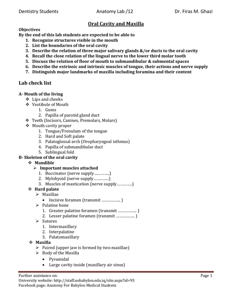
Dentistry Students Anatomy Lab /12 Dr. Firas M. Ghazi Oral Cavity and Maxilla Objectives By the end of this lab students are expected to be able to 1. Recognize structures visible in the mouth 2. List the boundaries of the oral cavity 3. Describe the relation of three major salivary glands &/or ducts to the oral cavity 4. Recall the close relation of the lingual nerve to the lower third molar tooth 5. Discuss the relation of floor of mouth to submandibular & submental spaces 6. Describe the extrinsic and intrinsic muscles of tongue, their actions and nerve supply 7. Distinguish major landmarks of maxilla including foramina and their content Lab check list A- Mouth of the living Lips and cheeks Vestibule of Mouth 1. Gums 2. Papilla of parotid gland duct Teeth (Incisors, Canines, Premolars, Molars) Mouth cavity proper 1. Tongue/Frenulum of the tongue 2. Hard and Soft palate 3. Palatoglossal arch (Oropharyngeal isthmus) 4. Papilla of submandibular duct 5. Sublingual fold B- Skeleton of the oral cavity Mandible Important muscles attached 1. Buccinator nerve supply………….. 2. Mylohyoid nerve supply………….. 3. Muscles of mastication nerve supply………….. Hard palate Maxillae Incisive foramen transmit …………….. Palatine bone 1. Greater palatine foramen transmit …………….. 2. Lesser palatine foramen transmit …………….. Sutures 1. Intermaxillary 2. Interpalatine 3. Palatomaxillary Maxilla Paired (upper jaw is formed by two maxillae) Body of the Maxilla Pyramidal Large cavity inside (maxillary air sinus) Further assistance on: University website: http://staff.uobabylon.edu.iq/site.aspx?id=93 Facebook page: Anatomy For Babylon Medical Students Page 1 Dentistry Students Anatomy Lab /12 Dr. Firas M. Ghazi Anterior surface Infraorbital foramen transmit…………….. Incisive fossa: above the incisor teeth Canine eminence: produced by canine root Canine fossa: lateral to canine eminence Nasal surface (base) Part of lateral wall of nasal cavity Maxillary hiatus: leads into maxillary sinus Orbital surface: Floor of orbital cavity Infratemporal surface: (Anterior boundary of …………… Few foramina for posterior superior alveolar nerve and vessels Processes of the Maxilla 1. Frontal process: projects upwards/articulates with …….. 2. Zygomatic process: extends laterally/articulate with ………… 3. Alveolar process: carries the upper teeth 4. Palatine process: part of hard palate Blood supply of maxillary teeth by branches maxillary artery Posterior, Middle, and Anterior-superior alveolar Arteries C- Boundaries Roof Hard and Soft Palate Floor Horseshoe-shaped region situated beneath the anterior two-third of the tongue Mucus membrane Sublingual salivary gland Submandibular duct Genoglossus muscle Geniohyoid muscle Mylohyoid muscle D- Tongue Parts: Anterior 2/3rd (oral part) Posterior 1/3rd (pharyngeal part) Root Important landmarks: Sulcus terminalis Foramen caecum Muscles (Intrinsic and Extrinsic) Vascular supply Lingual artery Lingual and Deep lingual veins Nerve supply: Motor supply all by X)) except …………… Sensory supply: Anterior 2/3rd: …………………….. Posterior 1/3rd: ………………………….. Taste fibers: Anterior 2/3rd: ……………………..Posterior 1/3rd: ………………………….. Further assistance on: University website: http://staff.uobabylon.edu.iq/site.aspx?id=93 Facebook page: Anatomy For Babylon Medical Students Page 2 Dentistry Students Anatomy Lab /12 Dr. Firas M. Ghazi Comprehension questions: On lying down, what prevent the tongue from falling back into the oropharynx? Home work (1): An eight-year-old girl presented with swelling in the floor of her mouth (Asterisk in figure 1) that began 10 days prior. She denied any recent fever or illness. Examination revealed a non tender, sublingual mass with no obvious extension into the neck. No cervical lymph node enlargement was present. Diagnosis: Ranula Question 1- Which one of the following structures is the most likely cause? A. Submandibular gland B. Sublingual salivary gland C. Lingual vein D. submental lymph node 4 1 3 2 * Figure 1: Source of Image and case: http://www.aafp.org/afp/2011/0815/p441.html 2- Identify the numbered structures in the figure Home work (2): 1. Discuss the anatomical aspects of Ludwig’s angina 2. Define the eye tooth and discuss its relation to facial vein Further assistance on: University website: http://staff.uobabylon.edu.iq/site.aspx?id=93 Facebook page: Anatomy For Babylon Medical Students Page 3
