Anti-Nogo A+B antibody ab47085 Product datasheet 9 Abreviews 3 Images
advertisement
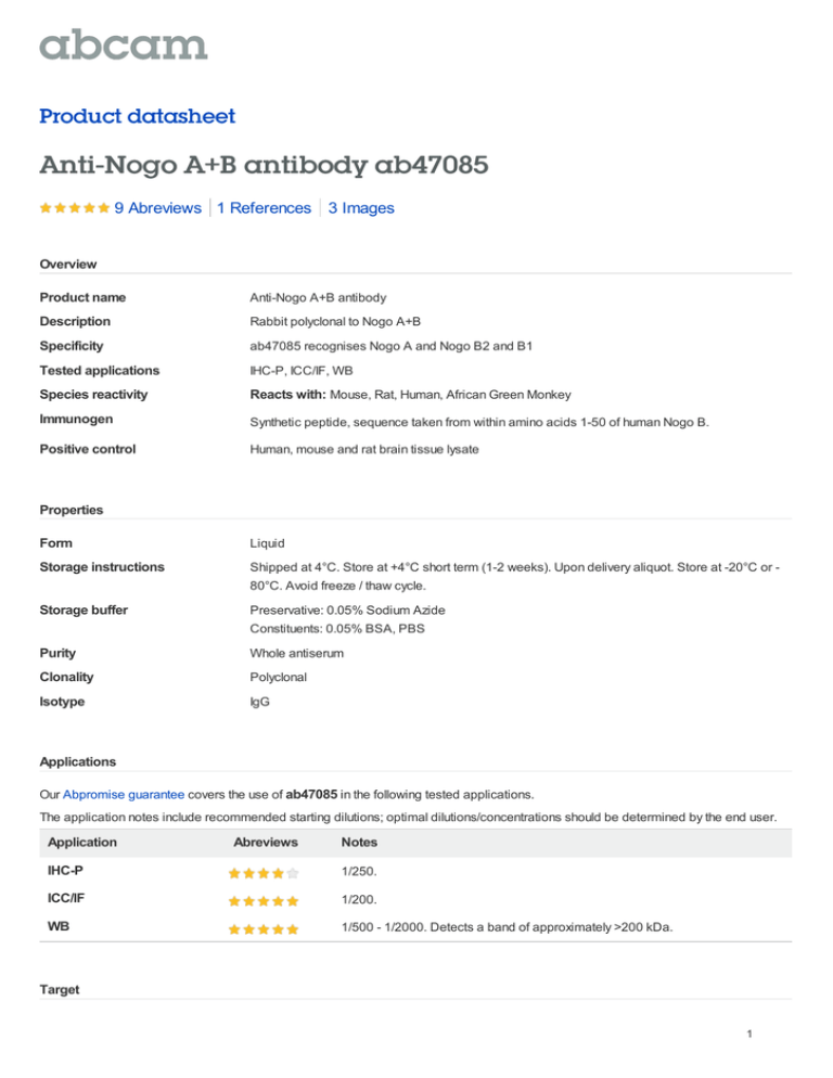
Product datasheet Anti-Nogo A+B antibody ab47085 9 Abreviews 1 References 3 Images Overview Product name Anti-Nogo A+B antibody Description Rabbit polyclonal to Nogo A+B Specificity ab47085 recognises Nogo A and Nogo B2 and B1 Tested applications IHC-P, ICC/IF, WB Species reactivity Reacts with: Mouse, Rat, Human, African Green Monkey Immunogen Synthetic peptide, sequence taken from within amino acids 1-50 of human Nogo B. Positive control Human, mouse and rat brain tissue lysate Properties Form Liquid Storage instructions Shipped at 4°C. Store at +4°C short term (1-2 weeks). Upon delivery aliquot. Store at -20°C or 80°C. Avoid freeze / thaw cycle. Storage buffer Preservative: 0.05% Sodium Azide Constituents: 0.05% BSA, PBS Purity Whole antiserum Clonality Polyclonal Isotype IgG Applications Our Abpromise guarantee covers the use of ab47085 in the following tested applications. The application notes include recommended starting dilutions; optimal dilutions/concentrations should be determined by the end user. Application Abreviews Notes IHC-P 1/250. ICC/IF 1/200. WB 1/500 - 1/2000. Detects a band of approximately >200 kDa. Target 1 Relevance NOGO is a potent neurite outgrowth inhibitor which may also help block the regeneration of the central nervous system in higher vertebrates. Adult mammalian axon regeneration is generally successful in the peripheral nervous system but poor in the central nervous system. Inhibition results from physical barriers imposed by glial scars, a lack of neurotrophic factors, and growthinhibitory molecules associated with myelin, the insulating axon sheath. These molecules include NI35, myelin-associated glycoprotein (159460), and Nogo. Several isoforms (A-E) of NOGO exist. Cellular localization Endoplasmic reticulum; endoplasmic reticulum membrane; multi-pass membrane protein. Note=Anchored to the membrane of the endoplasmic reticulum through 2 putative transmembrane domains. Anti-Nogo A+B antibody images All lanes : Anti-Nogo A+B antibody (ab47085) at 1/2000 dilution Lane 1 : Human brain tissue lysate Lane 2 : Mouse brain tissue lysate Lane 3 : Rat brain tissue lysate Observed band size : 41,43,48-50,>200 kDa Western blot - Nogo A/B antibody (ab47085) ab47085 staining Nogo A+B in African Green Monkey COS-7 cells by ICC/IF (Immunocytochemistry/immunofluorescence). Cells were fixed with paraformaldehyde, permeabilized with 0.5% Triton X-100 and blocked with 3% BSA for 1 hour at 23°C. Samples were incubated with primary antibody (1/500 in PBS-BSA) for 1 hour at 23°C. An Alexa Fluor® 488-conjugated Goat anti-rabbit IgG polyclonal (1/1000) was used as the secondary antibody. Immunocytochemistry/ Immunofluorescence Anti-Nogo A+B antibody (ab47085) This image is courtesy of an anonymous Abreview 2 Immunocytochemistry stainning for Nogo A+B in Cor1 neural stem cells from mouse using Rabbit polyclonal to Nogo A+B (ab47085; 1/200 incubated for 2h at RT); Immunoreactivity was detected in cell-body as well as in neurites. Immunocytochemistry/ Immunofluorescence Nogo A+B antibody (ab47085) Carl Hobbs, King`s College London, United Kingdom Please note: All products are "FOR RESEARCH USE ONLY AND ARE NOT INTENDED FOR DIAGNOSTIC OR THERAPEUTIC USE" Our Abpromise to you: Quality guaranteed and expert technical support Replacement or refund for products not performing as stated on the datasheet Valid for 12 months from date of delivery Response to your inquiry within 24 hours We provide support in Chinese, English, French, German, Japanese and Spanish Extensive multi-media technical resources to help you We investigate all quality concerns to ensure our products perform to the highest standards If the product does not perform as described on this datasheet, we will offer a refund or replacement. For full details of the Abpromise, please visit http://www.abcam.com/abpromise or contact our technical team. Terms and conditions Guarantee only valid for products bought direct from Abcam or one of our authorized distributors 3
