Anti-Lgi1 antibody ab30868 Product datasheet 5 Abreviews 4 Images
advertisement
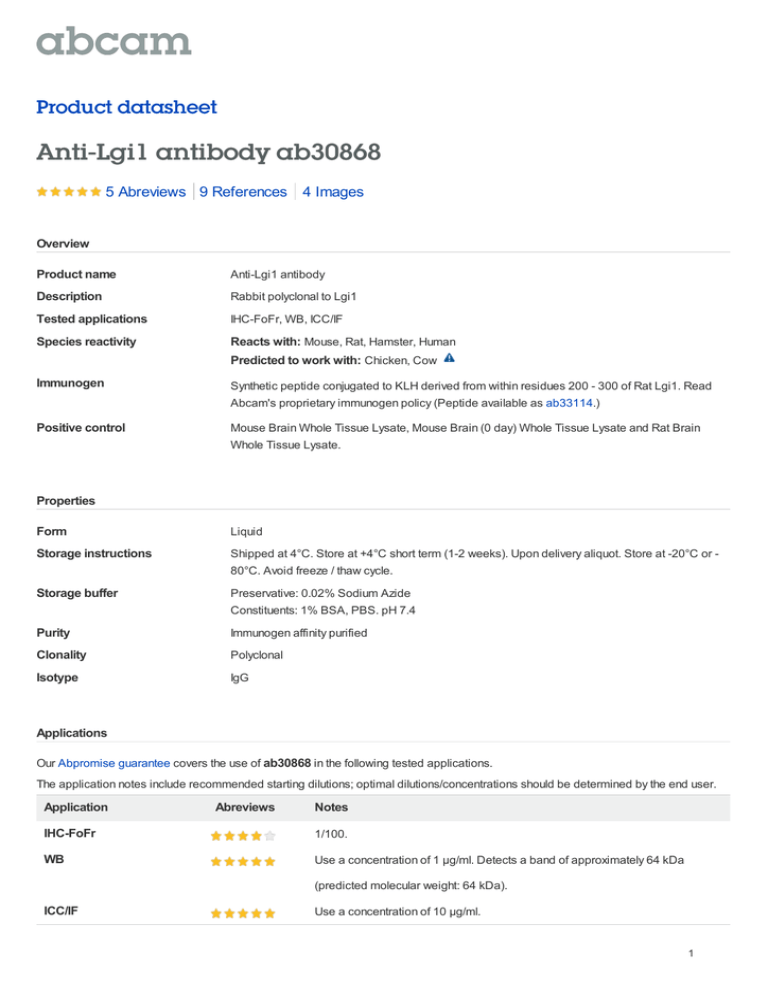
Product datasheet Anti-Lgi1 antibody ab30868 5 Abreviews 9 References 4 Images Overview Product name Anti-Lgi1 antibody Description Rabbit polyclonal to Lgi1 Tested applications IHC-FoFr, WB, ICC/IF Species reactivity Reacts with: Mouse, Rat, Hamster, Human Predicted to work with: Chicken, Cow Immunogen Synthetic peptide conjugated to KLH derived from within residues 200 - 300 of Rat Lgi1. Read Abcam's proprietary immunogen policy (Peptide available as ab33114.) Positive control Mouse Brain Whole Tissue Lysate, Mouse Brain (0 day) Whole Tissue Lysate and Rat Brain Whole Tissue Lysate. Properties Form Liquid Storage instructions Shipped at 4°C. Store at +4°C short term (1-2 weeks). Upon delivery aliquot. Store at -20°C or 80°C. Avoid freeze / thaw cycle. Storage buffer Preservative: 0.02% Sodium Azide Constituents: 1% BSA, PBS. pH 7.4 Purity Immunogen affinity purified Clonality Polyclonal Isotype IgG Applications Our Abpromise guarantee covers the use of ab30868 in the following tested applications. The application notes include recommended starting dilutions; optimal dilutions/concentrations should be determined by the end user. Application Abreviews Notes IHC-FoFr 1/100. WB Use a concentration of 1 µg/ml. Detects a band of approximately 64 kDa (predicted molecular weight: 64 kDa). ICC/IF Use a concentration of 10 µg/ml. 1 Target Function Regulates voltage-gated potassium channels assembled from KCNA1, KCNA4 and KCNAB1. It slows down channel inactivation by precluding channel closure mediated by the KCNAB1 subunit. Ligand for ADAM22 that positively regulates synaptic transmission mediated by AMPAtype glutamate receptors (By similarity). Plays a role in suppressing the production of MMP1/3 through the phosphatidylinositol 3-kinase/ERK pathway. May play a role in the control of neuroblastoma cell survival. Tissue specificity Predominantly expressed in neural tissues, especially in brain. Expression is reduced in lowgrade brain tumors and significantly reduced or absent in malignant gliomas. Isoform 1 is absent in the cerebellum and is detectable in the occipital cortex and hippocampus; higher amounts are observed in the parietal and frontal cortices, putamen, and, particularly, in the temporal neocortex, where it is 3.5 times more abundant than in the hippocampus (at protein level). Isoform 3 shows the highest expression in the occipital cortex and the lowest in the hippocampus (at protein level). Involvement in disease Defects in LGI1 are the cause of lateral temporal lobe epilepsy autosomal dominant (ADLTE) [MIM:600512]; also known as autosomal dominant partial epilepsy with auditory features (ADPEAF). ADLTE is a form of epilepsy characterized by partial seizures, usually preceded by auditory signs. Sequence similarities Contains 7 EAR repeats. Contains 3 LRR (leucine-rich) repeats. Contains 1 LRRCT domain. Contains 1 LRRNT domain. Post-translational modifications Glycosylated. Cellular localization Secreted. Cell junction > synapse. Isoform 1 but not isoform 2 is secreted. Isoform 1 is enriched in the Golgi apparatus while isoform 2 accumulates in the endoplasmic reticulum. Anti-Lgi1 antibody images 2 Anti-Lgi1 antibody (ab30868) at 1/1000 dilution + Rat brain tissue lysate at 30 µg Secondary HRP-conjugate Goat anti-rabbit IgG polyclonal at 1/5000 dilution developed using the ECL technique Performed under reducing conditions. Predicted band size : 64 kDa Western blot - Anti-Lgi1 antibody (ab30868) Observed band size : 64 kDa This image is courtesy of an anonymous Abreview Exposure time : 5 minutes This image is courtesy of an anonymous Abreview Lane 1 : Marker Lanes 2 - 4 : Anti-Lgi1 antibody (ab30868) at 1 µg/ml Lane 1 : As above Lane 2 : Mouse Brain Whole Tissue Lysate at 20 µg Lane 3 : Brain (Mouse) Tissue Lysate - 0 days old (ab7188) at 20 µg Lane 4 : Rat Brain Whole Tissue Lysate at 20 µg Western blot - Lgi1 antibody (ab30868) Secondary Lanes 2 - 4 : IR Dye 680 Conjugated Goat anti-Rabbit IgG (H+L) at 1/15000 dilution Performed under reducing conditions. Predicted band size : 64 kDa Observed band size : 64 kDa 3 ab30868 at 10ug/ml staining hamster CHO cells by ICC/IF. The cells were paraformaldehyde fixed, permeabilized and blocked before incubation with the antibody for 16 hours. An Alexa Fluor ® 488 conjugated goat polyclonal antibody was used as the secondary. Immunocytochemistry/ Immunofluorescence Lgi1 antibody (ab30868) This image is courtesy of an anonymous Abreview IHC-FoFr image of Lgi1 (ab30868) staining in rat spinal cord sections. The sections used came from animals perfused fixed with Paraformaldehyde 4%, in phosphate buffer 0.2M. Following postfixation in the same fixative overnight, the tissues were Immunohistochemistry (PFA perfusion fixed frozen sections) - Lgi1 antibody (ab30868) Sophie Pezet, ESPCI, France cryoprotected in sucrose 30% overnight. Tissues were then cut using a cryostat and the immunostainings were preformed using the ‘free floating’ technique. Image B is a higher power magnification of the Image A, at the level of the interface between the white and grey matter in the lamina V. Numbers in image A are Rexed's laminae numbers. Please note: All products are "FOR RESEARCH USE ONLY AND ARE NOT INTENDED FOR DIAGNOSTIC OR THERAPEUTIC USE" Our Abpromise to you: Quality guaranteed and expert technical support Replacement or refund for products not performing as stated on the datasheet Valid for 12 months from date of delivery Response to your inquiry within 24 hours We provide support in Chinese, English, French, German, Japanese and Spanish Extensive multi-media technical resources to help you We investigate all quality concerns to ensure our products perform to the highest standards If the product does not perform as described on this datasheet, we will offer a refund or replacement. For full details of the Abpromise, please visit http://www.abcam.com/abpromise or contact our technical team. Terms and conditions Guarantee only valid for products bought direct from Abcam or one of our authorized distributors 4
