Anti-CUG-BP1 antibody ab69583 Product datasheet 2 Images
advertisement

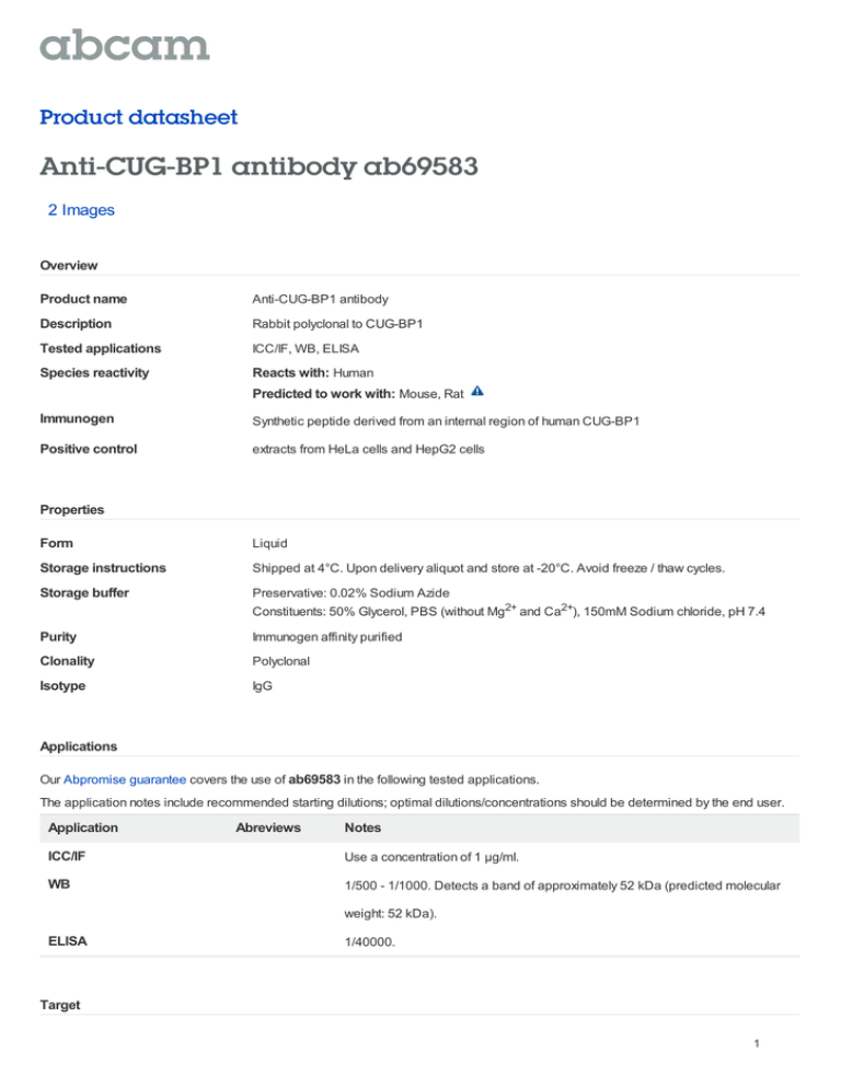
Product datasheet Anti-CUG-BP1 antibody ab69583 2 Images Overview Product name Anti-CUG-BP1 antibody Description Rabbit polyclonal to CUG-BP1 Tested applications ICC/IF, WB, ELISA Species reactivity Reacts with: Human Predicted to work with: Mouse, Rat Immunogen Synthetic peptide derived from an internal region of human CUG-BP1 Positive control extracts from HeLa cells and HepG2 cells Properties Form Liquid Storage instructions Shipped at 4°C. Upon delivery aliquot and store at -20°C. Avoid freeze / thaw cycles. Storage buffer Preservative: 0.02% Sodium Azide Constituents: 50% Glycerol, PBS (without Mg2+ and Ca2+), 150mM Sodium chloride, pH 7.4 Purity Immunogen affinity purified Clonality Polyclonal Isotype IgG Applications Our Abpromise guarantee covers the use of ab69583 in the following tested applications. The application notes include recommended starting dilutions; optimal dilutions/concentrations should be determined by the end user. Application Abreviews Notes ICC/IF Use a concentration of 1 µg/ml. WB 1/500 - 1/1000. Detects a band of approximately 52 kDa (predicted molecular weight: 52 kDa). ELISA 1/40000. Target 1 Function RNA-binding protein implicated in the regulation of several post-transcriptional events. Involved in pre-mRNA alternative splicing, mRNA translation and stability. Mediates exon inclusion and/or exclusion in pre-mRNA that are subject to tissue-specific and developmentally regulated alternative splicing. Specifically activates exon 5 inclusion of cardiac isoforms of TNNT2 during heart remodeling at the juvenile to adult transition. Acts as both an activator and repressor of a pair of coregulated exons: promotes inclusion of the smooth muscle (SM) exon but exclusion of the non-muscle (NM) exon in actinin pre-mRNAs. Activates SM exon 5 inclusion by antagonizing the repressive effect of PTB. Promotes exclusion of exon 11 of the INSR pre-mRNA. Inhibits, together with HNRNPH1, insulin receptor (IR) pre-mRNA exon 11 inclusion in myoblast. Increases translation and controls the choice of translation initiation codon of CEBPB mRNA. Increases mRNA translation of CEBPB in aging liver (By similarity). Increases translation of CDKN1A mRNA by antagonizing the repressive effect of CALR3. Mediates rapid cytoplasmic mRNA deadenylation. Recruits the deadenylase PARN to the poly(A) tail of EDEN-containing mRNAs to promote their deadenylation. Required for completion of spermatogenesis (By similarity). Binds to (CUG)n triplet repeats in the 3'-UTR of transcripts such as DMPK and to Bruno response elements (BREs). Binds to muscle-specific splicing enhancer (MSE) intronic sites flanking the alternative exon 5 of TNNT2 pre-mRNA. Binds to AU-rich sequences (AREs or EDEN-like) localized in the 3'-UTR of JUN and FOS mRNAs. Binds to the IR RNA. Binds to the 5'-region of CDKN1A and CEBPB mRNAs. Binds with the 5'-region of CEBPB mRNA in aging liver. Tissue specificity Ubiquitous. Sequence similarities Belongs to the CELF/BRUNOL family. Contains 3 RRM (RNA recognition motif) domains. Post-translational modifications Phosphorylated. Its phosphorylation status increases in senescent cells. Cellular localization Nucleus. Cytoplasm. RNA-binding activity is detected in both nuclear and cytoplasmic compartments. Anti-CUG-BP1 antibody images All lanes : Anti-CUG-BP1 antibody (ab69583) at 1/500 dilution Lane 1 : extracts from HeLa cells Lane 2 : extracts from HepG2 cells Lane 3 : extracts from HeLa cells with immunising peptide at 5 µg Lysates/proteins at 5 µg per lane. Western blot - CUG-BP1 antibody (ab69583) Predicted band size : 52 kDa Observed band size : 52 kDa 2 ICC/IF image of ab69583 stained HeLa cells. The cells were 4% PFA fixed (10 min) and then incubated in 1%BSA / 10% normal goat serum / 0.3M glycine in 0.1% PBS-Tween for 1h to permeabilise the cells and block nonspecific protein-protein interactions. The cells were then incubated with the antibody (ab69583, 1µg/ml) overnight at +4°C. The secondary antibody (green) was Alexa Fluor® 488 goat anti-rabbit IgG (H+L) used at a 1/1000 dilution for 1h. Alexa Fluor® 594 WGA Immunocytochemistry/ Immunofluorescence- was used to label plasma membranes (red) at CUG-BP1 antibody(ab69583) a 1/200 dilution for 1h. DAPI was used to stain the cell nuclei (blue) at a concentration of 1.43µM. Please note: All products are "FOR RESEARCH USE ONLY AND ARE NOT INTENDED FOR DIAGNOSTIC OR THERAPEUTIC USE" Our Abpromise to you: Quality guaranteed and expert technical support Replacement or refund for products not performing as stated on the datasheet Valid for 12 months from date of delivery Response to your inquiry within 24 hours We provide support in Chinese, English, French, German, Japanese and Spanish Extensive multi-media technical resources to help you We investigate all quality concerns to ensure our products perform to the highest standards If the product does not perform as described on this datasheet, we will offer a refund or replacement. For full details of the Abpromise, please visit http://www.abcam.com/abpromise or contact our technical team. Terms and conditions Guarantee only valid for products bought direct from Abcam or one of our authorized distributors 3


![Anti-QK1 antibody [EPR7306] ab126742 Product datasheet 1 Abreviews 2 Images](http://s2.studylib.net/store/data/012617058_1-a0b458c5816746a0092a778b9b599a81-300x300.png)