Initial results with fully implanted Neurostep FES system for foot drop
advertisement
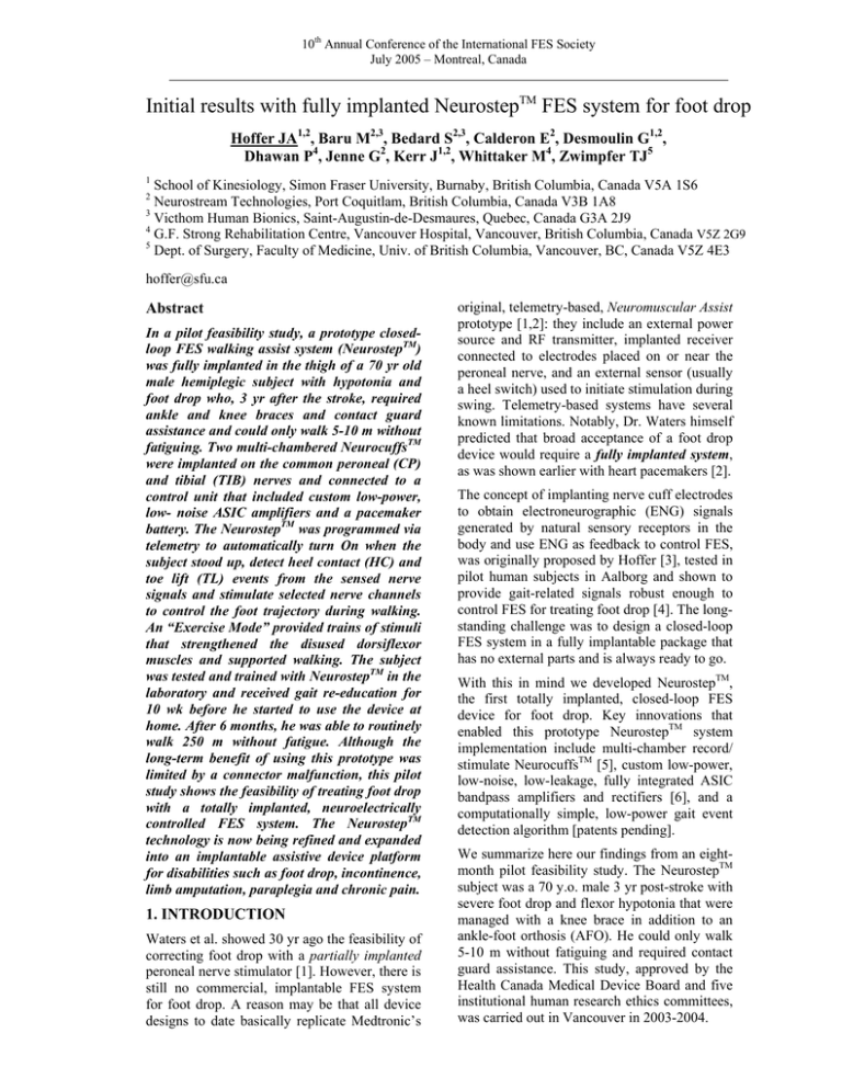
10th Annual Conference of the International FES Society July 2005 – Montreal, Canada Initial results with fully implanted NeurostepTM FES system for foot drop Hoffer JA1,2, Baru M2,3, Bedard S2,3, Calderon E2, Desmoulin G1,2, Dhawan P4, Jenne G2, Kerr J1,2, Whittaker M4, Zwimpfer TJ5 1 School of Kinesiology, Simon Fraser University, Burnaby, British Columbia, Canada V5A 1S6 Neurostream Technologies, Port Coquitlam, British Columbia, Canada V3B 1A8 3 Victhom Human Bionics, Saint-Augustin-de-Desmaures, Quebec, Canada G3A 2J9 4 G.F. Strong Rehabilitation Centre, Vancouver Hospital, Vancouver, British Columbia, Canada V5Z 2G9 5 Dept. of Surgery, Faculty of Medicine, Univ. of British Columbia, Vancouver, BC, Canada V5Z 4E3 2 hoffer@sfu.ca Abstract In a pilot feasibility study, a prototype closedloop FES walking assist system (NeurostepTM) was fully implanted in the thigh of a 70 yr old male hemiplegic subject with hypotonia and foot drop who, 3 yr after the stroke, required ankle and knee braces and contact guard assistance and could only walk 5-10 m without fatiguing. Two multi-chambered NeurocuffsTM were implanted on the common peroneal (CP) and tibial (TIB) nerves and connected to a control unit that included custom low-power, low- noise ASIC amplifiers and a pacemaker battery. The NeurostepTM was programmed via telemetry to automatically turn On when the subject stood up, detect heel contact (HC) and toe lift (TL) events from the sensed nerve signals and stimulate selected nerve channels to control the foot trajectory during walking. An “Exercise Mode” provided trains of stimuli that strengthened the disused dorsiflexor muscles and supported walking. The subject was tested and trained with NeurostepTM in the laboratory and received gait re-education for 10 wk before he started to use the device at home. After 6 months, he was able to routinely walk 250 m without fatigue. Although the long-term benefit of using this prototype was limited by a connector malfunction, this pilot study shows the feasibility of treating foot drop with a totally implanted, neuroelectrically controlled FES system. The NeurostepTM technology is now being refined and expanded into an implantable assistive device platform for disabilities such as foot drop, incontinence, limb amputation, paraplegia and chronic pain. 1. INTRODUCTION Waters et al. showed 30 yr ago the feasibility of correcting foot drop with a partially implanted peroneal nerve stimulator [1]. However, there is still no commercial, implantable FES system for foot drop. A reason may be that all device designs to date basically replicate Medtronic’s original, telemetry-based, Neuromuscular Assist prototype [1,2]: they include an external power source and RF transmitter, implanted receiver connected to electrodes placed on or near the peroneal nerve, and an external sensor (usually a heel switch) used to initiate stimulation during swing. Telemetry-based systems have several known limitations. Notably, Dr. Waters himself predicted that broad acceptance of a foot drop device would require a fully implanted system, as was shown earlier with heart pacemakers [2]. The concept of implanting nerve cuff electrodes to obtain electroneurographic (ENG) signals generated by natural sensory receptors in the body and use ENG as feedback to control FES, was originally proposed by Hoffer [3], tested in pilot human subjects in Aalborg and shown to provide gait-related signals robust enough to control FES for treating foot drop [4]. The longstanding challenge was to design a closed-loop FES system in a fully implantable package that has no external parts and is always ready to go. With this in mind we developed NeurostepTM, the first totally implanted, closed-loop FES device for foot drop. Key innovations that enabled this prototype NeurostepTM system implementation include multi-chamber record/ stimulate NeurocuffsTM [5], custom low-power, low-noise, low-leakage, fully integrated ASIC bandpass amplifiers and rectifiers [6], and a computationally simple, low-power gait event detection algorithm [patents pending]. We summarize here our findings from an eightmonth pilot feasibility study. The NeurostepTM subject was a 70 y.o. male 3 yr post-stroke with severe foot drop and flexor hypotonia that were managed with a knee brace in addition to an ankle-foot orthosis (AFO). He could only walk 5-10 m without fatiguing and required contact guard assistance. This study, approved by the Health Canada Medical Device Board and five institutional human research ethics committees, was carried out in Vancouver in 2003-2004. 10th Annual Conference of the International FES Society July 2005 – Montreal, Canada 2. METHODS Figure 1 shows the NeurostepTM prototype and its dimensions. Circuitry, battery and telemetry antenna are sealed in a pacemaker-like welded titanium case. Two 30 mm NeurocuffsTM attach to the device header via 25 cm long flexible leads and 3-pole modified IS-1 connectors. the TIB NeurocuffTM (“HC”) were measured by an internal NeurostepTM circuit and telemetered out of the body. Impedances initially fluctuated and stabilized about 3 weeks after NeurocuffTM implant (Fig. 3). The lower impedance in Ch. 2 is attributed to shorts in the header connectors. Stimulation of each CP channel with brief trains of 100 µs x 150-250 µA pulses elicited tingling sensations that the subject consistently referred to specific locations of the left leg. Perceptual thresholds (Fig. 4) slowly doubled in Ch. 3 and 4 over the first nine weeks. The rise in Ch. 2 threshold was attributed to a shorted connector. In a first surgery, through a 6 cm posterior thigh incision (Fig. 2; arrow) the surgeon isolated the common peroneal (CP) and tibial (TIB) nerves, measured nerve perimeters with a flexible ruler, implanted suitably sized, multi-chambered TIB and CP NeurocuffsTM, and sealed each cuff with a suture through interdigitated closing elements [7]. The cuff lead connectors were inserted in a dummy device placed in a subcutaneous pocket in the medial thigh and the incision was closed. The dummy device had a percutaneous cable to allow external recordings of signals during the first 10 d. After the signals were evaluated, the incision was re-opened and the dummy device and its percutaneous wires were removed and replaced with an implanted NeurostepTM unit. The NeurostepTM was turned On or Off with a magnet or telemetry programming interface and programmed to automatically wake up when the subject stood up, detect heel contact (HC) and toe lift (TL) events from the nerve signals sensed while walking and stimulate selected CP channels to control foot trajectory during swing. Ch.1 600 Ch.2 400 Ch.3 200 Ch.4 0 0 10 20 30 40 50 60 70 Figure 4. Sensory stimulation thresholds vs. time. As expected, motor thresholds were higher than sensory thresholds. Motor thresholds also rose gradually for Ch. 3 & 4 and suddenly for Ch. 2 (Fig. 5). Stimulation of Ch. 3 and 4 produced strong dorsiflexion, indicating greater proximity to deep peroneal nerve axons. Stimulation of Ch. 1 sometimes elicited a limb flexion reflex. Motor Thresholds (ua*100usec) 1000 800 Ch.1 600 Ch.2 400 Ch.3 200 Ch.4 0 0 10 20 30 40 50 60 70 Days after 1st Implant (Day#) Figure 5. Motor stimulation thresholds vs. time. Stimulation Strength vs. Days after Implant 80 Im p e d a n c e s S u b je c t 1 2 -7 4 -2 8 7 R (kOhm) 800 Days after 1st Implant (Day #) Stimulation Intensity (uA) Figure 2 6 C H1 5 C H2 4 C H3 3 C H4 2 HC 1 Dorsiflexion Force (N) Figure 1 Stimulation Intensity (uA) Sensory Thresholds (uA*100usec) 1000 70 60 50 Average of initial four Stim Peaks (N) 40 30 20 10 0 0 20 40 60 T ime (Days after implant) 0 0 5 10 17 21 26 31 35 40 47 54 59 D ay Figure 3. Resistive part of electrode impedances plotted vs. days after NeurocuffTM implant. 3. RESULTS Impedances between the recording/stimulating electrode in each CP NeurocuffTM channel with respect to its indifferent electrode, and between the recording and the indifferent electrodes in Figure 6. Improvement in dorsiflexion force elicited by similar Ch. 3 and Ch. 4 stimulation, vs. time. During the second month of NeurostepTM mediated stimulation the ankle dorsiflexor force elicited by CP stimulation of standard intensity greatly increased (Fig. 6), indicating a reversal of the disuse atrophy in the paralyzed muscles. The fatigue resistance in the ankle dorsiflexors also improved (Fig. 7). 10th Annual Conference of the International FES Society July 2005 – Montreal, Canada Measurement of Fatigue vs. Days after Implant 80 70 60 Time to reach 50 70% of initial 40 averaged force (sec) 30 20 10 0 0 10 20 30 40 50 60 Time (Days after Implant) Figure 7. Improvement in resistance to fatigue during Ch. 3 and Ch. 4 stimulation vs. time. The NeurostepTM circuitry amplified, rectified, smoothed, and could transmit to the external telemetry interface the digitally sampled ENG signal from any selected nerve channel. Fig. 8 shows ENG signals acquired during walking on day 19 together with foot pressure information recorded by shoe insole sensors (F-Scan). The TIB ENG profile closely resembled foot contact force during stance phase. The NeurostepTM event detection algorithm identified HC and TL events from the ENG signals (shown for one step by dotted lines in the example of Fig. 8). Unfortunately, the successes achieved in this pilot study were marred by internal shorts in the connectors that caused progressive degradation of cuff impedances, amplitudes of recorded signals and threshold stimulation currents. Even though the functional benefit available from the NeurostepTM prototype was reduced by leaks, the subject no longer needed a knee brace after using the implanted system for 10 wk, and his gait had markedly improved. He continued to use the device in Exercise Mode at home, which proved very useful. Six months after implant, the subject reported that he often walked the length of his driveway and back (250 m) without fatigue and his balance control had greatly improved. The CP stimulation was never painful, nor unpleasant. The subject was appreciative of how much using the device had improved his mobility, balance and lifestyle until its battery ran out after eight months. Left Heel Force This pilot study shows that subjects with foot drop can benefit from a fully implanted system with neuroelectric control. The NeurostepTM technology is now being refined and expanded into an implantable assistive device platform for disabilities such as foot drop, incontinence, limb amputation, paraplegia and chronic pain. Left Toe Force References Left Tibial ENG (NeurostepTM) Error! Left Foot Force (total force) Right Foot Force (total force) Figure 8. Example of tibial nerve signals recorded during walking (top trace) and F-Scan recordings of Heel, Toe and total force for same steps (day 19). In early walking trials (day 19-24 post-implant) the ENG signal quality SQ, defined as (average signal modulation amplitude)/(average noise ripple), ranged from 1.3 to 1.7, and TL and HC event detection success rates ranged from 72 to 90%. With progressive connector failures, SQs declined further and success rates worsened. In previous animal data with SQ > 2, success rates for detecting comparable events were 99-100%. 4. DISCUSSION AND CONCLUSIONS In this NeurostepTM prototype pilot feasibility study, the following positive findings emerged: 1. nerve signals can be used to control a fully implanted closed-loop FES system for walking. 2. multi-channel CP nerve stimulation produced correct ankle dorsiflexion and could recruit hip and knee flexion reflexes that assist walking. 3. use of Exercise Mode substantially increased paralyzed muscle force and fatigue resistance. 4. automatic On/Standby feature worked well. [1] Waters RL, McNeal D, Perry J. Experimental correction of footdrop by electrical stimulation of the peroneal nerve. J Bone Joint Surg Am. 57: 1047-1054, 1975. [2] Waters RL, McNeal DR, Faloon W, Clifford B. Functional electrical stimulation of the peroneal nerve for hemiplegia. Long-term clinical followup. J Bone Joint Surg Am. 67:792-3, 1985. [3] Hoffer JA. Closed-loop, implanted-sensor, functional electrical stimulation system for partial restoration of motor functions. US Patent 4,750,499, June 14, 1988. [4] Hansen M, Haugland MK, Sinkjaer T. Evaluating robustness of gait event detection based on machine learning and natural sensors. IEEE Trans Neur Syst Rehab Eng 12:81-8, 2004. [5] Hoffer JA, Chen Y, Strange K, Christensen P. Nerve cuff having one or more isolated chambers. U.S. Patent 5,824,027, Oct. 20, 1998. [6] Baru M. Implantable signal amplifying circuit for electroneurographic recording. Canadian Patent 2,382,500, April 6, 2004. [7] Kallesøe K, Hoffer JA, Strange K, Valenzuela I. Implantable Cuff having Improved Closure. U.S. Patent 5,487,756, Jan. 30, 1996. Acknowledgements NeurocuffTM & NeurostepTM prototype development was funded in part by grants from British Columbia Science Council and National Research Council of Canada to Neurostream Technologies (Hoffer, P.I.).
