Cerebral palsy
advertisement
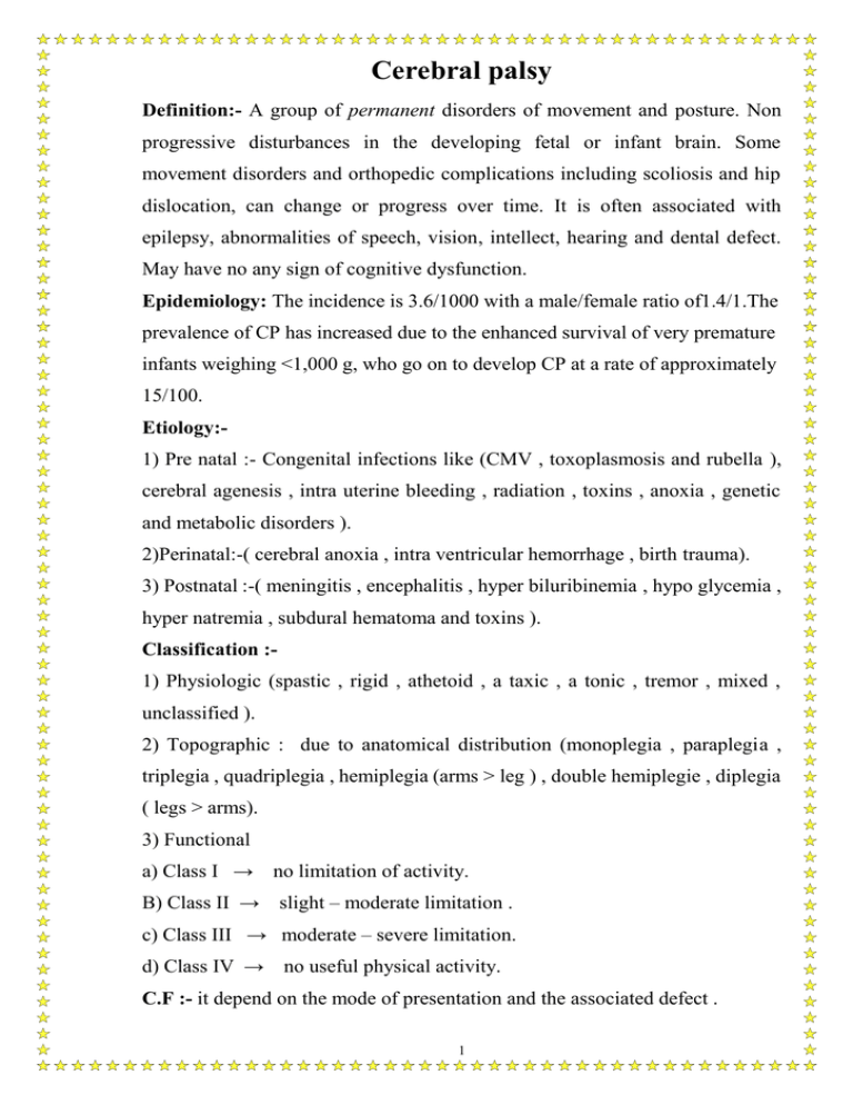
Cerebral palsy Definition:- A group of permanent disorders of movement and posture. Non progressive disturbances in the developing fetal or infant brain. Some movement disorders and orthopedic complications including scoliosis and hip dislocation, can change or progress over time. It is often associated with epilepsy, abnormalities of speech, vision, intellect, hearing and dental defect. May have no any sign of cognitive dysfunction. Epidemiology: The incidence is 3.6/1000 with a male/female ratio of1.4/1.The prevalence of CP has increased due to the enhanced survival of very premature infants weighing <1,000 g, who go on to develop CP at a rate of approximately 15/100. Etiology:1) Pre natal :- Congenital infections like (CMV , toxoplasmosis and rubella ), cerebral agenesis , intra uterine bleeding , radiation , toxins , anoxia , genetic and metabolic disorders ). 2)Perinatal:-( cerebral anoxia , intra ventricular hemorrhage , birth trauma). 3) Postnatal :-( meningitis , encephalitis , hyper biluribinemia , hypo glycemia , hyper natremia , subdural hematoma and toxins ). Classification :1) Physiologic (spastic , rigid , athetoid , a taxic , a tonic , tremor , mixed , unclassified ). 2) Topographic : due to anatomical distribution (monoplegia , paraplegia , triplegia , quadriplegia , hemiplegia (arms > leg ) , double hemiplegie , diplegia ( legs > arms). 3) Functional a) Class I → no limitation of activity. B) Class II → slight – moderate limitation . c) Class III → moderate – severe limitation. d) Class IV → no useful physical activity. C.F :- it depend on the mode of presentation and the associated defect . 1 Spastic hemiplegia (25%) :- Decreased spontaneous movement on the effected side and show hand preference at a very early age, delayed walking until ( 18 – 24 )m , circumductional gait, dystonia on running, growth arrest of affected side. Equina varus, walking on tip to, ankle clonus, increase DTR, babinski sign. About one third of patients have a seizure disorder that usually develops in first year or two, 25% have cognitive abnormalities including MR. A CT scan or MRI study may show an atrophic cerebral hemisphere with a dilated lateral ventricle contra lateral to the side of affected extremities Spastic diplegia (35%):- Commando crawl, difficult application of diaper because of excessive adduction of the hips, scissoring posture of lower extremities, it is the commonest type and it is always congenital. Seizure in minority of cases, the prognosis for normal intellectual development is excellent. Spastic quadriplegia (20%) :- Most severe type, high association with seizure and MR, bulbar palsy leading to swallowing difficulties and aspiration pneumonia. Athetoid (choreoathetoid, extrapyramidal, or dyskinetic ) (15%):Relatively rare .Mostly caused by birth asphyxia, and can also caused by kernicterus. Characterized by hypotonia, feeding difficulties, athetoid movement may not become evident until one year of age, speech typically affected, UMN signs aren’t present. Seizure is uncommon and intellect is preserved in most patients. D.D:- Myasthenia gravis, chorea, ataxia telengectasia, hypothyroidism, Down`s syndrome, degenerative diseases, spinal cord tumors, muscles dystrophy. Diagnosis: - History, physical exam, EEG, CT scan, MRI, tests for hearing and visual functions, Genetic evaluation in patients with congenital malformations or evidence of metabolic disorder. Complication: - Acquired dislocation of the hip, scoliosis, epilepsy, speech defect due to high tone deafness or associated with MR, eye problems ( squint, 2 nystagmus, blindness ), dental problems ( tooth grinding, malocclusion, gingivitis ), hearing loss and MR. Treatment :- In the ( C.P. clinic ) in which a team of physicians from different specialty as well as occupational and physiotherapist, speech pathologist, social worker, educational, developmental psychologist.The aim of the treatment is to make use of patient abilities as effectively as possible by : 1) Physiotherapy. 2) Occupational therapy. 3) Education of the parents of how to handle their child in daily activities. 4) Use of adaptive equipment (motorized wheel chair, special feeding device). 5) Surgery: for CDH. ( adductor tenotomy and psoas transfer and release), rhizotomy procedure ( division roots of spinal nerve) which used in severe spastic diplegia, tenotomy for Achilles tendon. 6) Care for lower urinary tract dysfunctions. 7) Treatment of severe spasticity by: oral diazepam, oral baclofen, dantrolene, intrathecal baclofen, and botulinum toxin injected into specific muscle groups. 8) Botulinum toxin could be injected into salivary glands to reduce the severity of drooling that is seen in 10-30% of patients with CP. 9) Reserpine or tetrabenzine can be useful for hyperkinetic movement disorders including athetosis or chorea. 10)Treatment of dystonia by small doses of levodopa . Artane and Carbamazapine is sometimes useful 11) Hearing aids. 12) Special education. 13) Treatment of seizures by anticonvulsant. 14) Enhancement of communication skills. 3 Prevention :1) Good pre natal care. 2) Prevention of kernicterus. 3) Care for LBW infants. 4) Treatment of apneic episodes. 5) Prenatal treatment of the mothers with magnesium Prognosis:- depend on : 1) Severity of the condition. 2) Associated intellectual defect. 3) Adequate education and available facilities. 4) Centers that treats these conditions. 5) Type of C.P. Ataxias Ataxia is the inability to make smooth, accurate , and coordinated movements, usually due to a disorder of the cerebellum and/ or sensory pathways in the posterior columns of the spinal cord. Ataxias may be generalized or primarily affect gait or the hands and arms. They may be acute or chronic. Causes of acute or recurrent ataxia 1- Brain tumors. 2- Drugs : piperazine, phenytoin, alcohol. 3- Encephalitis (brain stem). 4- Migraine: Basilar, and Benign paroxysmal vertigo. 5- Post infectious/ immune: a- Acute cerebellar ataxia: occurs primarily in children between 1-3 years of age and is a diagnosis by exclusion. It often follows a viral infection like ( varicella, coxsackie virus, or echo virus) infection by 2-3 weeks. b- Cerebellar abscess. c- Acute labyrinthitis. 6- Trauma. 4 7- Vascular disorders: Cerebellar hemorrhage, and Kawasaki disease. 8- Pseudo ataxia ( Epileptic). 9- Genetic disorders: ( Maple syrup disease, and Pyruvate dehydrogenase deficiency). Causes of Chronic or Progressive Ataxia 1- Brain tumors: Cerebellar tumors, Medulloblastoma, Ependymoma, Supra tentorial tumors, Neuroblasoma. 2- Congenital malformation: Dandy-Walker malformation, Chiare malformation. 3- Hereditary ataxia: Abetalipoproteinemia, Ataxia- telangectasia, Friedreich ataxia. Causes of floppy baby syndrome 1) Brain→ hypotonic C.P., Brain stem stroke, Brain stem encephalitis. 2) Spinal cord→ werdnig – hoffman disease, (hypotonia, a reflexia, tongue fasciculation and repeated chest infection). 3) Peripheral nerve: Guillian – Barre disease, lead poisoning. 4) Neuromuscular junction: myasthenia gravis. 5) Muscles→ myopathy. 5
