Staphylococcus .aureus dentoalveolar infections AL- Amidi , B.H.H
advertisement
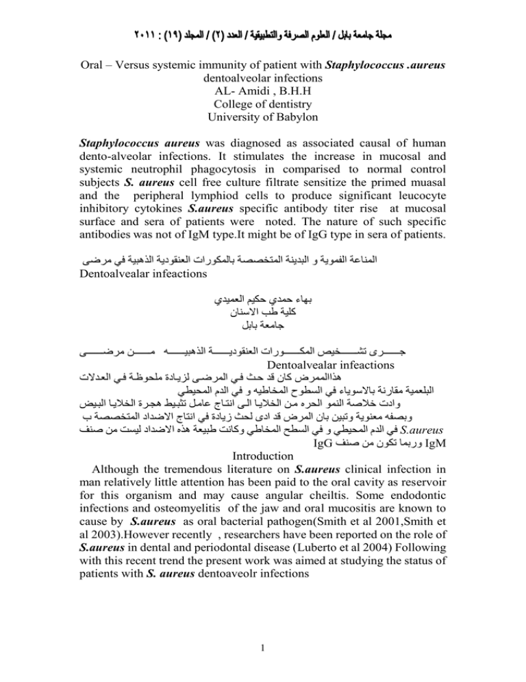
Oral – Versus systemic immunity of patient with Staphylococcus .aureus dentoalveolar infections AL- Amidi , B.H.H College of dentistry University of Babylon Staphylococcus aureus was diagnosed as associated causal of human dento-alveolar infections. It stimulates the increase in mucosal and systemic neutrophil phagocytosis in comparised to normal control subjects S. aureus cell free culture filtrate sensitize the primed muasal and the peripheral lymphiod cells to produce significant leucocyte inhibitory cytokines S.aureus specific antibody titer rise at mucosal surface and sera of patients were noted. The nature of such specific antibodies was not of IgM type.It might be of IgG type in sera of patients. المناعة الفموية و البدينة المتخصصة بالمكورات العنقودية الذهبية في مرضى Dentoalvealar infeactions بهاء حمدي حكيم العميدي كلية طب االسنان جامعة بابل مررررررررررر مرضرررررررررررى جرررررررررررم وررررررررررررخيا المكرررررررررررورات العنقوديررررررررررة الذهبيررررررررررر Dentoalvealar infeactions هذاالممرض كان قد حر فري المرضرى لديراد ملحوظرة فري العردالت البلعمية مقارنة باالسوياء في السطوح المخاطي و في الدم المحيطي وادت خالصة النمو الحره مر الخاليرا الرى انترام عامرل وهبري هخرر الخاليرا البري وبصف معنوية ووبي بان المرض قد ادم لح زياد في انتام االضداد المتخصصة ب في الدم المحيطي و في السطح المخاطي وكانت طبيعة هذه االضداد ليست م صنفS.aureus IgG وربما وكون م صنفIgM Introduction Although the tremendous literature on S.aureus clinical infection in man relatively little attention has been paid to the oral cavity as reservoir for this organism and may cause angular cheiltis. Some endodontic infections and osteomyelitis of the jaw and oral mucositis are known to cause by S.aureus as oral bacterial pathogen(Smith et al 2001,Smith et al 2003).However recently , researchers have been reported on the role of S.aureus in dental and periodontal disease (Luberto et al 2004) Following with this recent trend the present work was aimed at studying the status of patients with S. aureus dentoaveolr infections 1 Materials and Methods 1. Patients: three groups of disease entities included in the dentoalveolar infection were clinically diagnosed as peridontitis 7gingivitis 5 and chronic pulpitis 3 (smraranayke et al 2002). Ten apparently normal human subjects were included as a control. 2. sample processing: The dentoaloveolar material of 15 patients was swabbed by sterile cotton swabs into which three mls of sterile normal saline was added. Through mixing was dons for these samples 3. Bacteriology : Loopful inocula were taken from the swab saline mixtures and quadrate streaked onto blood agar and nutrient agar plates, then incubated at 37C for an overnight period under aerobic conditions. Biochemical isolates as Maffidin 2000. 4. Immunoglobulin separation: the through mixed swab saline becomes slightly opaque due to protein & cellular contents ( Item three) the suspension was centrifuged at 3000 R.P.M for five minutes. Supernats were aspirate into sterile clean centrifuged tube which 3 ml of PEG6000 6% solution was added in refrigerator at 4C for 1hr (Item two) precipitation was scattered at bottom centrifuged at5000 R.P.M for 15 min. discard supernate and keep precipitation redissolution into 0.5% formal saline (Johnaston and Thorap 1982) 5. mucosal leucocytes separation : the deposite of primery swab-saline suspension (item three) was washed once with saline and resuspended in 3 ml saline. To this washed cell suspension three ml of 3% dextrane was added the ◦ mixture left at 250 c (room temp) for 20 min. dextrian – leucocyte apper layer was aspirated tubbed into plane tube ,centrifuged at 3000 R.P.M for 15 min. The supernate was discarded,pellet saved and resuspended into saline and washed twice , the resuspended into original volume in sterile saline 6. blood : blood with and without anticoagulant in three ml . amounts were collected from 15 patients and 10 normal subjects . 7. standard tube agglutination test (STAT): The STAT was done as in Garvey et al 1977. 8. N.B.T test : N.B.T on mucosal and peripheral blood leucocytes was done as in Park et al 1968 9. LIF test: 2 LIF on mucosal and systemic was perfomed by capillary method as in Soberg, 1968 Result 1- The infectous Agents The primary plate culture of the patients dentoalveolar materials anto blood agar showed colonies of staphylococcal like morphotypes with golden endo pigmentation. The pure isolates were gram positive cocci in clusters, catalase and coagulase positive. Salt tolerant and phenol agar. These criteria are consistent with S.aureus 2- Dentoalvelar Infections: The s.aureus was found associated with seven cases of peridontitits five cases of gingivitis and three cases of chronic pulpitis 3- Neutrophil phagocytosis : The phagocytic activity as tested by N.B.T were of higher percentages both for mucosal and in peripheral neutrophils in comparison to normal control subjects (Table 1-3) 4- Leuconte inhibitory factors: Significant leucocytes inhibition noted both for mucosal and peripheral leucocytes indicating that the sensitizar stimulate primed leucocyte for production of LIF cytokine by patient in comparison to non significant LIF in control subjects (Tables 1-3) 5- Mucosal antibody : The range of mean values of mucosal antibody titers specific to S.aureus 14.6 – 42.7 as compared to 2 in normal control subjects mucosal antibodies were 2ME resistant (table 1-3) 6- Serum antibodies : The range of mean values of serum antibody titers specific for S.aureus in patients were 146-427 as compared to 10 in normal control subjects. Serum antibodies were 2ME resistant (Tables 1-3) 3 Table 1: Immune status of S.aureus associated peridontits (7cases) Mean Medan Range MIGC 0.75 gm/L 0.28 0.6- 0.82 N.S means 0.299 STPC 71.3 gm/L 73.5 68 – 73.5 66.43 SGC 39.4 gm/L 38.7 36- 48.2 36.22 2ME – 14.6 16 8- 32 2 2ME + SIGT 14.6 16 8- 32 2 2ME – 146 320 80- 320 10 2ME + LIF 146 320 80- 320 10 M 54.4 56 48 – 58 0.94 S NBT 62.3 62 52- 68 0.95 M 39.6 % 42 33- 448 % 9.8 S 34.3% 34 32- 39 % 10.8 MIGT * N.S= Normal subject MIGC=Mucosal lmmunoglobulin concentration STPC =Serum total protein concentration SGC=Serum globulin concentration MIGT= Mucosal lmmunoglobulin titer SIGT =Serum lmmunoglobulin titer LIF = Lecocytes lnhibitory Factor. M = Mucosal S= Systemic N.B.T=Nitroblue tetrazolium reduction Test . 4 Table 2: Immune status of S.aureus associated gingivitis (five cases) Mean Medan Range MIGC 0.75 0.73 0.64- 0.85 N.S means 0.299 STPC 67.2 69.5 39.3– 80.3 66.43 SGC 42.1 36.2 36- 48.2 36.22 2ME – 30.4 16 8- 32 2 2ME + SIGT 30.4 16 8- 32 2 2ME – 304 160 80- 320 10 2ME + LIF 304 160 80- 320 10 M 49.4 53 42 – 51 0.96 S NBT 55.6 62 36 - 67 0.95 M 42 % 45 35- 45 9.8 S 37.2% 37 34- 44 10.8 MIGT * N.S= Normal subject MIGC=Mucosal lmmunoglobulin concentration STPC =Serum total protein concentration SGC=Serum globulin concentration MIGT= Mucosal lmmunoglobulin titer SIGT =Serum lmmunoglobulin titer LIF = Lecocytes lnhibitory Factor. M = Mucosal S= Systemic N.B.T=Nitroblue tetrazolium reduction Test . 5 Table 3: Immune stats of S.aureus associated chronic pulpitise (three cases) Mean Medan Range MIGC 0.99 0.78 0.78- 1.4 N.S means 0.299 STPC 75.3 78.1 70.2 – 82.4 66.43 SGC 43.7 40.2 40.2- 46.3 36.22 2ME – 42.7 32 8- 32 2 2ME + SIGT 42.7 32 32- 64 2 2ME – 427 320 320 - 640 10 2ME + LIF 427 320 320 – 640 10 M 57.3 55 55 – 57 0.96 S NBT 64.3 63 63 - 66 0.95 M 45 % 41 41 - 48 9.8 S 37.7 % 36 36 - 39 10.8 MIGT * N.S= Normal subject MIGC=Mucosal lmmunoglobulin concentration STPC =Serum total protein concentration SGC=Serum globulin concentration MIGT= Mucosal lmmunoglobulin titer SIGT =Serum lmmunoglobulin titer LIF = Lecocytes lnhibitory Factor. M = Mucosal S= Systemic N.B.T=Nitroblue tetrazolium reduction Test . 6 Discussion Staphylococcus aureus is diagnosed (table 1-3) in association with dento-alveolar infections (Smith et al 2001; Samaranyake et al 2002; Loberto et al 2004). The S. aureus antibodies specific rise in patients sera and dentoaveolar protein can be induced by B cell dependent and / or Th2 dependent epitope(s) (Zubler, 1998; Delves et al 2006). The LIF significant result can be induced by Th1 or T dth dependent epitopes (Male et al, 2006). There might be an epitope in the antigenic make up of S. aureus that activate nutrophile phagocytosis N.B.T in patients Tolllike receptors and specific pattern recognition structures perhaps involved in such increase (Delves et al 2006; Male et al 2006) The 2ME resistant mucosal antibody means that such antibody is of secretary type (Tomasi,1976 ) while 2ME resistant serum antibodies can be of non IgM type possibly of IgG type(Male et al 2006). References 1- Delves, P.J.; Martin, S.J; Burton, D.R and Roitt, I. 2006. Roitts Essential Immunolaye . 11th ed. Blackwell scientific publishing. U.K. 2- Fritschi, Z. Albert- Kiszely and Persson, G.R 2008. staphylococcus and other bacteria in untreated peridontitis. J. Dent. Res. 87(6): 589593. 3- Gravey J.S.; Cremer, N.E and Sussdorf, DH 1977. Methods In Immunology 3rd .ed. W.A. Benjamin , INC, London. 4- Johnstone , A and Thorpe, R. 1982 Immunochemistry in practice. Blackwell scientific publications 5- Luberto, J.C.S.; Paiva Martins, C.A.D.; Dos santos, S.S.F.; Cortelli, J.R. and Jorge A.O.C, 2004. staphylococcus spp. In the oral cavity and periodontal pokts of chromic perdontitis patients. Braz . J. Microbial 35: 46- 68 6- Macfaddin , J.F. 2000. Biochemical Identification of Medical Bacteria 2 nd .ed. Williams and Wilkins company co. 7- Male, D.; Brostoff, I.; Roth, D.B. and Roitt, I. 2006. immunology 7 ed. ed. mosby, U.K. 8- Samaranayake L.P.; Jones, B.M. And scully, C,2002. Essential Microbiology for Dentistry. 2nd .ed Churchill- Livingstone, London. 9- Smith, A.J.; Jackson, M.S and Bagg, J. 2001. the Ecology of Staphylocaus species in the oral cavity. J. Med . Microbial 50: 940946. 7 10- Smith A.J.; Robertson D.; Tang, MK.; Jackson, M.S.; Mackenzie, D and Bagg, J., 2003. Staphylococcus aureus in the oral cavity. Brit. Dent. J. 195(12) : 701-703. 11- Soberg,M.Act.Med.Scand.1968.184:235. 12- Tomasi, T. 1976. the immune system of secretions. Prinitice- Hall I.N.C, USA. 8
